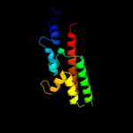
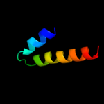
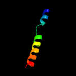
1 c2ra8A_
99.7
45
PDB header: structural genomics, unknown functionChain: A: PDB Molecule: uncharacterized protein q64v53_bacfr;PDBTitle: crystal structure of the q64v53_bacfr protein from bacteroides2 fragilis. northeast structural genomics consortium target bfr43
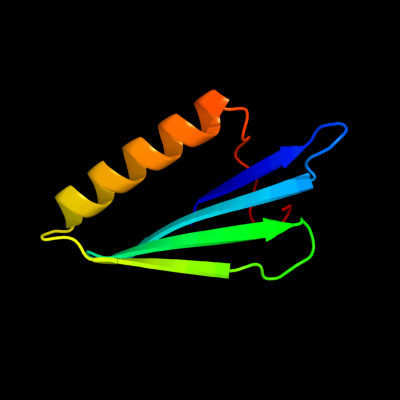
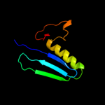
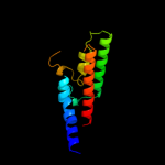
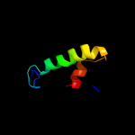
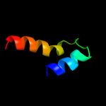
2 c2eocA_
99.6
25
PDB header: transferaseChain: A: PDB Molecule: poly [adp-ribose] polymerase 3;PDBTitle: solution structure of the wgr domain from human poly [adp-2 ribose] polymerase-3
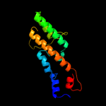
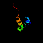
3 d2cr9a1
99.4
17
Fold: WGR domain-likeSuperfamily: WGR domain-likeFamily: WGR domain4 d1efya1
99.4
13
Fold: Domain of poly(ADP-ribose) polymeraseSuperfamily: Domain of poly(ADP-ribose) polymeraseFamily: Domain of poly(ADP-ribose) polymerase5 d2rd6a1
99.3
15
Fold: Domain of poly(ADP-ribose) polymeraseSuperfamily: Domain of poly(ADP-ribose) polymeraseFamily: Domain of poly(ADP-ribose) polymerase6 d1gs0a1
99.2
12
Fold: Domain of poly(ADP-ribose) polymeraseSuperfamily: Domain of poly(ADP-ribose) polymeraseFamily: Domain of poly(ADP-ribose) polymerase7 c3c4hA_
99.1
14
PDB header: transferaseChain: A: PDB Molecule: poly(adp-ribose) polymerase 3;PDBTitle: human poly(adp-ribose) polymerase 3, catalytic fragment in complex2 with an inhibitor dr2313
8 c1gs0B_
99.0
13
PDB header: transferaseChain: B: PDB Molecule: poly (adp-ribose) polymerase-2;PDBTitle: crystal structure of the catalytic fragment of murine poly2 (adp-ribose) polymerase-2
9 c2paxA_
99.0
15
PDB header: transferaseChain: A: PDB Molecule: poly(adp-ribose) polymerase;PDBTitle: the catalytic fragment of poly(adp-ribose) polymerase2 complexed with 4-amino-1,8-naphthalimide
10 c2l9vA_
22.1
11
PDB header: rna binding proteinChain: A: PDB Molecule: pre-mrna-processing factor 40 homolog a;PDBTitle: nmr structure of the ff domain l24a mutant's folding transition state
11 c1nohB_
19.4
18
PDB header: viral proteinChain: B: PDB Molecule: head morphogenesis protein;PDBTitle: the structure of bacteriophage phi29 scaffolding protein2 gp7 after prohead assembly
12 c1zp9A_
14.0
16
PDB header: transferaseChain: A: PDB Molecule: rio1 kinase;PDBTitle: crystal structure of full-legnth a.fulgidus rio1 serine kinase bound2 to atp and mn2+ ions.
13 c1oxzA_
13.7
10
PDB header: membrane proteinChain: A: PDB Molecule: adp-ribosylation factor binding protein gga1;PDBTitle: crystal structure of the human gga1 gat domain
14 d1oxza_
13.7
10
Fold: Spectrin repeat-likeSuperfamily: GAT-like domainFamily: GAT domain15 c1nafA_
12.7
10
PDB header: signaling protein, membrane proteinChain: A: PDB Molecule: adp-ribosylation factor binding protein gga1;PDBTitle: crystal structure of the human gga1 gat domain
16 c3mtuE_
11.5
27
PDB header: contractile proteinChain: E: PDB Molecule: head morphogenesis protein, tropomyosin alpha-1 chain;PDBTitle: structure of the tropomyosin overlap complex from chicken smooth2 muscle
17 d1vdla_
11.2
15
Fold: RuvA C-terminal domain-likeSuperfamily: UBA-likeFamily: UBA domain18 c3bsuF_
10.3
14
PDB header: hydrolase/rna/dnaChain: F: PDB Molecule: ribonuclease h1;PDBTitle: hybrid-binding domain of human rnase h1 in complex with 12-2 mer rna/dna
19 d1dp7p_
10.0
22
Fold: DNA/RNA-binding 3-helical bundleSuperfamily: "Winged helix" DNA-binding domainFamily: P4 origin-binding domain-like20 d1vbka2
9.0
24
Fold: THUMP domainSuperfamily: THUMP domain-likeFamily: THUMP domain21 c2zfhA_
not modelled
8.9
15
PDB header: structural genomics, unknown functionChain: A: PDB Molecule: cuta;PDBTitle: crystal structure of putative cuta1 from homo sapiens at 2.05a2 resolution
22 d1kr4a_
not modelled
8.9
35
Fold: Ferredoxin-likeSuperfamily: GlnB-likeFamily: Divalent ion tolerance proteins CutA (CutA1)23 c3godA_
not modelled
8.7
12
PDB header: immune systemChain: A: PDB Molecule: cas1;PDBTitle: structural basis for dnase activity of a conserved protein2 implicated in crispr-mediated antiviral defense
24 d1p1la_
not modelled
8.5
20
Fold: Ferredoxin-likeSuperfamily: GlnB-likeFamily: Divalent ion tolerance proteins CutA (CutA1)25 d1uzca_
not modelled
8.2
14
Fold: Another 3-helical bundleSuperfamily: FF domainFamily: FF domain26 d1naqa_
not modelled
8.1
25
Fold: Ferredoxin-likeSuperfamily: GlnB-likeFamily: Divalent ion tolerance proteins CutA (CutA1)27 d1qkia1
not modelled
8.1
28
Fold: NAD(P)-binding Rossmann-fold domainsSuperfamily: NAD(P)-binding Rossmann-fold domainsFamily: Glyceraldehyde-3-phosphate dehydrogenase-like, N-terminal domain28 d1ugja_
not modelled
7.9
21
Fold: PRC-barrel domainSuperfamily: PRC-barrel domainFamily: RIKEN cDNA 2310057j16 protein (KIAA1543)29 d2f76x1
not modelled
7.5
12
Fold: Retroviral matrix proteinsSuperfamily: Retroviral matrix proteinsFamily: Mason-Pfizer monkey virus matrix protein30 c1xk8A_
not modelled
7.5
15
PDB header: structural genomics, unknown functionChain: A: PDB Molecule: divalent cation tolerant protein cuta;PDBTitle: divalent cation tolerant protein cuta from homo sapiens2 o60888
31 d2zfha1
not modelled
7.5
15
Fold: Ferredoxin-likeSuperfamily: GlnB-likeFamily: Divalent ion tolerance proteins CutA (CutA1)32 c3c4mC_
not modelled
7.5
40
PDB header: membrane proteinChain: C: PDB Molecule: parathyroid hormone;PDBTitle: structure of human parathyroid hormone in complex with the2 extracellular domain of its g-protein-coupled receptor (pth1r)
33 c3c4mD_
not modelled
7.5
40
PDB header: membrane proteinChain: D: PDB Molecule: parathyroid hormone;PDBTitle: structure of human parathyroid hormone in complex with the2 extracellular domain of its g-protein-coupled receptor (pth1r)
34 c1zwgA_
not modelled
7.0
40
PDB header: hormoneChain: A: PDB Molecule: parathyroid hormone;PDBTitle: succinyl human parathyroid hormone 4-37, nmr, 10 structures
35 c2zomC_
not modelled
7.0
25
PDB header: unknown functionChain: C: PDB Molecule: protein cuta, chloroplast, putative, expressed;PDBTitle: crystal structure of cuta1 from oryza sativa
36 d1ukua_
not modelled
6.9
20
Fold: Ferredoxin-likeSuperfamily: GlnB-likeFamily: Divalent ion tolerance proteins CutA (CutA1)37 c1vbkA_
not modelled
6.9
24
PDB header: structural genomics, unknown functionChain: A: PDB Molecule: hypothetical protein ph1313;PDBTitle: crystal structure of ph1313 from pyrococcus horikoshii ot3
38 d1osce_
not modelled
6.8
15
Fold: Ferredoxin-likeSuperfamily: GlnB-likeFamily: Divalent ion tolerance proteins CutA (CutA1)39 c2nuhA_
not modelled
6.8
5
PDB header: unknown functionChain: A: PDB Molecule: periplasmic divalent cation tolerance protein;PDBTitle: crystal structure of cuta from the phytopathgen bacterium xylella2 fastidiosa
40 c2bnlE_
not modelled
6.5
18
PDB header: stress-responseChain: E: PDB Molecule: modulator protein rsbr;PDBTitle: the structure of the n-terminal domain of rsbr
41 c3ejbC_
not modelled
6.4
23
PDB header: oxidoreductase/lipid transportChain: C: PDB Molecule: acyl carrier protein;PDBTitle: crystal structure of p450bioi in complex with tetradecanoic2 acid ligated acyl carrier protein
42 c3do6B_
not modelled
6.4
32
PDB header: ligaseChain: B: PDB Molecule: formate--tetrahydrofolate ligase;PDBTitle: crystal structure of putative formyltetrahydrofolate2 synthetase (tm1766) from thermotoga maritima at 1.85 a3 resolution
43 d1vhfa_
not modelled
6.3
35
Fold: Ferredoxin-likeSuperfamily: GlnB-likeFamily: Divalent ion tolerance proteins CutA (CutA1)44 d1nzaa_
not modelled
6.3
20
Fold: Ferredoxin-likeSuperfamily: GlnB-likeFamily: Divalent ion tolerance proteins CutA (CutA1)45 c3ahpA_
not modelled
6.2
20
PDB header: electron transportChain: A: PDB Molecule: cuta1;PDBTitle: crystal structure of stable protein, cuta1, from a psychrotrophic2 bacterium shewanella sp. sib1
46 c2r0qF_
not modelled
5.7
28
PDB header: recombination/dnaChain: F: PDB Molecule: putative transposon tn552 dna-invertase bin3;PDBTitle: crystal structure of a serine recombinase- dna regulatory2 complex
47 d1eg7a_
not modelled
5.6
32
Fold: P-loop containing nucleoside triphosphate hydrolasesSuperfamily: P-loop containing nucleoside triphosphate hydrolasesFamily: Nitrogenase iron protein-like48 c2diuA_
not modelled
5.4
26
PDB header: rna binding proteinChain: A: PDB Molecule: kiaa0430 protein;PDBTitle: solution structure of the rrm domain of kiaa0430 protein
49 c2xskA_
not modelled
5.3
55
PDB header: chaperoneChain: A: PDB Molecule: csgc;PDBTitle: e. coli curli protein csgc - secys