| 1 |
|
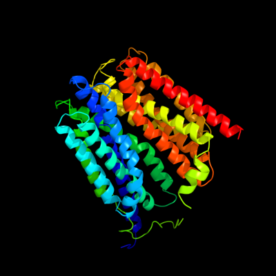
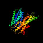
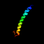
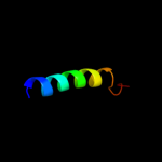
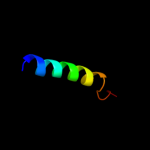
PDB 1pw4 chain A
Region: 3 - 509
Aligned: 408
Modelled: 409
Confidence: 100.0%
Identity: 13%
Fold: MFS general substrate transporter
Superfamily: MFS general substrate transporter
Family: Glycerol-3-phosphate transporter
Phyre2
| 2 |
|
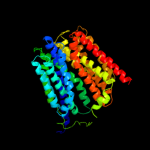
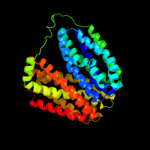
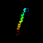
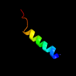
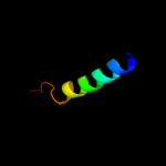
PDB 2gfp chain A
Region: 19 - 431
Aligned: 356
Modelled: 358
Confidence: 100.0%
Identity: 16%
PDB header:membrane protein
Chain: A: PDB Molecule:multidrug resistance protein d;
PDBTitle: structure of the multidrug transporter emrd from2 escherichia coli
Phyre2
| 3 |
|
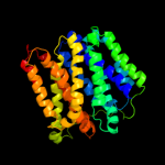
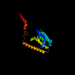
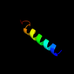
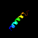
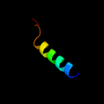
PDB 3o7p chain A
Region: 15 - 504
Aligned: 406
Modelled: 416
Confidence: 100.0%
Identity: 9%
PDB header:transport protein
Chain: A: PDB Molecule:l-fucose-proton symporter;
PDBTitle: crystal structure of the e.coli fucose:proton symporter, fucp (n162a)
Phyre2
| 4 |
|
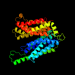
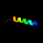
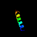
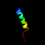
PDB 2xut chain C
Region: 21 - 498
Aligned: 435
Modelled: 435
Confidence: 100.0%
Identity: 14%
PDB header:transport protein
Chain: C: PDB Molecule:proton/peptide symporter family protein;
PDBTitle: crystal structure of a proton dependent oligopeptide (pot)2 family transporter.
Phyre2
| 5 |
|
PDB 1pv7 chain A
Region: 16 - 508
Aligned: 399
Modelled: 403
Confidence: 100.0%
Identity: 12%
Fold: MFS general substrate transporter
Superfamily: MFS general substrate transporter
Family: LacY-like proton/sugar symporter
Phyre2
| 6 |
|
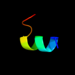
PDB 2g9p chain A
Region: 72 - 85
Aligned: 14
Modelled: 14
Confidence: 17.9%
Identity: 21%
PDB header:antimicrobial protein
Chain: A: PDB Molecule:antimicrobial peptide latarcin 2a;
PDBTitle: nmr structure of a novel antimicrobial peptide, latarcin 2a,2 from spider (lachesana tarabaevi) venom
Phyre2
| 7 |
|
PDB 3b9y chain A
Region: 20 - 243
Aligned: 216
Modelled: 224
Confidence: 10.0%
Identity: 11%
PDB header:transport protein
Chain: A: PDB Molecule:ammonium transporter family rh-like protein;
PDBTitle: crystal structure of the nitrosomonas europaea rh protein
Phyre2
| 8 |
|
PDB 1wrg chain A
Region: 464 - 510
Aligned: 46
Modelled: 47
Confidence: 7.9%
Identity: 15%
PDB header:membrane protein
Chain: A: PDB Molecule:light-harvesting protein b-880, beta chain;
PDBTitle: light-harvesting complex 1 beta subunit from wild-type2 rhodospirillum rubrum
Phyre2
| 9 |
|
PDB 1fft chain B domain 2
Region: 463 - 511
Aligned: 49
Modelled: 49
Confidence: 7.6%
Identity: 14%
Fold: Transmembrane helix hairpin
Superfamily: Cytochrome c oxidase subunit II-like, transmembrane region
Family: Cytochrome c oxidase subunit II-like, transmembrane region
Phyre2
| 10 |
|
PDB 3kzi chain T
Region: 484 - 507
Aligned: 24
Modelled: 24
Confidence: 7.3%
Identity: 17%
PDB header:electron transport
Chain: T: PDB Molecule:photosystem ii reaction center protein t;
PDBTitle: crystal structure of monomeric form of cyanobacterial photosystem ii
Phyre2
| 11 |
|
PDB 2axt chain T domain 1
Region: 484 - 507
Aligned: 24
Modelled: 24
Confidence: 7.3%
Identity: 17%
Fold: Single transmembrane helix
Superfamily: Photosystem II reaction center protein T, PsbT
Family: PsbT-like
Phyre2
| 12 |
|
PDB 2axt chain T
Region: 484 - 507
Aligned: 24
Modelled: 24
Confidence: 7.3%
Identity: 17%
PDB header:electron transport
Chain: T: PDB Molecule:photosystem ii reaction center t protein;
PDBTitle: crystal structure of photosystem ii from thermosynechococcus elongatus
Phyre2
| 13 |
|
PDB 3a0b chain T
Region: 484 - 507
Aligned: 24
Modelled: 24
Confidence: 7.3%
Identity: 17%
PDB header:electron transport
Chain: T: PDB Molecule:photosystem ii reaction center protein t;
PDBTitle: crystal structure of br-substituted photosystem ii complex
Phyre2
| 14 |
|
PDB 3a0h chain T
Region: 484 - 507
Aligned: 24
Modelled: 24
Confidence: 7.3%
Identity: 17%
PDB header:electron transport
Chain: T: PDB Molecule:photosystem ii reaction center protein t;
PDBTitle: crystal structure of i-substituted photosystem ii complex
Phyre2
| 15 |
|
PDB 3arc chain T
Region: 484 - 507
Aligned: 24
Modelled: 24
Confidence: 7.3%
Identity: 17%
PDB header:electron transport, photosynthesis
Chain: T: PDB Molecule:photosystem ii reaction center protein t;
PDBTitle: crystal structure of oxygen-evolving photosystem ii at 1.9 angstrom2 resolution
Phyre2
| 16 |
|
PDB 2axt chain T
Region: 484 - 507
Aligned: 24
Modelled: 24
Confidence: 7.3%
Identity: 17%
PDB header:electron transport
Chain: T: PDB Molecule:photosystem ii reaction center t protein;
PDBTitle: crystal structure of photosystem ii from thermosynechococcus elongatus
Phyre2
| 17 |
|
PDB 3a0h chain T
Region: 484 - 507
Aligned: 24
Modelled: 24
Confidence: 7.3%
Identity: 17%
PDB header:electron transport
Chain: T: PDB Molecule:photosystem ii reaction center protein t;
PDBTitle: crystal structure of i-substituted photosystem ii complex
Phyre2
| 18 |
|
PDB 1s5l chain T
Region: 484 - 507
Aligned: 24
Modelled: 24
Confidence: 7.3%
Identity: 17%
PDB header:photosynthesis
Chain: T: PDB Molecule:photosystem ii psbt protein;
PDBTitle: architecture of the photosynthetic oxygen evolving center
Phyre2
| 19 |
|
PDB 1s5l chain T
Region: 484 - 507
Aligned: 24
Modelled: 24
Confidence: 7.3%
Identity: 17%
PDB header:photosynthesis
Chain: T: PDB Molecule:photosystem ii psbt protein;
PDBTitle: architecture of the photosynthetic oxygen evolving center
Phyre2
| 20 |
|
PDB 3bz1 chain T
Region: 484 - 507
Aligned: 24
Modelled: 24
Confidence: 7.2%
Identity: 17%
PDB header:electron transport
Chain: T: PDB Molecule:photosystem ii reaction center protein t;
PDBTitle: crystal structure of cyanobacterial photosystem ii (part 12 of 2). this file contains first monomer of psii dimer
Phyre2
| 21 |
|
| 22 |
|
| 23 |
|