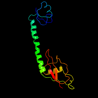
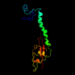
1 c2qbcH_
100.0
100
PDB header: ribosomeChain: H: PDB Molecule: 50s ribosomal protein l9;PDBTitle: crystal structure of the bacterial ribosome from2 escherichia coli in complex with gentamicin. this file3 contains the 50s subunit of the second 70s ribosome, with4 gentamicin bound. the entire crystal structure contains5 two 70s ribosomes and is described in remark 400.
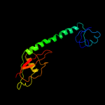
2 c2b66I_
100.0
40
PDB header: ribosomeChain: I: PDB Molecule: 50s ribosomal protein l9;PDBTitle: 50s ribosomal subunit from a crystal structure of release factor rf1,2 trnas and mrna bound to the ribosome. this file contains the 50s3 subunit from a crystal structure of release factor rf1, trnas and4 mrna bound to the ribosome and is described in remark 400
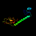
3 c2v49I_
100.0
41
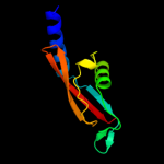
PDB header: ribosomeChain: I: PDB Molecule: 50s ribosomal protein l9;PDBTitle: structure of the ribosome recycling factor bound to the2 thermus thermophilus 70s ribosome with mrna, asl-phe and3 trna-fmet (part 4 of 4). this file contains the 50s4 subunit of molecule 2.
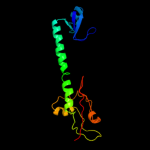
4 c3bboJ_
100.0
32
PDB header: ribosomeChain: J: PDB Molecule: ribosomal protein l9;PDBTitle: homology model for the spinach chloroplast 50s subunit2 fitted to 9.4a cryo-em map of the 70s chlororibosome
5 d2gycf1
99.9
100
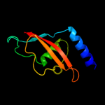
Fold: Ribosomal protein L9 C-domainSuperfamily: Ribosomal protein L9 C-domainFamily: Ribosomal protein L9 C-domain6 d1diva1
99.9
38
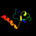
Fold: Ribosomal protein L9 C-domainSuperfamily: Ribosomal protein L9 C-domainFamily: Ribosomal protein L9 C-domain7 d2gycf2
99.8
100
Fold: MbtH/L9 domain-likeSuperfamily: L9 N-domain-likeFamily: Ribosomal protein L9 N-domain8 d2j01i2
99.8
51
Fold: MbtH/L9 domain-likeSuperfamily: L9 N-domain-likeFamily: Ribosomal protein L9 N-domain9 d2hbaa1
99.8
45
Fold: MbtH/L9 domain-likeSuperfamily: L9 N-domain-likeFamily: Ribosomal protein L9 N-domain10 d1cqua_
99.8
45
Fold: MbtH/L9 domain-likeSuperfamily: L9 N-domain-likeFamily: Ribosomal protein L9 N-domain11 c1pnyF_
99.8
55
PDB header: ribosomeChain: F: PDB Molecule: 50s ribosomal protein l9;PDBTitle: crystal structure of the wild type ribosome from e. coli,2 50s subunit of 70s ribosome. this file, 1pny, contains3 only molecules of the 50s ribosomal subunit. the 30s4 subunit is in the pdb file 1pnx.
12 c1sm1F_
99.8
55
PDB header: ribosome/antibioticChain: F: PDB Molecule: 50s ribosomal protein l9;PDBTitle: complex of the large ribosomal subunit from deinococcus radiodurans2 with quinupristin and dalfopristin
13 d2j01i1
99.8
35
Fold: Ribosomal protein L9 C-domainSuperfamily: Ribosomal protein L9 C-domainFamily: Ribosomal protein L9 C-domain14 c3rbgB_
32.8
26
PDB header: immune systemChain: B: PDB Molecule: cytotoxic and regulatory t-cell molecule;PDBTitle: crystal structure analysis of class-i mhc restricted t-cell associated2 molecule
15 c3ctuB_
31.7
30
PDB header: structural genomics, unknown functionChain: B: PDB Molecule: cbs domain protein;PDBTitle: crystal structure of cbs domain protein from streptococcus2 pneumoniae tigr4
16 c3ddjA_
30.4
22
PDB header: amp-binding proteinChain: A: PDB Molecule: cbs domain-containing protein;PDBTitle: crystal structure of a cbs domain-containing protein in complex with2 amp (sso3205) from sulfolobus solfataricus at 1.80 a resolution
17 d3ddja1
30.2
22
Fold: CBS-domain pairSuperfamily: CBS-domain pairFamily: CBS-domain pair18 d1vr9a3
28.3
26
Fold: CBS-domain pairSuperfamily: CBS-domain pairFamily: CBS-domain pair19 c3lqnA_
26.4
17
PDB header: structural genomics, unknown functionChain: A: PDB Molecule: cbs domain protein;PDBTitle: crystal structure of cbs domain-containing protein of2 unknown function from bacillus anthracis str. ames ancestor
20 d1o50a3
26.2
17
Fold: CBS-domain pairSuperfamily: CBS-domain pairFamily: CBS-domain pair21 c1vr9B_
not modelled
26.2
26
PDB header: unknown functionChain: B: PDB Molecule: cbs domain protein/act domain protein;PDBTitle: crystal structure of a cbs domain pair/act domain protein (tm0892)2 from thermotoga maritima at 1.70 a resolution
22 c3lhhA_
not modelled
25.4
30
PDB header: membrane proteinChain: A: PDB Molecule: cbs domain protein;PDBTitle: the crystal structure of cbs domain protein from shewanella2 oneidensis mr-1.
23 d2yzia1
not modelled
24.8
23
Fold: CBS-domain pairSuperfamily: CBS-domain pairFamily: CBS-domain pair24 c3gbyA_
not modelled
23.6
17
PDB header: structural genomics, unknown functionChain: A: PDB Molecule: uncharacterized protein ct1051;PDBTitle: crystal structure of a protein with unknown function ct10512 from chlorobium tepidum
25 c3kpbA_
not modelled
23.6
20
PDB header: unknown functionChain: A: PDB Molecule: uncharacterized protein mj0100;PDBTitle: crystal structure of the cbs domain pair of protein mj01002 in complex with 5 -methylthioadenosine and s-adenosyl-l-3 methionine.
26 c2emqA_
not modelled
22.5
17
PDB header: structural genomics, unknown functionChain: A: PDB Molecule: hypothetical conserved protein;PDBTitle: hypothetical conserved protein (gk1048) from geobacillus kaustophilus
27 c2k50A_
not modelled
21.7
23
PDB header: structural genomics, unknown functionChain: A: PDB Molecule: replication factor a related protein;PDBTitle: solution nmr structure of the replication factor a related2 protein from methanobacterium thermoautotrophicum.3 northeast structural genomics target tr91a.
28 d2v8qe1
not modelled
20.7
21
Fold: CBS-domain pairSuperfamily: CBS-domain pairFamily: CBS-domain pair29 c2or7A_
not modelled
20.6
17
PDB header: immune systemChain: A: PDB Molecule: t-cell immunoglobulin and mucin domain-PDBTitle: tim-2
30 d2j9la1
not modelled
20.2
21
Fold: CBS-domain pairSuperfamily: CBS-domain pairFamily: CBS-domain pair31 c3kxrA_
not modelled
20.1
26
PDB header: transport proteinChain: A: PDB Molecule: magnesium transporter, putative;PDBTitle: structure of the cystathionine beta-synthase pair domain of the2 putative mg2+ transporter so5017 from shewanella oneidensis mr-1.
32 c2p9mD_
not modelled
19.5
27
PDB header: structural genomics, unknown functionChain: D: PDB Molecule: hypothetical protein mj0922;PDBTitle: crystal structure of conserved hypothetical protein mj0922 from2 methanocaldococcus jannaschii dsm 2661
33 d2ef7a1
not modelled
19.3
41
Fold: CBS-domain pairSuperfamily: CBS-domain pairFamily: CBS-domain pair34 c3i8nB_
not modelled
19.1
20
PDB header: structural genomics, unknown functionChain: B: PDB Molecule: uncharacterized protein vp2912;PDBTitle: a domain of a conserved functionally known protein from2 vibrio parahaemolyticus rimd 2210633.
35 c3nqrD_
not modelled
19.0
20
PDB header: transport proteinChain: D: PDB Molecule: magnesium and cobalt efflux protein corc;PDBTitle: a putative cbs domain-containing protein from salmonella typhimurium2 lt2
36 d2ooxe2
not modelled
18.5
25
Fold: CBS-domain pairSuperfamily: CBS-domain pairFamily: CBS-domain pair37 c3fioB_
not modelled
17.7
28
PDB header: nucleotide binding protein, metal bindinChain: B: PDB Molecule: a cystathionine beta-synthase domain proteinPDBTitle: crystal structure of a fragment of a cystathionine beta-2 synthase domain protein fused to a zn-ribbon-like domain
38 d1yava3
not modelled
17.6
17
Fold: CBS-domain pairSuperfamily: CBS-domain pairFamily: CBS-domain pair39 d1nksa_
not modelled
17.6
14
Fold: P-loop containing nucleoside triphosphate hydrolasesSuperfamily: P-loop containing nucleoside triphosphate hydrolasesFamily: Nucleotide and nucleoside kinases40 c2v8qE_
not modelled
17.5
21
PDB header: transferaseChain: E: PDB Molecule: 5'-amp-activated protein kinase subunit gamma-1;PDBTitle: crystal structure of the regulatory fragment of mammalian2 ampk in complexes with amp
41 c3m45D_
not modelled
17.2
15
PDB header: cell adhesionChain: D: PDB Molecule: cell adhesion molecule 2;PDBTitle: crystal structure of ig1 domain of mouse syncam 2
42 c2ouxB_
not modelled
16.2
23
PDB header: transport proteinChain: B: PDB Molecule: magnesium transporter;PDBTitle: crystal structure of the soluble part of a magnesium transporter
43 c3ocmA_
not modelled
16.2
25
PDB header: membrane proteinChain: A: PDB Molecule: putative membrane protein;PDBTitle: the crystal structure of a domain from a possible membrane protein of2 bordetella parapertussis
44 c2yvxD_
not modelled
16.1
30
PDB header: transport proteinChain: D: PDB Molecule: mg2+ transporter mgte;PDBTitle: crystal structure of magnesium transporter mgte
45 c2o8kA_
not modelled
15.9
26
PDB header: transcription/dnaChain: A: PDB Molecule: rna polymerase sigma factor rpon;PDBTitle: nmr structure of the sigma-54 rpon domain bound to the-242 promoter element
46 c3lfrB_
not modelled
15.5
15
PDB header: transport proteinChain: B: PDB Molecule: putative metal ion transporter;PDBTitle: the crystal structure of a cbs domain from a putative metal2 ion transporter bound to amp from pseudomonas syringae to3 1.55a
47 c3e0eA_
not modelled
15.4
36
PDB header: replicationChain: A: PDB Molecule: replication protein a;PDBTitle: crystal structure of a domain of replication protein a from2 methanococcus maripaludis. northeast structural genomics3 targe mrr110b
48 c1yavB_
not modelled
14.6
17
PDB header: structural genomics, unknown functionChain: B: PDB Molecule: hypothetical protein bsu14130;PDBTitle: crystal structure of cbs domain-containing protein ykul2 from bacillus subtilis
49 c2p9rA_
not modelled
14.4
18
PDB header: signaling proteinChain: A: PDB Molecule: alpha-2-macroglobulin;PDBTitle: human alpha2-macroglogulin is composed of multiple domains,2 as predicted by homology with complement component c3
50 d1suxa_
not modelled
14.0
20
Fold: TIM beta/alpha-barrelSuperfamily: Triosephosphate isomerase (TIM)Family: Triosephosphate isomerase (TIM)51 c3jtfB_
not modelled
13.9
15
PDB header: transport proteinChain: B: PDB Molecule: magnesium and cobalt efflux protein;PDBTitle: the cbs domain pair structure of a magnesium and cobalt efflux protein2 from bordetella parapertussis in complex with amp
52 d1zfja4
not modelled
13.7
29
Fold: CBS-domain pairSuperfamily: CBS-domain pairFamily: CBS-domain pair53 d1jjcb1
not modelled
13.5
11
Fold: Putative DNA-binding domainSuperfamily: Putative DNA-binding domainFamily: Domains B1 and B5 of PheRS-beta, PheT54 d1k1xa1
not modelled
13.1
43
Fold: immunoglobulin/albumin-binding domain-likeSuperfamily: Families 57/38 glycoside transferase middle domainFamily: 4-alpha-glucanotransferase, domain 255 d1o7ia_
not modelled
12.9
21
Fold: OB-foldSuperfamily: Nucleic acid-binding proteinsFamily: Single strand DNA-binding domain, SSB56 c2pfiA_
not modelled
12.4
11
PDB header: transport proteinChain: A: PDB Molecule: chloride channel protein clc-ka;PDBTitle: crystal structure of the cytoplasmic domain of the human2 chloride channel clc-ka
57 c3ocoB_
not modelled
12.4
20
PDB header: structural genomics, unknown functionChain: B: PDB Molecule: hemolysin-like protein containing cbs domains;PDBTitle: the crystal structure of a hemolysin-like protein containing cbs2 domain of oenococcus oeni psu
58 d1ttja_
not modelled
12.3
25
Fold: TIM beta/alpha-barrelSuperfamily: Triosephosphate isomerase (TIM)Family: Triosephosphate isomerase (TIM)59 c3hf7A_
not modelled
12.3
20
PDB header: structural genomics, unknown functionChain: A: PDB Molecule: uncharacterized cbs-domain protein;PDBTitle: the crystal structure of a cbs-domain pair with bound amp from2 klebsiella pneumoniae to 2.75a
60 c2yvzA_
not modelled
12.2
30
PDB header: transport proteinChain: A: PDB Molecule: mg2+ transporter mgte;PDBTitle: crystal structure of magnesium transporter mgte cytosolic domain,2 mg2+-free form
61 d1nunb2
not modelled
12.1
15
Fold: Immunoglobulin-like beta-sandwichSuperfamily: ImmunoglobulinFamily: I set domains62 c1z9mA_
not modelled
11.9
26
PDB header: cell adhesionChain: A: PDB Molecule: gapa225;PDBTitle: crystal structure of nectin-like molecule-1 protein domain 1
63 d2d4za3
not modelled
11.9
25
Fold: CBS-domain pairSuperfamily: CBS-domain pairFamily: CBS-domain pair64 c1x37A_
not modelled
11.8
25
PDB header: hydrolaseChain: A: PDB Molecule: atp-dependent protease la 1;PDBTitle: structure of bacillus subtilis lon protease ssd domain
65 c2auvA_
not modelled
11.6
11
PDB header: oxidoreductaseChain: A: PDB Molecule: potential nad-reducing hydrogenase subunit;PDBTitle: solution structure of hndac : a thioredoxin-like [2fe-2s]2 ferredoxin involved in the nadp-reducing hydrogenase3 complex
66 d2nyca1
not modelled
11.5
30
Fold: CBS-domain pairSuperfamily: CBS-domain pairFamily: CBS-domain pair67 d1kv5a_
not modelled
11.5
25
Fold: TIM beta/alpha-barrelSuperfamily: Triosephosphate isomerase (TIM)Family: Triosephosphate isomerase (TIM)68 d1dkga1
not modelled
11.4
22
Fold: Head domain of nucleotide exchange factor GrpESuperfamily: Head domain of nucleotide exchange factor GrpEFamily: Head domain of nucleotide exchange factor GrpE69 d1m55a_
not modelled
11.4
30
Fold: Origin of replication-binding domain, RBD-likeSuperfamily: Origin of replication-binding domain, RBD-likeFamily: Replication protein Rep, nuclease domain70 d2riha1
not modelled
11.3
25
Fold: CBS-domain pairSuperfamily: CBS-domain pairFamily: CBS-domain pair71 d1u2ca1
not modelled
11.2
19
Fold: Immunoglobulin-like beta-sandwichSuperfamily: Cadherin-likeFamily: Dystroglycan, N-terminal domain72 d2ouxa2
not modelled
10.8
23
Fold: CBS-domain pairSuperfamily: CBS-domain pairFamily: CBS-domain pair73 c2qstB_
not modelled
10.7
14
PDB header: cell adhesionChain: B: PDB Molecule: carcinoembryonic antigen-related cell adhesionPDBTitle: crystal structure of the v39c mutant of the n-terminal2 domain of carcinoembryonic antigen (cea)
74 d2p5ka1
not modelled
10.6
15
Fold: DNA/RNA-binding 3-helical bundleSuperfamily: "Winged helix" DNA-binding domainFamily: Arginine repressor (ArgR), N-terminal DNA-binding domain75 d2yzqa2
not modelled
10.5
25
Fold: CBS-domain pairSuperfamily: CBS-domain pairFamily: CBS-domain pair76 d1b4aa1
not modelled
10.4
18
Fold: DNA/RNA-binding 3-helical bundleSuperfamily: "Winged helix" DNA-binding domainFamily: Arginine repressor (ArgR), N-terminal DNA-binding domain77 c2ds4A_
not modelled
10.4
19
PDB header: protein bindingChain: A: PDB Molecule: tripartite motif protein 45;PDBTitle: solution structure of the filamin domain from human2 tripartite motif protein 45
78 d2yvxa2
not modelled
10.4
32
Fold: CBS-domain pairSuperfamily: CBS-domain pairFamily: CBS-domain pair79 d1ixla_
not modelled
10.3
42
Fold: Thioesterase/thiol ester dehydrase-isomeraseSuperfamily: Thioesterase/thiol ester dehydrase-isomeraseFamily: PaaI/YdiI-like80 d2e9ia1
not modelled
10.1
19
Fold: Immunoglobulin-like beta-sandwichSuperfamily: E set domainsFamily: Filamin repeat (rod domain)81 d1qzma_
not modelled
10.1
38
Fold: P-loop containing nucleoside triphosphate hydrolasesSuperfamily: P-loop containing nucleoside triphosphate hydrolasesFamily: Extended AAA-ATPase domain82 d1ncna_
not modelled
9.9
17
Fold: Immunoglobulin-like beta-sandwichSuperfamily: ImmunoglobulinFamily: V set domains (antibody variable domain-like)83 c2dp3A_
not modelled
9.7
15
PDB header: isomeraseChain: A: PDB Molecule: triosephosphate isomerase;PDBTitle: crystal structure of a double mutant (c202a/a198v) of triosephosphate2 isomerase from giardia lamblia
84 c2ptvA_
not modelled
9.6
15
PDB header: immune systemChain: A: PDB Molecule: cd48 antigen;PDBTitle: structure of nk cell receptor ligand cd48
85 c3ocmB_
not modelled
9.5
25
PDB header: membrane proteinChain: B: PDB Molecule: putative membrane protein;PDBTitle: the crystal structure of a domain from a possible membrane protein of2 bordetella parapertussis
86 d1n55a_
not modelled
9.4
25
Fold: TIM beta/alpha-barrelSuperfamily: Triosephosphate isomerase (TIM)Family: Triosephosphate isomerase (TIM)87 c2k75A_
not modelled
9.4
19
PDB header: dna binding proteinChain: A: PDB Molecule: uncharacterized protein ta0387;PDBTitle: solution nmr structure of the ob domain of ta0387 from2 thermoplasma acidophilum. northeast structural genomics3 consortium target tar80b.
88 d1m2da_
not modelled
9.3
17
Fold: Thioredoxin foldSuperfamily: Thioredoxin-likeFamily: Thioredoxin-like 2Fe-2S ferredoxin89 c2ednA_
not modelled
9.3
17
PDB header: contractile proteinChain: A: PDB Molecule: myosin-binding protein c, fast-type;PDBTitle: solution structure of the first ig-like domain from human2 myosin-binding protein c, fast-type
90 d1f9na1
not modelled
9.0
15
Fold: DNA/RNA-binding 3-helical bundleSuperfamily: "Winged helix" DNA-binding domainFamily: Arginine repressor (ArgR), N-terminal DNA-binding domain91 d1sw3a_
not modelled
8.9
20
Fold: TIM beta/alpha-barrelSuperfamily: Triosephosphate isomerase (TIM)Family: Triosephosphate isomerase (TIM)92 d2b78a1
not modelled
8.9
20
Fold: PUA domain-likeSuperfamily: PUA domain-likeFamily: Hypothetical RNA methyltransferase domain (HRMD)93 d1mo0a_
not modelled
8.8
20
Fold: TIM beta/alpha-barrelSuperfamily: Triosephosphate isomerase (TIM)Family: Triosephosphate isomerase (TIM)94 d2rc3a1
not modelled
8.8
28
Fold: CBS-domain pairSuperfamily: CBS-domain pairFamily: CBS-domain pair95 c3rghA_
not modelled
8.7
15
PDB header: cell adhesionChain: A: PDB Molecule: filamin-a;PDBTitle: structure of filamin a immunoglobulin-like repeat 10 from homo sapiens
96 c2d4zB_
not modelled
8.6
25
PDB header: transport proteinChain: B: PDB Molecule: chloride channel protein;PDBTitle: crystal structure of the cytoplasmic domain of the chloride channel2 clc-0
97 d2btma_
not modelled
8.4
25
Fold: TIM beta/alpha-barrelSuperfamily: Triosephosphate isomerase (TIM)Family: Triosephosphate isomerase (TIM)98 c3lv9A_
not modelled
8.2
20
PDB header: membrane proteinChain: A: PDB Molecule: putative transporter;PDBTitle: crystal structure of cbs domain of a putative transporter from2 clostridium difficile 630
99 c1s1i0_
not modelled
8.1
38
PDB header: ribosomeChain: 0: PDB Molecule: 60s ribosomal protein l32;PDBTitle: structure of the ribosomal 80s-eef2-sordarin complex from2 yeast obtained by docking atomic models for rna and protein3 components into a 11.7 a cryo-em map. this file, 1s1i,4 contains 60s subunit. the 40s ribosomal subunit is in file5 1s1h.