| Secondary structure and disorder prediction | |
| | |
1 | . | . | . | . | . | . | . | . | 10 | . | . | . | . | . | . | . | . | . | 20 | . | . | . | . | . | . | . | . | . | 30 | . | . | . | . | . | . | . | . | . | 40 | . | . | . | . | . | . | . | . | . | 50 | . | . | . | . | . | . | . | . | . | 60 |
| Sequence | |
M | L | Y | I | F | R | L | I | I | T | V | I | Y | S | I | L | V | C | V | F | G | S | I | Y | C | L | F | S | P | R | N | P | K | H | V | A | T | F | G | H | M | F | G | R | L | A | P | L | F | G | L | K | V | E | C | R | K | P | T | D |
| Secondary structure | |
|  | | | | | | | | | | | | | | | | | | | | | | | | |
|
|
|
|
| | | | | | | | | | | | | | | | | | |
|
|  | | | | |  |
|
|
|
| SS confidence | |
|
|
|
|
|
|
|
|
|
|
|
|
|
|
|
|
|
|
|
|
|
|
|
|
|
|
|
|
|
|
|
|
|
|
|
|
|
|
|
|
|
|
|
|
|
|
|
|
|
|
|
|
|
|
|
|
|
|
|
|
| Disorder | |
? | ? |
|
|
|
|
|
|
|
|
|
|
|
|
|
|
|
|
|
|
|
|
|
|
|
|
|
|
|
|
|
|
|
|
|
|
|
|
|
|
|
|
|
|
|
|
|
|
|
|
|
|
|
|
|
|
|
|
|
|
| Disorder confidence | |
|
|
|
|
|
|
|
|
|
|
|
|
|
|
|
|
|
|
|
|
|
|
|
|
|
|
|
|
|
|
|
|
|
|
|
|
|
|
|
|
|
|
|
|
|
|
|
|
|
|
|
|
|
|
|
|
|
|
|
|
| |
| | |
. | . | . | . | . | . | . | . | . | 70 | . | . | . | . | . | . | . | . | . | 80 | . | . | . | . | . | . | . | . | . | 90 | . | . | . | . | . | . | . | . | . | 100 | . | . | . | . | . | . | . | . | . | 110 | . | . | . | . | . | . | . | . | . | 120 |
| Sequence | |
A | E | S | Y | G | N | A | I | Y | I | A | N | H | Q | N | N | Y | D | M | V | T | A | S | N | I | V | Q | P | P | T | V | T | V | G | K | K | S | L | L | W | I | P | F | F | G | Q | L | Y | W | L | T | G | N | L | L | I | D | R | N | N |
| Secondary structure | |
|
|
|
|
|
| | | | | | |
|
|
| | | | | | | | | | |
|
|
|
| | | | | | | | | | |
|
| | | | | | | | | | |
|
| | | | |
|
|
|
| SS confidence | |
|
|
|
|
|
|
|
|
|
|
|
|
|
|
|
|
|
|
|
|
|
|
|
|
|
|
|
|
|
|
|
|
|
|
|
|
|
|
|
|
|
|
|
|
|
|
|
|
|
|
|
|
|
|
|
|
|
|
|
|
| Disorder | |
|
|
|
|
|
|
|
|
|
|
|
|
|
|
|
|
|
|
|
|
|
|
|
|
|
|
|
|
|
|
|
|
|
|
|
|
|
|
|
|
|
|
|
|
|
|
|
|
|
|
|
|
|
|
|
|
|
| ? |
|
| Disorder confidence | |
|
|
|
|
|
|
|
|
|
|
|
|
|
|
|
|
|
|
|
|
|
|
|
|
|
|
|
|
|
|
|
|
|
|
|
|
|
|
|
|
|
|
|
|
|
|
|
|
|
|
|
|
|
|
|
|
|
|
|
|
| |
| | |
. | . | . | . | . | . | . | . | . | 130 | . | . | . | . | . | . | . | . | . | 140 | . | . | . | . | . | . | . | . | . | 150 | . | . | . | . | . | . | . | . | . | 160 | . | . | . | . | . | . | . | . | . | 170 | . | . | . | . | . | . | . | . | . | 180 |
| Sequence | |
R | T | K | A | H | G | T | I | A | E | V | V | N | H | F | K | K | R | R | I | S | I | W | M | F | P | E | G | T | R | S | R | G | R | G | L | L | P | F | K | T | G | A | F | H | A | A | I | A | A | G | V | P | I | I | P | V | C | V | S |
| Secondary structure | |
| | | | | | | | | | | | | | | | |
|
|
| | | | | |
|
|
| | |
|
|
|
|
|
|
|
|
| | | | | | | | | | |
|
|
|
| | | | | | | |
| SS confidence | |
|
|
|
|
|
|
|
|
|
|
|
|
|
|
|
|
|
|
|
|
|
|
|
|
|
|
|
|
|
|
|
|
|
|
|
|
|
|
|
|
|
|
|
|
|
|
|
|
|
|
|
|
|
|
|
|
|
|
|
|
| Disorder | |
|
|
|
|
|
|
|
|
|
|
|
|
|
|
|
|
|
|
|
|
|
|
|
|
|
|
|
|
|
|
|
| ? | ? | ? | ? |
|
|
|
|
|
|
|
|
|
|
|
|
|
|
|
|
|
|
|
|
|
|
|
|
| Disorder confidence | |
|
|
|
|
|
|
|
|
|
|
|
|
|
|
|
|
|
|
|
|
|
|
|
|
|
|
|
|
|
|
|
|
|
|
|
|
|
|
|
|
|
|
|
|
|
|
|
|
|
|
|
|
|
|
|
|
|
|
|
|
| |
| | |
. | . | . | . | . | . | . | . | . | 190 | . | . | . | . | . | . | . | . | . | 200 | . | . | . | . | . | . | . | . | . | 210 | . | . | . | . | . | . | . | . | . | 220 | . | . | . | . | . | . | . | . | . | 230 | . | . | . | . | . | . | . | . | . | 240 |
| Sequence | |
T | T | S | N | K | I | N | L | N | R | L | H | N | G | L | V | I | V | E | M | L | P | P | I | D | V | S | Q | Y | G | K | D | Q | V | R | E | L | A | A | H | C | R | S | I | M | E | Q | K | I | A | E | L | D | K | E | V | A | E | R | E |
| Secondary structure | |
| | | | |
|
|
|
|
|
|
|
| | | | | | | | |
|
|
|
| | | |
|
| | | | | | | | | | | | | | | | | | | | | | | | | | | | | | |
| SS confidence | |
|
|
|
|
|
|
|
|
|
|
|
|
|
|
|
|
|
|
|
|
|
|
|
|
|
|
|
|
|
|
|
|
|
|
|
|
|
|
|
|
|
|
|
|
|
|
|
|
|
|
|
|
|
|
|
|
|
|
|
|
| Disorder | |
|
|
|
|
|
| ? | ? | ? | ? | ? |
|
|
|
|
|
|
|
|
|
|
|
|
|
|
|
|
| ? | ? |
|
|
|
|
|
|
|
|
|
|
|
|
|
|
|
|
|
|
|
|
|
|
| ? | ? |
| ? | ? | ? | ? |
| Disorder confidence | |
|
|
|
|
|
|
|
|
|
|
|
|
|
|
|
|
|
|
|
|
|
|
|
|
|
|
|
|
|
|
|
|
|
|
|
|
|
|
|
|
|
|
|
|
|
|
|
|
|
|
|
|
|
|
|
|
|
|
|
|
| |
| | |
. | . | . | . | . |
| Sequence | |
A | A | G | K | V |
| Secondary structure | |
|
|
|
|
|
| SS confidence | |
|
|
|
|
|
| Disorder | |
? | ? | ? | ? | ? |
| Disorder confidence | |
|
|
|
|
|
| |
| Confidence Key |
| High(9) | |
|
|
|
|
|
|
|
|
|
Low (0) |
| ? | Disordered |
| Alpha helix |
| Beta strand |
Hover over an aligned region to see model and summary info
Please note, only up to the top 20 hits are modelled to reduce computer load
|
| 1 |
|
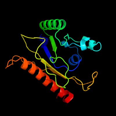
PDB 1iuq chain A
Region: 59 - 243
Aligned: 185
Modelled: 185
Confidence: 98.6%
Identity: 16%
Fold: Glycerol-3-phosphate (1)-acyltransferase
Superfamily: Glycerol-3-phosphate (1)-acyltransferase
Family: Glycerol-3-phosphate (1)-acyltransferase
Phyre2
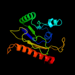
| 2 |
|
PDB 2ke4 chain A
Region: 203 - 244
Aligned: 42
Modelled: 42
Confidence: 16.7%
Identity: 26%
PDB header:membrane protein
Chain: A: PDB Molecule:cdc42-interacting protein 4;
PDBTitle: the nmr structure of the tc10 and cdc42 interacting domain2 of cip4
Phyre2
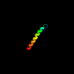
| 3 |
|
PDB 1ybx chain A
Region: 204 - 239
Aligned: 36
Modelled: 36
Confidence: 7.6%
Identity: 17%
PDB header:structural genomics, unknown function
Chain: A: PDB Molecule:conserved hypothetical protein;
PDBTitle: conserved hypothetical protein cth-383 from clostridium thermocellum
Phyre2
|
| Detailed template information | |
Due to computational demand, binding site predictions are not run for batch jobs
If you want to predict binding sites, please manually submit your model of choice to 3DLigandSite
Phyre is for academic use only
| Please cite: Protein structure prediction on
the web: a case study using the Phyre server |
| Kelley LA and Sternberg MJE. Nature Protocols
4, 363 - 371 (2009) [pdf] [Import into BibTeX] |
| |
| If you use the binding site
predictions from 3DLigandSite, please also cite: |
| 3DLigandSite: predicting ligand-binding sites using similar structures. |
| Wass MN, Kelley LA and Sternberg
MJ Nucleic Acids Research 38, W469-73 (2010) [PubMed] |
| |
|
|
|
|