Phyre Home Retrieve Phyre Job Id
Emaill.a.kelley@imperial.ac.uk
DescriptionP28911
DateThu Jan 5 11:45:22 GMT 2012
Unique Job IDeb85fd122e621475

Summary 

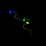
Top model
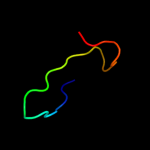
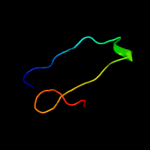
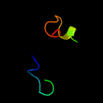
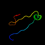
Image coloured by rainbow N → C terminus
Model (left) based on template c3kk7B_
Top template information
PDB header:cell invasion
Chain: B: PDB Molecule:putative cell invasion protein with mac/perforin domain;
PDBTitle: crystal structure of putative cell invasion protein with mac/perforin2 domain (np_812351.1) from bacteriodes thetaiotaomicron vpi-5482 at3 2.46 a resolution
Confidence and coverage
Confidence: 51.3% Coverage: 48%
61 residues ( 48% of your sequence) have been modelled with 51.3% confidence by the single highest scoring template.
You may wish to submit your sequence to Phyrealarm. This will automatically scan your sequence every week for new potential templates as they appear in the Phyre2 library.
Please note: You must be registered and logged in to use Phyrealarm.
3D viewing
Interactive 3D view in Jmol

Sequence analysis 

Secondary structure and disorder prediction 

   1........10.........20.........30.........40.........50.........60
Sequence  MSAEFMVICKKILFRNCVIVSLFVFTYNTWAQCNNNIKIMRKYESEGKYTVRNLVKNKAI
Secondary structure 


SS confidence 


Disorder  ????


??


?


Disorder confidence 


   .........70.........80.........90.........100.........110.........120
Sequence  ALELAEIYVKNRYGQDAAEEEKPYEITELTTSWVVEGTIHSDQIAGGVFIIEIGKNDGRI
Secondary structure 


SS confidence 


Disorder 


???????


Disorder confidence 


   .......
Sequence  LNFGHGK
Secondary structure 

SS confidence 


Disorder 


???
Disorder confidence 


Confidence Key
High(9)                    Low (0)
?Disordered
Alpha helix
Beta strand

Domain analysis 

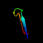
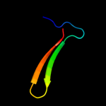
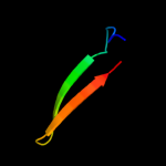
Hover over an aligned region to see model and summary info

Please note, only up to the top 20 hits are modelled to reduce computer load

RankAligned region

PDB 3kk7 chain B

3D model

Region: 27 - 87
Aligned: 61
Modelled: 61
Confidence: 51.3%
Identity: 23%
PDB header:cell invasion
Chain: B: PDB Molecule:putative cell invasion protein with mac/perforin domain;
PDBTitle: crystal structure of putative cell invasion protein with mac/perforin2 domain (np_812351.1) from bacteriodes thetaiotaomicron vpi-5482 at3 2.46 a resolution

Phyre2

PDB 1f0c chain B

3D model

Region: 109 - 126
Aligned: 18
Modelled: 18
Confidence: 25.0%
Identity: 44%
PDB header:viral protein
Chain: B: PDB Molecule:ice inhibitor;
PDBTitle: structure of the viral serpin crma

Phyre2

PDB 2epg chain B

3D model

Region: 102 - 126
Aligned: 25
Modelled: 25
Confidence: 24.3%
Identity: 24%
PDB header:structural genomics, unknown function
Chain: B: PDB Molecule:hypothetical protein ttha1785;
PDBTitle: crystal structure of ttha1785

Phyre2

PDB 1uc2 chain A

3D model

Region: 102 - 126
Aligned: 25
Modelled: 25
Confidence: 21.9%
Identity: 24%
Fold: Hypothetical protein PH1602
Superfamily: Hypothetical protein PH1602
Family: Hypothetical protein PH1602

Phyre2

PDB 2obk chain E

3D model

Region: 98 - 121
Aligned: 19
Modelled: 24
Confidence: 18.7%
Identity: 32%
PDB header:structural genomics, unknown function
Chain: E: PDB Molecule:selt/selw/selh selenoprotein domain;
PDBTitle: x-ray structure of the putative se binding protein from pseudomonas2 fluorescens. northeast structural genomics consortium target plr6.

Phyre2

PDB 2fa8 chain A domain 1

3D model

Region: 98 - 121
Aligned: 19
Modelled: 24
Confidence: 18.2%
Identity: 37%
Fold: Thioredoxin fold
Superfamily: Thioredoxin-like
Family: Selenoprotein W-related

Phyre2

PDB 2r32 chain A

3D model

Region: 50 - 126
Aligned: 71
Modelled: 75
Confidence: 15.0%
Identity: 23%
PDB header:immune system
Chain: A: PDB Molecule:gcn4-pii/tumor necrosis factor ligand
PDBTitle: crystal structure of human gitrl variant

Phyre2

PDB 1ibx chain A

3D model

Region: 78 - 92
Aligned: 15
Modelled: 15
Confidence: 12.4%
Identity: 13%
Fold: beta-Grasp (ubiquitin-like)
Superfamily: CAD & PB1 domains
Family: CAD domain

Phyre2

PDB 3dex chain A

3D model

Region: 94 - 121
Aligned: 23
Modelled: 28
Confidence: 11.6%
Identity: 26%
PDB header:structural genomics, unknown function
Chain: A: PDB Molecule:sav_2001;
PDBTitle: crystal structure of sav_2001 protein from streptomyces2 avermitilis, northeast structural genomics consortium3 target svr107.

Phyre2

PDB 3c0n chain A domain 1

3D model

Region: 84 - 98
Aligned: 15
Modelled: 15
Confidence: 11.3%
Identity: 33%
Fold: C-type lectin-like
Superfamily: C-type lectin-like
Family: Aerolysin/Pertussis toxin (APT) domain

Phyre2

PDB 1ept chain A

3D model

Region: 74 - 103
Aligned: 30
Modelled: 30
Confidence: 11.3%
Identity: 23%
PDB header:hydrolase (serine protease)
Chain: A: PDB Molecule:porcine e-trypsin;
PDBTitle: refined 1.8 angstroms resolution crystal structure of2 porcine epsilon-trypsin

Phyre2

PDB 1tm9 chain A

3D model

Region: 35 - 74
Aligned: 40
Modelled: 40
Confidence: 10.8%
Identity: 28%
Fold: Hypothetical protein MG354
Superfamily: Hypothetical protein MG354
Family: Hypothetical protein MG354

Phyre2

PDB 2gvs chain A

3D model

Region: 28 - 52
Aligned: 25
Modelled: 25
Confidence: 10.8%
Identity: 52%
PDB header:lipid binding protein
Chain: A: PDB Molecule:chemosensory protein csp-sg4;
PDBTitle: nmr solution structure of cspsg4

Phyre2

PDB 2oai chain A domain 1

3D model

Region: 81 - 98
Aligned: 18
Modelled: 18
Confidence: 10.1%
Identity: 28%
Fold: FAD-binding/transporter-associated domain-like
Superfamily: FAD-binding/transporter-associated domain-like
Family: CorC/HlyC domain-like

Phyre2

PDB 2ojl chain B

3D model

Region: 98 - 121
Aligned: 19
Modelled: 24
Confidence: 9.6%
Identity: 37%
PDB header:structural genomics, unknown function
Chain: B: PDB Molecule:hypothetical protein;
PDBTitle: crystal structure of q7waf1_borpa from bordetella parapertussis.2 northeast structural genomics target bpr68.

Phyre2

PDB 2p0g chain B

3D model

Region: 94 - 121
Aligned: 23
Modelled: 28
Confidence: 9.4%
Identity: 30%
PDB header:structural genomics, unknown function
Chain: B: PDB Molecule:selenoprotein w-related protein;
PDBTitle: crystal structure of selenoprotein w-related protein from2 vibrio cholerae. northeast structural genomics target vcr75

Phyre2

PDB 2npb chain A

3D model

Region: 90 - 122
Aligned: 28
Modelled: 33
Confidence: 8.9%
Identity: 14%
PDB header:oxidoreductase
Chain: A: PDB Molecule:selenoprotein w;
PDBTitle: nmr solution structure of mouse selw

Phyre2

PDB 3lzn chain A

3D model

Region: 84 - 110
Aligned: 27
Modelled: 27
Confidence: 8.3%
Identity: 22%
PDB header:transport protein
Chain: A: PDB Molecule:p19 protein;
PDBTitle: crystal structure analysis of the apo p19 protein from campylobacter2 jejuni at 1.59 a at ph 9

Phyre2

PDB 2v31 chain A

3D model

Region: 66 - 110
Aligned: 44
Modelled: 45
Confidence: 7.6%
Identity: 20%
PDB header:ligase
Chain: A: PDB Molecule:ubiquitin-activating enzyme e1 x;
PDBTitle: structure of first catalytic cysteine half-domain of mouse2 ubiquitin-activating enzyme

Phyre2

PDB 3bq9 chain A

3D model

Region: 13 - 22
Aligned: 10
Modelled: 10
Confidence: 7.5%
Identity: 60%
PDB header:structural genomics, unknown function
Chain: A: PDB Molecule:predicted rossmann fold nucleotide-binding domain-
PDBTitle: crystal structure of predicted nucleotide-binding protein from2 idiomarina baltica os145

Phyre2
1

c3kk7B_
2

c1f0cB_
3

c2epgB_
4

d1uc2a_
5

c2obkE_
6

d2fa8a1
7

c2r32A_
8

d1ibxa_
9

c3dexA_
10

d3c0na1
11

c1eptA_
12

d1tm9a_
13

c2gvsA_
14

d2oaia1
15

c2ojlB_
16

c2p0gB_
17

c2npbA_
18

c3lznA_
19

c2v31A_
20

c3bq9A_
21


22


23


24


25


Detailed template information 

#
Template Alignment Coverage3D Model Confidence
% i.d. Template Information
1c3kk7B_


51.3 23 PDB header:cell invasion
Chain: B: PDB Molecule:putative cell invasion protein with mac/perforin domain;
PDBTitle: crystal structure of putative cell invasion protein with mac/perforin2 domain (np_812351.1) from bacteriodes thetaiotaomicron vpi-5482 at3 2.46 a resolution
2c1f0cB_


25.0 44 PDB header:viral protein
Chain: B: PDB Molecule:ice inhibitor;
PDBTitle: structure of the viral serpin crma
3c2epgB_


24.3 24 PDB header:structural genomics, unknown function
Chain: B: PDB Molecule:hypothetical protein ttha1785;
PDBTitle: crystal structure of ttha1785
4d1uc2a_


21.9 24 Fold:Hypothetical protein PH1602
Superfamily:Hypothetical protein PH1602
Family:Hypothetical protein PH1602
5c2obkE_


18.7 32 PDB header:structural genomics, unknown function
Chain: E: PDB Molecule:selt/selw/selh selenoprotein domain;
PDBTitle: x-ray structure of the putative se binding protein from pseudomonas2 fluorescens. northeast structural genomics consortium target plr6.
6d2fa8a1


18.2 37 Fold:Thioredoxin fold
Superfamily:Thioredoxin-like
Family:Selenoprotein W-related
7c2r32A_


15.0 23 PDB header:immune system
Chain: A: PDB Molecule:gcn4-pii/tumor necrosis factor ligand
PDBTitle: crystal structure of human gitrl variant
8d1ibxa_


12.4 13 Fold:beta-Grasp (ubiquitin-like)
Superfamily:CAD & PB1 domains
Family:CAD domain
9c3dexA_


11.6 26 PDB header:structural genomics, unknown function
Chain: A: PDB Molecule:sav_2001;
PDBTitle: crystal structure of sav_2001 protein from streptomyces2 avermitilis, northeast structural genomics consortium3 target svr107.
10d3c0na1


11.3 33 Fold:C-type lectin-like
Superfamily:C-type lectin-like
Family:Aerolysin/Pertussis toxin (APT) domain
11c1eptA_


11.3 23 PDB header:hydrolase (serine protease)
Chain: A: PDB Molecule:porcine e-trypsin;
PDBTitle: refined 1.8 angstroms resolution crystal structure of2 porcine epsilon-trypsin
12d1tm9a_


10.8 28 Fold:Hypothetical protein MG354
Superfamily:Hypothetical protein MG354
Family:Hypothetical protein MG354
13c2gvsA_


10.8 52 PDB header:lipid binding protein
Chain: A: PDB Molecule:chemosensory protein csp-sg4;
PDBTitle: nmr solution structure of cspsg4
14d2oaia1


10.1 28 Fold:FAD-binding/transporter-associated domain-like
Superfamily:FAD-binding/transporter-associated domain-like
Family:CorC/HlyC domain-like
15c2ojlB_


9.6 37 PDB header:structural genomics, unknown function
Chain: B: PDB Molecule:hypothetical protein;
PDBTitle: crystal structure of q7waf1_borpa from bordetella parapertussis.2 northeast structural genomics target bpr68.
16c2p0gB_


9.4 30 PDB header:structural genomics, unknown function
Chain: B: PDB Molecule:selenoprotein w-related protein;
PDBTitle: crystal structure of selenoprotein w-related protein from2 vibrio cholerae. northeast structural genomics target vcr75
17c2npbA_


8.9 14 PDB header:oxidoreductase
Chain: A: PDB Molecule:selenoprotein w;
PDBTitle: nmr solution structure of mouse selw
18c3lznA_


8.3 22 PDB header:transport protein
Chain: A: PDB Molecule:p19 protein;
PDBTitle: crystal structure analysis of the apo p19 protein from campylobacter2 jejuni at 1.59 a at ph 9
19c2v31A_


7.6 20 PDB header:ligase
Chain: A: PDB Molecule:ubiquitin-activating enzyme e1 x;
PDBTitle: structure of first catalytic cysteine half-domain of mouse2 ubiquitin-activating enzyme
20c3bq9A_


7.5 60 PDB header:structural genomics, unknown function
Chain: A: PDB Molecule:predicted rossmann fold nucleotide-binding domain-
PDBTitle: crystal structure of predicted nucleotide-binding protein from2 idiomarina baltica os145
21c2latA_


not modelled 6.6 46 PDB header:membrane protein
Chain: A: PDB Molecule:dolichyl-diphosphooligosaccharide--protein
PDBTitle: solution structure of a human minimembrane protein ost4
22d2p13a1


not modelled 6.4 22 Fold:FAD-binding/transporter-associated domain-like
Superfamily:FAD-binding/transporter-associated domain-like
Family:CorC/HlyC domain-like
23c2hgoA_


not modelled 5.2 33 PDB header:toxin
Chain: A: PDB Molecule:cassiicolin;
PDBTitle: nmr structure of cassiicolin
24c2o62A_


not modelled 5.0 19 PDB header:unknown function
Chain: A: PDB Molecule:hypothetical protein;
PDBTitle: crystal structure of a protein with unknown function from duf35982 family (npun_r4044) from nostoc punctiforme pcc 73102 at 1.75 a3 resolution
25c3iufA_


not modelled 5.0 43 PDB header:protein binding
Chain: A: PDB Molecule:zinc finger protein ubi-d4;
PDBTitle: crystal structure of the c2h2-type zinc finger domain of2 human ubi-d4

Binding site prediction 

Due to computational demand, binding site predictions are not run for batch jobs

If you want to predict binding sites, please manually submit your model of choice to 3DLigandSite


Phyre is for academic use only

Please cite: Protein structure prediction on the web: a case study using the Phyre server
Kelley LA and Sternberg MJE. Nature Protocols 4, 363 - 371 (2009) [pdf] [Import into BibTeX]
 
If you use the binding site predictions from 3DLigandSite, please also cite:
3DLigandSite: predicting ligand-binding sites using similar structures.
Wass MN, Kelley LA and Sternberg MJ Nucleic Acids Research 38, W469-73 (2010) [PubMed]
 
© Structural Bioinformatics Group
Imperial College London
Lawrence Kelley, Benjamin Jefferys
Disclaimer
Terms and Conditions
Component software
Template detection: HHpred 1.51
Secondary structure prediction: Psi-pred 2.5
Disorder prediction: Disopred 2.4
Transmembrane prediction: Memsat_SVM
Multi-template modelling and ab initio: Poing 1.0