| 1 |
|
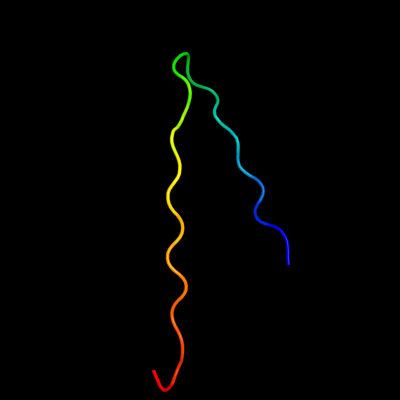
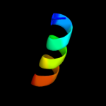
PDB 2ii8 chain F
Region: 26 - 49
Aligned: 24
Modelled: 24
Confidence: 30.6%
Identity: 13%
PDB header:signaling protein
Chain: F: PDB Molecule:anabaena sensory rhodopsin transducer protein;
PDBTitle: anabaena sensory rhodopsin transducer
Phyre2
| 2 |
|
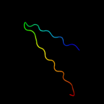
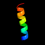
PDB 1nc7 chain A
Region: 25 - 49
Aligned: 25
Modelled: 25
Confidence: 29.4%
Identity: 16%
Fold: Hypothetical protein TM1070
Superfamily: Hypothetical protein TM1070
Family: Hypothetical protein TM1070
Phyre2
| 3 |
|
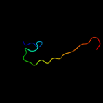
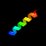
PDB 2wj8 chain N
Region: 8 - 21
Aligned: 14
Modelled: 14
Confidence: 13.2%
Identity: 36%
PDB header:rna binding protein/rna
Chain: N: PDB Molecule:nucleoprotein;
PDBTitle: respiratory syncitial virus ribonucleoprotein
Phyre2
| 4 |
|
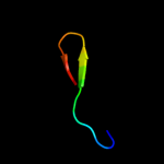
PDB 3b8e chain B
Region: 5 - 18
Aligned: 14
Modelled: 14
Confidence: 12.8%
Identity: 21%
PDB header:hydrolase/transport protein
Chain: B: PDB Molecule:sodium/potassium-transporting atpase subunit
PDBTitle: crystal structure of the sodium-potassium pump
Phyre2
| 5 |
|
PDB 3ixz chain B
Region: 5 - 22
Aligned: 18
Modelled: 18
Confidence: 12.3%
Identity: 28%
PDB header:hydrolase
Chain: B: PDB Molecule:potassium-transporting atpase subunit beta;
PDBTitle: pig gastric h+/k+-atpase complexed with aluminium fluoride
Phyre2
| 6 |
|
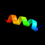
PDB 1xme chain C domain 1
Region: 4 - 24
Aligned: 21
Modelled: 21
Confidence: 9.4%
Identity: 57%
Fold: Single transmembrane helix
Superfamily: Bacterial ba3 type cytochrome c oxidase subunit IIa
Family: Bacterial ba3 type cytochrome c oxidase subunit IIa
Phyre2
| 7 |
|
PDB 2krx chain A
Region: 25 - 39
Aligned: 15
Modelled: 15
Confidence: 7.6%
Identity: 27%
PDB header:structural genomics, unknown function
Chain: A: PDB Molecule:asl3597 protein;
PDBTitle: solution nmr structure of asl3597 from nostoc sp. pcc7120. northeast2 structural genomics consortium target id nsr244.
Phyre2
| 8 |
|
PDB 3arc chain L
Region: 9 - 24
Aligned: 16
Modelled: 16
Confidence: 7.0%
Identity: 13%
PDB header:electron transport, photosynthesis
Chain: L: PDB Molecule:photosystem ii reaction center protein l;
PDBTitle: crystal structure of oxygen-evolving photosystem ii at 1.9 angstrom2 resolution
Phyre2
| 9 |
|
PDB 1zll chain E
Region: 10 - 23
Aligned: 14
Modelled: 14
Confidence: 6.8%
Identity: 21%
PDB header:membrane protein/signaling protein
Chain: E: PDB Molecule:cardiac phospholamban;
PDBTitle: nmr structure of unphosphorylated human phospholamban2 pentamer
Phyre2