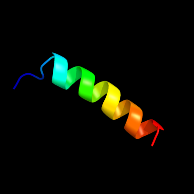
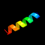
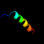
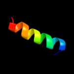
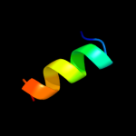
1 c3dxrA_
33.2
26
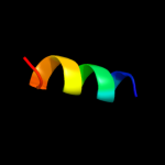
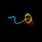
PDB header: protein transportChain: A: PDB Molecule: mitochondrial import inner membrane translocasePDBTitle: crystal structure of the yeast inter-membrane space2 chaperone assembly tim9.10
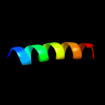
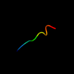
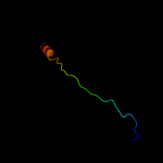
2 c3dxrB_
32.1
33
PDB header: protein transportChain: B: PDB Molecule: mitochondrial import inner membrane translocasePDBTitle: crystal structure of the yeast inter-membrane space2 chaperone assembly tim9.10
3 d2bska1
28.1
32
Fold: Tim10-likeSuperfamily: Tim10-likeFamily: Tim10/DDP4 c3cjhJ_
28.1
32
PDB header: protein transportChain: J: PDB Molecule: mitochondrial import inner membrane translocase subunitPDBTitle: tim8-tim13 complex
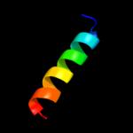
5 c2bskD_
22.2
37
PDB header: protein transportChain: D: PDB Molecule: mitochondrial import inner membrane translocasePDBTitle: crystal structure of the tim9 tim10 hexameric complex
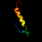
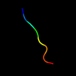
6 c2k19A_
21.9
39
PDB header: antimicrobial proteinChain: A: PDB Molecule: putative piscicolin 126 immunity protein;PDBTitle: nmr solution structure of pisi
7 c2iqcA_
16.9
38
PDB header: protein bindingChain: A: PDB Molecule: fanconi anemia group f protein;PDBTitle: crystal structure of human fancf protein that functions in2 the assembly of a dna damage signaling complex
8 d2cvea1
14.0
75
Fold: Ribosomal protein S5 domain 2-likeSuperfamily: Ribosomal protein S5 domain 2-likeFamily: YigZ N-terminal domain-like9 d2bskb1
12.7
33
Fold: Tim10-likeSuperfamily: Tim10-likeFamily: Tim10/DDP10 c2xglB_
12.4
29
PDB header: antibioticChain: B: PDB Molecule: colicin-m immunity protein;PDBTitle: the x-ray structure of the escherichia coli colicin m immunity2 protein demonstrates the presence of a disulphide bridge, which is3 functionally essential
11 d1vi7a1
11.1
50
Fold: Ribosomal protein S5 domain 2-likeSuperfamily: Ribosomal protein S5 domain 2-likeFamily: YigZ N-terminal domain-like12 d2incc1
11.1
53
Fold: beta-Grasp (ubiquitin-like)Superfamily: TmoB-likeFamily: TmoB-like13 c3hgkE_
11.0
33
PDB header: transferaseChain: E: PDB Molecule: effector protein hopab2;PDBTitle: crystal structure of effect protein avrptob complexed with2 kinase pto
14 c3ol4B_
11.0
40
PDB header: unknown functionChain: B: PDB Molecule: putative uncharacterized protein;PDBTitle: crystal structure of a putative uncharacterized protein from2 mycobacterium smegmatis, an ortholog of rv0543c
15 d1x4pa1
10.6
53
Fold: Surp module (SWAP domain)Superfamily: Surp module (SWAP domain)Family: Surp module (SWAP domain)16 c2k48A_
10.6
36
PDB header: viral proteinChain: A: PDB Molecule: nucleoprotein;PDBTitle: nmr structure of the n-terminal coiled coil domain of the2 andes hantavirus nucleocapsid protein
17 d2tpta2
9.6
21
Fold: Nucleoside phosphorylase/phosphoribosyltransferase catalytic domainSuperfamily: Nucleoside phosphorylase/phosphoribosyltransferase catalytic domainFamily: Nucleoside phosphorylase/phosphoribosyltransferase catalytic domain18 c2cveA_
8.6
75
PDB header: structural genomics, unknown functionChain: A: PDB Molecule: hypothetical protein ttha1053;PDBTitle: crystal structure of a conserved hypothetical protein tt1547 from2 thermus thermophilus hb8
19 c3sviA_
8.5
27
PDB header: signaling proteinChain: A: PDB Molecule: type iii effector hopab2;PDBTitle: structure of the pto-binding domain of hoppmal generated by limited2 thermolysin digestion
20 c1vi7A_
8.3
50
PDB header: structural genomics, unknown functionChain: A: PDB Molecule: hypothetical protein yigz;PDBTitle: crystal structure of an hypothetical protein
21 c2kvcA_
not modelled
7.8
60
PDB header: unknown functionChain: A: PDB Molecule: putative uncharacterized protein;PDBTitle: solution structure of the mycobacterium tuberculosis protein rv0543c,2 a member of the duf3349 superfamily. seattle structural genomics3 center for infectious disease target mytud.17112.a
22 d1ci3m2
not modelled
7.7
39
Fold: Barrel-sandwich hybridSuperfamily: Rudiment single hybrid motifFamily: Cytochrome f, small domain23 c3p43A_
not modelled
7.5
36
PDB header: hydrolaseChain: A: PDB Molecule: putative uncharacterized protein;PDBTitle: structure and activities of archaeal members of the ligd 3'2 phosphoesterase dna repair enzyme superfamily
24 c2lkyA_
not modelled
7.4
50
PDB header: structural genomics, unknown functionChain: A: PDB Molecule: uncharacterized protein;PDBTitle: solution structure of msmeg_1053, the second duf3349 annotated protein2 in the genome of mycobacterium smegmatis, seattle structural genomics3 center for infectious disease target mysma.17112.b
25 d1zvsa1
not modelled
7.1
24
Fold: Immunoglobulin-like beta-sandwichSuperfamily: ImmunoglobulinFamily: C1 set domains (antibody constant domain-like)26 c2xksA_
not modelled
7.0
20
PDB header: immune systemChain: A: PDB Molecule: beta-2-microglobulin;PDBTitle: prion-like conversion during amyloid formation at atomic2 resolution
27 c3kxeD_
not modelled
6.8
35
PDB header: protein bindingChain: D: PDB Molecule: antitoxin protein pard-1;PDBTitle: a conserved mode of protein recognition and binding in a2 pard-pare toxin-antitoxin complex
28 c1xvlC_
not modelled
6.8
30
PDB header: metal transportChain: C: PDB Molecule: mn transporter;PDBTitle: the three-dimensional structure of mntc from synechocystis2 6803
29 d1ffkw_
not modelled
6.6
50
Fold: Rubredoxin-likeSuperfamily: Zn-binding ribosomal proteinsFamily: Ribosomal protein L37ae30 d1wgna_
not modelled
6.4
67
Fold: RuvA C-terminal domain-likeSuperfamily: UBA-likeFamily: UBA domain31 d1qo3a1
not modelled
6.1
29
Fold: Immunoglobulin-like beta-sandwichSuperfamily: ImmunoglobulinFamily: C1 set domains (antibody constant domain-like)32 c1s1i0_
not modelled
6.1
38
PDB header: ribosomeChain: 0: PDB Molecule: 60s ribosomal protein l32;PDBTitle: structure of the ribosomal 80s-eef2-sordarin complex from2 yeast obtained by docking atomic models for rna and protein3 components into a 11.7 a cryo-em map. this file, 1s1i,4 contains 60s subunit. the 40s ribosomal subunit is in file5 1s1h.
33 c4a1cX_
not modelled
6.1
55
PDB header: ribosomeChain: X: PDB Molecule: 60s ribosomal protein l32;PDBTitle: t.thermophila 60s ribosomal subunit in complex with2 initiation factor 6. this file contains 5s rrna,3 5.8s rrna and proteins of molecule 4.
34 d1ydpa1
not modelled
6.0
24
Fold: Immunoglobulin-like beta-sandwichSuperfamily: ImmunoglobulinFamily: C1 set domains (antibody constant domain-like)35 c3ardB_
not modelled
5.9
13
PDB header: immune systemChain: B: PDB Molecule: beta-2-microglobulin;PDBTitle: ternary crystal structure of the mouse nkt tcr-cd1d-3'deoxy-alpha-2 galactosylceramide
36 c3areB_
not modelled
5.9
13
PDB header: immune systemChain: B: PDB Molecule: beta-2-microglobulin;PDBTitle: ternary crystal structure of the mouse nkt tcr-cd1d-4'deoxy-alpha-2 galactosylceramide
37 c2zkry_
not modelled
5.6
75
PDB header: ribosomal protein/rnaChain: Y: PDB Molecule: 5s ribosomal rna;PDBTitle: structure of a mammalian ribosomal 60s subunit within an2 80s complex obtained by docking homology models of the rna3 and proteins into an 8.7 a cryo-em map
38 d1a6tb2
not modelled
5.3
19
Fold: Immunoglobulin-like beta-sandwichSuperfamily: ImmunoglobulinFamily: C1 set domains (antibody constant domain-like)39 d1sm3h2
not modelled
5.2
24
Fold: Immunoglobulin-like beta-sandwichSuperfamily: ImmunoglobulinFamily: C1 set domains (antibody constant domain-like)40 d1de4a1
not modelled
5.2
17
Fold: Immunoglobulin-like beta-sandwichSuperfamily: ImmunoglobulinFamily: C1 set domains (antibody constant domain-like)41 c3cceY_
not modelled
5.2
50
PDB header: ribosomeChain: Y: PDB Molecule: 50s ribosomal protein l32e;PDBTitle: structure of anisomycin resistant 50s ribosomal subunit: 23s rrna2 mutation u2535a
42 d1vqoy1
not modelled
5.2
50
Fold: Barstar-likeSuperfamily: Ribosomal protein L32eFamily: Ribosomal protein L32e43 c1dfwA_
not modelled
5.1
36
PDB header: immune systemChain: A: PDB Molecule: lung surfactant protein b;PDBTitle: conformational mapping of the n-terminal segment of2 surfactant protein b in lipid using 13c-enhanced fourier3 transform infrared spectroscopy (ftir)