Phyre Home Retrieve Phyre Job Id
Emaill.a.kelley@imperial.ac.uk
DescriptionP76208
DateThu Jan 5 12:20:33 GMT 2012
Unique Job IDe16c5b5a14d236cc

Summary 

Top model
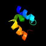
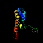
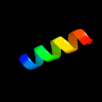
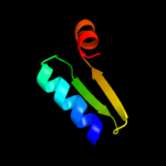
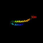
Image coloured by rainbow N → C terminus
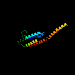
Model (left) based on template c2ejsA_
Top template information
PDB header:ligase
Chain: A: PDB Molecule:autocrine motility factor receptor, isoform 2;
PDBTitle: solution structure of ruh-076, a human cue domain
Confidence and coverage
Confidence: 38.2% Coverage: 23%
41 residues ( 23% of your sequence) have been modelled with 38.2% confidence by the single highest scoring template.
You may wish to submit your sequence to Phyrealarm. This will automatically scan your sequence every week for new potential templates as they appear in the Phyre2 library.
Please note: You must be registered and logged in to use Phyrealarm.
3D viewing
Interactive 3D view in Jmol

Sequence analysis 

Secondary structure and disorder prediction 

   1........10.........20.........30.........40.........50.........60
Sequence  MTYQQAGRIAVLKRILGWVIFIPALISTLISLLKFMNTRQENKEGINAVMLDFTHVMIDM
Secondary structure 


SS confidence 


Disorder  ??


????


Disorder confidence 


   .........70.........80.........90.........100.........110.........120
Sequence  MQANTPFLNLFWYNSPTPNFNGGVNVMFWVIFILIFVGLALQDSGARMSRQARFLREGVE
Secondary structure 


SS confidence 


Disorder 


????


?


Disorder confidence 


   .........130.........140.........150.........160.........170........
Sequence  DQLILEKAKGEEGLTREQIESRIVVPHHTIFLQFFSLYILPVICIAAGYVFFSLLGFI
Secondary structure 


SS confidence 


Disorder 


???????


??
????


???
Disorder confidence 


Confidence Key
High(9)                    Low (0)
?Disordered
Alpha helix
Beta strand

Domain analysis 

Hover over an aligned region to see model and summary info

Please note, only up to the top 20 hits are modelled to reduce computer load

RankAligned region

PDB 2ejs chain A

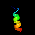
3D model

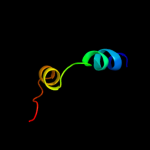
Region: 106 - 146
Aligned: 41
Modelled: 41
Confidence: 38.2%
Identity: 29%
PDB header:ligase
Chain: A: PDB Molecule:autocrine motility factor receptor, isoform 2;
PDBTitle: solution structure of ruh-076, a human cue domain

Phyre2

PDB 1t3j chain A

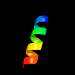
3D model

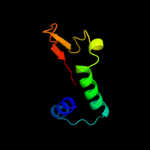
Region: 106 - 129
Aligned: 24
Modelled: 24
Confidence: 25.2%
Identity: 17%
PDB header:membrane protein
Chain: A: PDB Molecule:mitofusin 1;
PDBTitle: mitofusin domain hr2 v686m/i708m mutant

Phyre2

PDB 3i5q chain A

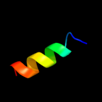
3D model

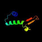
Region: 17 - 75
Aligned: 45
Modelled: 59
Confidence: 19.0%
Identity: 22%
PDB header:protein transport
Chain: A: PDB Molecule:nucleoporin nup170;
PDBTitle: nup170(aa1253-1502) at 2.2 a, s.cerevisiae

Phyre2

PDB 1r3j chain C

3D model

Region: 10 - 118
Aligned: 90
Modelled: 91
Confidence: 18.8%
Identity: 14%
Fold: Voltage-gated potassium channels
Superfamily: Voltage-gated potassium channels
Family: Voltage-gated potassium channels

Phyre2

PDB 3qsp chain B

3D model

Region: 25 - 132
Aligned: 108
Modelled: 108
Confidence: 18.1%
Identity: 20%
PDB header:hydrolase
Chain: B: PDB Molecule:putative uncharacterized protein;
PDBTitle: analysis of a new family of widely distributed metal-independent alpha2 mannosidases provides unique insight into the processing of n-linked3 glycans, streptococcus pneumoniae sp_2144 non-productive substrate4 complex with alpha-1,6-mannobiose

Phyre2

PDB 2bg9 chain B

3D model

Region: 18 - 176
Aligned: 134
Modelled: 145
Confidence: 16.8%
Identity: 12%
PDB header:ion channel/receptor
Chain: B: PDB Molecule:acetylcholine receptor protein, beta chain;
PDBTitle: refined structure of the nicotinic acetylcholine receptor2 at 4a resolution.

Phyre2

PDB 2e76 chain E

3D model

Region: 87 - 101
Aligned: 15
Modelled: 15
Confidence: 15.1%
Identity: 40%
PDB header:photosynthesis
Chain: E: PDB Molecule:cytochrome b6-f complex subunit 6;
PDBTitle: crystal structure of the cytochrome b6f complex with tridecyl-2 stigmatellin (tds) from m.laminosus

Phyre2

PDB 2e74 chain E domain 1

3D model

Region: 87 - 101
Aligned: 15
Modelled: 15
Confidence: 15.1%
Identity: 40%
Fold: Single transmembrane helix
Superfamily: PetL subunit of the cytochrome b6f complex
Family: PetL subunit of the cytochrome b6f complex

Phyre2

PDB 2e74 chain E

3D model

Region: 87 - 101
Aligned: 15
Modelled: 15
Confidence: 15.1%
Identity: 40%
PDB header:photosynthesis
Chain: E: PDB Molecule:cytochrome b6-f complex subunit 6;
PDBTitle: crystal structure of the cytochrome b6f complex from m.laminosus

Phyre2

PDB 1vf5 chain R

3D model

Region: 87 - 101
Aligned: 15
Modelled: 15
Confidence: 15.1%
Identity: 40%
PDB header:photosynthesis
Chain: R: PDB Molecule:protein pet l;
PDBTitle: crystal structure of cytochrome b6f complex from m.laminosus

Phyre2

PDB 2e75 chain E

3D model

Region: 87 - 101
Aligned: 15
Modelled: 15
Confidence: 15.1%
Identity: 40%
PDB header:photosynthesis
Chain: E: PDB Molecule:cytochrome b6-f complex subunit 6;
PDBTitle: crystal structure of the cytochrome b6f complex with 2-nonyl-4-2 hydroxyquinoline n-oxide (nqno) from m.laminosus

Phyre2

PDB 1vf5 chain E

3D model

Region: 87 - 101
Aligned: 15
Modelled: 15
Confidence: 15.1%
Identity: 40%
PDB header:photosynthesis
Chain: E: PDB Molecule:protein pet l;
PDBTitle: crystal structure of cytochrome b6f complex from m.laminosus

Phyre2

PDB 1in0 chain A domain 2

3D model

Region: 102 - 143
Aligned: 41
Modelled: 42
Confidence: 12.4%
Identity: 15%
Fold: Ferredoxin-like
Superfamily: YajQ-like
Family: YajQ-like

Phyre2

PDB 1wf9 chain A domain 1

3D model

Region: 112 - 129
Aligned: 18
Modelled: 18
Confidence: 10.5%
Identity: 22%
Fold: beta-Grasp (ubiquitin-like)
Superfamily: Ubiquitin-like
Family: Ubiquitin-related

Phyre2

PDB 2oar chain A domain 1

3D model

Region: 43 - 129
Aligned: 85
Modelled: 87
Confidence: 10.0%
Identity: 9%
Fold: Gated mechanosensitive channel
Superfamily: Gated mechanosensitive channel
Family: Gated mechanosensitive channel

Phyre2

PDB 2ba3 chain A

3D model

Region: 119 - 135
Aligned: 16
Modelled: 17
Confidence: 8.9%
Identity: 56%
PDB header:dna binding protein
Chain: A: PDB Molecule:nika;
PDBTitle: nmr structure of nika n-terminal fragment

Phyre2

PDB 1f6g chain A

3D model

Region: 11 - 143
Aligned: 114
Modelled: 115
Confidence: 7.8%
Identity: 14%
Fold: Voltage-gated potassium channels
Superfamily: Voltage-gated potassium channels
Family: Voltage-gated potassium channels

Phyre2

PDB 2aor chain B

3D model

Region: 120 - 149
Aligned: 29
Modelled: 30
Confidence: 7.4%
Identity: 28%
PDB header:hydrolase/dna
Chain: B: PDB Molecule:dna mismatch repair protein muth;
PDBTitle: crystal structure of muth-hemimethylated dna complex

Phyre2

PDB 1cby chain A

3D model

Region: 6 - 88
Aligned: 62
Modelled: 64
Confidence: 7.4%
Identity: 21%
Fold: CytB endotoxin-like
Superfamily: CytB endotoxin-like
Family: CytB endotoxin-like

Phyre2

PDB 1cby chain A

3D model

Region: 6 - 88
Aligned: 62
Modelled: 64
Confidence: 7.4%
Identity: 21%
PDB header:toxin
Chain: A: PDB Molecule:delta-endotoxin cytb;
PDBTitle: delta-endotoxin

Phyre2
1

c2ejsA_
2

c1t3jA_
3

c3i5qA_
4

d1r3jc_
5

c3qspB_
6

c2bg9B_
7

c2e76E_
8

d2e74e1
9

c2e74E_
10

c1vf5R_
11

c2e75E_
12

c1vf5E_
13

d1in0a2
14

d1wf9a1
15

d2oara1
16

c2ba3A_
17

d1f6ga_
18

c2aorB_
19

d1cbya_
20

c1cbyA_
21


22


23


24


25


26


27


28


29


30


31


32


33


34


35


36


37


38


39


40


41


42


Detailed template information 

#
Template Alignment Coverage3D Model Confidence
% i.d. Template Information
1c2ejsA_


38.2 29 PDB header:ligase
Chain: A: PDB Molecule:autocrine motility factor receptor, isoform 2;
PDBTitle: solution structure of ruh-076, a human cue domain
2c1t3jA_


25.2 17 PDB header:membrane protein
Chain: A: PDB Molecule:mitofusin 1;
PDBTitle: mitofusin domain hr2 v686m/i708m mutant
3c3i5qA_


19.0 22 PDB header:protein transport
Chain: A: PDB Molecule:nucleoporin nup170;
PDBTitle: nup170(aa1253-1502) at 2.2 a, s.cerevisiae
4d1r3jc_


18.8 14 Fold:Voltage-gated potassium channels
Superfamily:Voltage-gated potassium channels
Family:Voltage-gated potassium channels
5c3qspB_


18.1 20 PDB header:hydrolase
Chain: B: PDB Molecule:putative uncharacterized protein;
PDBTitle: analysis of a new family of widely distributed metal-independent alpha2 mannosidases provides unique insight into the processing of n-linked3 glycans, streptococcus pneumoniae sp_2144 non-productive substrate4 complex with alpha-1,6-mannobiose
6c2bg9B_


16.8 12 PDB header:ion channel/receptor
Chain: B: PDB Molecule:acetylcholine receptor protein, beta chain;
PDBTitle: refined structure of the nicotinic acetylcholine receptor2 at 4a resolution.
7c2e76E_


15.1 40 PDB header:photosynthesis
Chain: E: PDB Molecule:cytochrome b6-f complex subunit 6;
PDBTitle: crystal structure of the cytochrome b6f complex with tridecyl-2 stigmatellin (tds) from m.laminosus
8d2e74e1


15.1 40 Fold:Single transmembrane helix
Superfamily:PetL subunit of the cytochrome b6f complex
Family:PetL subunit of the cytochrome b6f complex
9c2e74E_


15.1 40 PDB header:photosynthesis
Chain: E: PDB Molecule:cytochrome b6-f complex subunit 6;
PDBTitle: crystal structure of the cytochrome b6f complex from m.laminosus
10c1vf5R_


15.1 40 PDB header:photosynthesis
Chain: R: PDB Molecule:protein pet l;
PDBTitle: crystal structure of cytochrome b6f complex from m.laminosus
11c2e75E_


15.1 40 PDB header:photosynthesis
Chain: E: PDB Molecule:cytochrome b6-f complex subunit 6;
PDBTitle: crystal structure of the cytochrome b6f complex with 2-nonyl-4-2 hydroxyquinoline n-oxide (nqno) from m.laminosus
12c1vf5E_


15.1 40 PDB header:photosynthesis
Chain: E: PDB Molecule:protein pet l;
PDBTitle: crystal structure of cytochrome b6f complex from m.laminosus
13d1in0a2


12.4 15 Fold:Ferredoxin-like
Superfamily:YajQ-like
Family:YajQ-like
14d1wf9a1


10.5 22 Fold:beta-Grasp (ubiquitin-like)
Superfamily:Ubiquitin-like
Family:Ubiquitin-related
15d2oara1


10.0 9 Fold:Gated mechanosensitive channel
Superfamily:Gated mechanosensitive channel
Family:Gated mechanosensitive channel
16c2ba3A_


8.9 56 PDB header:dna binding protein
Chain: A: PDB Molecule:nika;
PDBTitle: nmr structure of nika n-terminal fragment
17d1f6ga_


7.8 14 Fold:Voltage-gated potassium channels
Superfamily:Voltage-gated potassium channels
Family:Voltage-gated potassium channels
18c2aorB_


7.4 28 PDB header:hydrolase/dna
Chain: B: PDB Molecule:dna mismatch repair protein muth;
PDBTitle: crystal structure of muth-hemimethylated dna complex
19d1cbya_


7.4 21 Fold:CytB endotoxin-like
Superfamily:CytB endotoxin-like
Family:CytB endotoxin-like
20c1cbyA_


7.4 21 PDB header:toxin
Chain: A: PDB Molecule:delta-endotoxin cytb;
PDBTitle: delta-endotoxin
21c2wh7A_


not modelled 7.3 33 PDB header:hydrolase
Chain: A: PDB Molecule:hyaluronidase-phage associated;
PDBTitle: the partial structure of a group a streptpcoccal phage-2 encoded tail fibre hyaluronate lyase hylp2
22c1in0B_


not modelled 7.1 15 PDB header:structural genomics, unknown function
Chain: B: PDB Molecule:yajq protein;
PDBTitle: yajq protein (hi1034)
23c3he5D_


not modelled 7.0 23 PDB header:de novo protein
Chain: D: PDB Molecule:synzip2;
PDBTitle: heterospecific coiled-coil pair synzip2:synzip1
24c1y4eA_


not modelled 7.0 50 PDB header:membrane protein
Chain: A: PDB Molecule:sodium/hydrogen exchanger 1;
PDBTitle: nmr structure of transmembrane segment iv of the nhe12 isoform of the na+/h+ exchanger
25c1o98A_


not modelled 6.9 40 PDB header:isomerase
Chain: A: PDB Molecule:2,3-bisphosphoglycerate-independent
PDBTitle: 1.4a crystal structure of phosphoglycerate mutase from2 bacillus stearothermophilus complexed with3 2-phosphoglycerate
26d1sfsa_


not modelled 6.6 21 Fold:TIM beta/alpha-barrel
Superfamily:(Trans)glycosidases
Family:1,4-beta-N-acetylmuraminidase
27c1sfsA_


not modelled 6.6 21 PDB header:structural genomics, unknown function
Chain: A: PDB Molecule:hypothetical protein;
PDBTitle: 1.07 a crystal structure of an uncharacterized b.2 stearothermophilus protein
28c2h09A_


not modelled 6.5 25 PDB header:transcription
Chain: A: PDB Molecule:transcriptional regulator mntr;
PDBTitle: crystal structure of diphtheria toxin repressor like protein2 from e. coli
29c1iflA_


not modelled 6.5 16 PDB header:virus
Chain: A: PDB Molecule:inovirus;
PDBTitle: molecular models and structural comparisons of native and2 mutant class i filamentous bacteriophages ff (fd, f1, m13),3 if1 and ike
30c2rmgA_


not modelled 6.5 27 PDB header:hormone
Chain: A: PDB Molecule:urocortin-2;
PDBTitle: human urocortin 2
31c2l16A_


not modelled 6.2 17 PDB header:protein transport
Chain: A: PDB Molecule:sec-independent protein translocase protein tatad;
PDBTitle: solution structure of bacillus subtilits tatad protein in dpc micelles
32c3f1bA_


not modelled 6.2 13 PDB header:transcription regulator
Chain: A: PDB Molecule:tetr-like transcriptional regulator;
PDBTitle: the crystal structure of a tetr-like transcriptional regulator from2 rhodococcus sp. rha1.
33c2d2cR_


not modelled 5.9 43 PDB header:photosynthesis
Chain: R: PDB Molecule:cytochrome b6-f complex subunit vi;
PDBTitle: crystal structure of cytochrome b6f complex with dbmib from2 m. laminosus
34c2d2cE_


not modelled 5.9 43 PDB header:photosynthesis
Chain: E: PDB Molecule:cytochrome b6-f complex subunit vi;
PDBTitle: crystal structure of cytochrome b6f complex with dbmib from2 m. laminosus
35d2dk5a1


not modelled 5.9 16 Fold:DNA/RNA-binding 3-helical bundle
Superfamily:"Winged helix" DNA-binding domain
Family:RPO3F domain-like
36d1na6a2


not modelled 5.7 32 Fold:Restriction endonuclease-like
Superfamily:Restriction endonuclease-like
Family:Type II restriction endonuclease catalytic domain
37c2oarA_


not modelled 5.6 11 PDB header:membrane protein
Chain: A: PDB Molecule:large-conductance mechanosensitive channel;
PDBTitle: mechanosensitive channel of large conductance (mscl)
38d1ug2a_


not modelled 5.4 26 Fold:DNA/RNA-binding 3-helical bundle
Superfamily:Homeodomain-like
Family:Myb/SANT domain
39d1tzyb_


not modelled 5.3 14 Fold:Histone-fold
Superfamily:Histone-fold
Family:Nucleosome core histones
40d1s4na_


not modelled 5.1 17 Fold:Nucleotide-diphospho-sugar transferases
Superfamily:Nucleotide-diphospho-sugar transferases
Family:Glycolipid 2-alpha-mannosyltransferase
41d1hs7a_


not modelled 5.1 28 Fold:STAT-like
Superfamily:t-snare proteins
Family:t-snare proteins
42d1kyqa2


not modelled 5.1 20 Fold:Siroheme synthase middle domains-like
Superfamily:Siroheme synthase middle domains-like
Family:Siroheme synthase middle domains-like

Binding site prediction 

Due to computational demand, binding site predictions are not run for batch jobs

If you want to predict binding sites, please manually submit your model of choice to 3DLigandSite


Phyre is for academic use only

Please cite: Protein structure prediction on the web: a case study using the Phyre server
Kelley LA and Sternberg MJE. Nature Protocols 4, 363 - 371 (2009) [pdf] [Import into BibTeX]
 
If you use the binding site predictions from 3DLigandSite, please also cite:
3DLigandSite: predicting ligand-binding sites using similar structures.
Wass MN, Kelley LA and Sternberg MJ Nucleic Acids Research 38, W469-73 (2010) [PubMed]
 
© Structural Bioinformatics Group
Imperial College London
Lawrence Kelley, Benjamin Jefferys
Disclaimer
Terms and Conditions
Component software
Template detection: HHpred 1.51
Secondary structure prediction: Psi-pred 2.5
Disorder prediction: Disopred 2.4
Transmembrane prediction: Memsat_SVM
Multi-template modelling and ab initio: Poing 1.0