Phyre Home Retrieve Phyre Job Id
Emaill.a.kelley@imperial.ac.uk
DescriptionQ9XB42
DateThu Jan 5 12:38:30 GMT 2012
Unique Job IDddd3c13c5b1f0cd8

Summary 

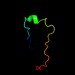
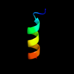
Top model
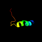
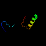
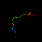
Image coloured by rainbow N → C terminus
Model (left) based on template d2cqma1
Top template information
Fold:Prokaryotic ribosomal protein L17
Superfamily:Prokaryotic ribosomal protein L17
Family:Prokaryotic ribosomal protein L17
Confidence and coverage
Confidence: 58.6% Coverage: 41%
30 residues ( 41% of your sequence) have been modelled with 58.6% confidence by the single highest scoring template.
You may wish to submit your sequence to Phyrealarm. This will automatically scan your sequence every week for new potential templates as they appear in the Phyre2 library.
Please note: You must be registered and logged in to use Phyrealarm.
3D viewing
Interactive 3D view in Jmol

Sequence analysis 

Secondary structure and disorder prediction 

   1........10.........20.........30.........40.........50.........60
Sequence  MKIISKRRAMTIYRQHPESRIFRYCTGKYQWHGSVCHYTGRDVPDIAGVLAVYAERRQDR
Secondary structure 


SS confidence 


Disorder  ???????


?


??
?
Disorder confidence 


   .........70...
Sequence  NGPYTCLMSITLN
Secondary structure 


SS confidence 


Disorder 


???
Disorder confidence 


Confidence Key
High(9)                    Low (0)
?Disordered
Alpha helix
Beta strand

Domain analysis 

Hover over an aligned region to see model and summary info

Please note, only up to the top 20 hits are modelled to reduce computer load

RankAligned region

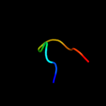
PDB 2cqm chain A domain 1

3D model

Region: 41 - 70
Aligned: 30
Modelled: 30
Confidence: 58.6%
Identity: 30%
Fold: Prokaryotic ribosomal protein L17
Superfamily: Prokaryotic ribosomal protein L17
Family: Prokaryotic ribosomal protein L17

Phyre2

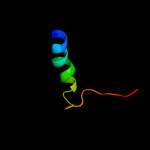
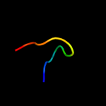
PDB 1gd8 chain A

3D model

Region: 41 - 70
Aligned: 30
Modelled: 30
Confidence: 53.1%
Identity: 30%
Fold: Prokaryotic ribosomal protein L17
Superfamily: Prokaryotic ribosomal protein L17
Family: Prokaryotic ribosomal protein L17

Phyre2

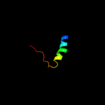
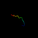
PDB 2zjr chain K domain 1

3D model

Region: 41 - 68
Aligned: 28
Modelled: 28
Confidence: 53.1%
Identity: 25%
Fold: Prokaryotic ribosomal protein L17
Superfamily: Prokaryotic ribosomal protein L17
Family: Prokaryotic ribosomal protein L17

Phyre2

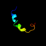
PDB 3bbo chain P

3D model

Region: 41 - 68
Aligned: 28
Modelled: 28
Confidence: 34.8%
Identity: 25%
PDB header:ribosome
Chain: P: PDB Molecule:ribosomal protein l17;
PDBTitle: homology model for the spinach chloroplast 50s subunit2 fitted to 9.4a cryo-em map of the 70s chlororibosome

Phyre2

PDB 2qam chain N domain 1

3D model

Region: 41 - 68
Aligned: 28
Modelled: 28
Confidence: 30.5%
Identity: 29%
Fold: Prokaryotic ribosomal protein L17
Superfamily: Prokaryotic ribosomal protein L17
Family: Prokaryotic ribosomal protein L17

Phyre2

PDB 1bnl chain A

3D model

Region: 6 - 39
Aligned: 34
Modelled: 34
Confidence: 15.5%
Identity: 18%
Fold: C-type lectin-like
Superfamily: C-type lectin-like
Family: Endostatin

Phyre2

PDB 2qh7 chain A

3D model

Region: 18 - 27
Aligned: 10
Modelled: 10
Confidence: 12.3%
Identity: 40%
PDB header:metal binding protein
Chain: A: PDB Molecule:zinc finger cdgsh-type domain 1;
PDBTitle: mitoneet is a uniquely folded 2fe-2s outer mitochondrial membrane2 protein stabilized by pioglitazone

Phyre2

PDB 3fnv chain B

3D model

Region: 18 - 27
Aligned: 10
Modelled: 10
Confidence: 12.3%
Identity: 40%
PDB header:metal binding protein
Chain: B: PDB Molecule:cdgsh iron sulfur domain-containing protein 2;
PDBTitle: crystal structure of miner1: the redox-active 2fe-2s protein causative2 in wolfram syndrome 2

Phyre2

PDB 2gyr chain B

3D model

Region: 16 - 28
Aligned: 13
Modelled: 13
Confidence: 11.2%
Identity: 38%
PDB header:hormone/growth factor
Chain: B: PDB Molecule:neurotrophic factor artemin, isoform 3;
PDBTitle: crystal structure of human artemin

Phyre2

PDB 1ir1 chain S

3D model

Region: 31 - 70
Aligned: 40
Modelled: 40
Confidence: 11.1%
Identity: 25%
Fold: RuBisCO, small subunit
Superfamily: RuBisCO, small subunit
Family: RuBisCO, small subunit

Phyre2

PDB 2j7q chain A domain 1

3D model

Region: 1 - 59
Aligned: 48
Modelled: 59
Confidence: 8.7%
Identity: 19%
Fold: Cysteine proteinases
Superfamily: Cysteine proteinases
Family: M48USP-like

Phyre2

PDB 3fka chain D

3D model

Region: 16 - 72
Aligned: 45
Modelled: 57
Confidence: 8.5%
Identity: 13%
PDB header:unknown function
Chain: D: PDB Molecule:uncharacterized ntf-2 like protein;
PDBTitle: crystal structure of a ntf-2 like protein of unknown function2 (spo1084) from silicibacter pomeroyi dss-3 at 1.69 a resolution

Phyre2

PDB 1koe chain A

3D model

Region: 8 - 39
Aligned: 32
Modelled: 32
Confidence: 8.4%
Identity: 19%
Fold: C-type lectin-like
Superfamily: C-type lectin-like
Family: Endostatin

Phyre2

PDB 1ztx chain E domain 1

3D model

Region: 21 - 37
Aligned: 17
Modelled: 17
Confidence: 8.2%
Identity: 12%
Fold: Immunoglobulin-like beta-sandwich
Superfamily: E set domains
Family: Class II viral fusion proteins C-terminal domain

Phyre2

PDB 2wsf chain N

3D model

Region: 6 - 61
Aligned: 55
Modelled: 56
Confidence: 7.3%
Identity: 20%
PDB header:photosynthesis
Chain: N: PDB Molecule:photosystem i-n subunit;
PDBTitle: improved model of plant photosystem i

Phyre2

PDB 3egp chain A

3D model

Region: 21 - 32
Aligned: 12
Modelled: 12
Confidence: 7.3%
Identity: 25%
PDB header:viral protein
Chain: A: PDB Molecule:envelope protein;
PDBTitle: crystal structure analysis of dengue-1 envelope protein2 domain iii

Phyre2

PDB 1ok8 chain A domain 1

3D model

Region: 21 - 37
Aligned: 17
Modelled: 17
Confidence: 6.8%
Identity: 24%
Fold: Immunoglobulin-like beta-sandwich
Superfamily: E set domains
Family: Class II viral fusion proteins C-terminal domain

Phyre2

PDB 2kzt chain A

3D model

Region: 42 - 56
Aligned: 15
Modelled: 15
Confidence: 6.7%
Identity: 20%
PDB header:apoptosis
Chain: A: PDB Molecule:programmed cell death protein 4;
PDBTitle: structure of the tandem ma-3 region of pdcd4

Phyre2

PDB 1hd2 chain A

3D model

Region: 27 - 51
Aligned: 25
Modelled: 25
Confidence: 6.6%
Identity: 12%
Fold: Thioredoxin fold
Superfamily: Thioredoxin-like
Family: Glutathione peroxidase-like

Phyre2

PDB 2o01 chain N

3D model

Region: 6 - 61
Aligned: 55
Modelled: 56
Confidence: 5.9%
Identity: 20%
PDB header:photosynthesis
Chain: N: PDB Molecule:photosystem i-n subunit;
PDBTitle: the structure of a plant photosystem i supercomplex at 3.42 angstrom resolution

Phyre2
1

d2cqma1
2

d1gd8a_
3

d2zjrk1
4

c3bboP_
5

d2qamn1
6

d1bnla_
7

c2qh7A_
8

c3fnvB_
9

c2gyrB_
10

d1ir1s_
11

d2j7qa1
12

c3fkaD_
13

d1koea_
14

d1ztxe1
15

c2wsfN_
16

c3egpA_
17

d1ok8a1
18

c2kztA_
19

d1hd2a_
20

c2o01N_
21


22


23


24


25


26


27


28


29


30


31


Detailed template information 

#
Template Alignment Coverage3D Model Confidence
% i.d. Template Information
1d2cqma1


58.6 30 Fold:Prokaryotic ribosomal protein L17
Superfamily:Prokaryotic ribosomal protein L17
Family:Prokaryotic ribosomal protein L17
2d1gd8a_


53.1 30 Fold:Prokaryotic ribosomal protein L17
Superfamily:Prokaryotic ribosomal protein L17
Family:Prokaryotic ribosomal protein L17
3d2zjrk1


53.1 25 Fold:Prokaryotic ribosomal protein L17
Superfamily:Prokaryotic ribosomal protein L17
Family:Prokaryotic ribosomal protein L17
4c3bboP_


34.8 25 PDB header:ribosome
Chain: P: PDB Molecule:ribosomal protein l17;
PDBTitle: homology model for the spinach chloroplast 50s subunit2 fitted to 9.4a cryo-em map of the 70s chlororibosome
5d2qamn1


30.5 29 Fold:Prokaryotic ribosomal protein L17
Superfamily:Prokaryotic ribosomal protein L17
Family:Prokaryotic ribosomal protein L17
6d1bnla_


15.5 18 Fold:C-type lectin-like
Superfamily:C-type lectin-like
Family:Endostatin
7c2qh7A_


12.3 40 PDB header:metal binding protein
Chain: A: PDB Molecule:zinc finger cdgsh-type domain 1;
PDBTitle: mitoneet is a uniquely folded 2fe-2s outer mitochondrial membrane2 protein stabilized by pioglitazone
8c3fnvB_


12.3 40 PDB header:metal binding protein
Chain: B: PDB Molecule:cdgsh iron sulfur domain-containing protein 2;
PDBTitle: crystal structure of miner1: the redox-active 2fe-2s protein causative2 in wolfram syndrome 2
9c2gyrB_


11.2 38 PDB header:hormone/growth factor
Chain: B: PDB Molecule:neurotrophic factor artemin, isoform 3;
PDBTitle: crystal structure of human artemin
10d1ir1s_


11.1 25 Fold:RuBisCO, small subunit
Superfamily:RuBisCO, small subunit
Family:RuBisCO, small subunit
11d2j7qa1


8.7 19 Fold:Cysteine proteinases
Superfamily:Cysteine proteinases
Family:M48USP-like
12c3fkaD_


8.5 13 PDB header:unknown function
Chain: D: PDB Molecule:uncharacterized ntf-2 like protein;
PDBTitle: crystal structure of a ntf-2 like protein of unknown function2 (spo1084) from silicibacter pomeroyi dss-3 at 1.69 a resolution
13d1koea_


8.4 19 Fold:C-type lectin-like
Superfamily:C-type lectin-like
Family:Endostatin
14d1ztxe1


8.2 12 Fold:Immunoglobulin-like beta-sandwich
Superfamily:E set domains
Family:Class II viral fusion proteins C-terminal domain
15c2wsfN_


7.3 20 PDB header:photosynthesis
Chain: N: PDB Molecule:photosystem i-n subunit;
PDBTitle: improved model of plant photosystem i
16c3egpA_


7.3 25 PDB header:viral protein
Chain: A: PDB Molecule:envelope protein;
PDBTitle: crystal structure analysis of dengue-1 envelope protein2 domain iii
17d1ok8a1


6.8 24 Fold:Immunoglobulin-like beta-sandwich
Superfamily:E set domains
Family:Class II viral fusion proteins C-terminal domain
18c2kztA_


6.7 20 PDB header:apoptosis
Chain: A: PDB Molecule:programmed cell death protein 4;
PDBTitle: structure of the tandem ma-3 region of pdcd4
19d1hd2a_


6.6 12 Fold:Thioredoxin fold
Superfamily:Thioredoxin-like
Family:Glutathione peroxidase-like
20c2o01N_


5.9 20 PDB header:photosynthesis
Chain: N: PDB Molecule:photosystem i-n subunit;
PDBTitle: the structure of a plant photosystem i supercomplex at 3.42 angstrom resolution
21c2riqA_


not modelled 5.9 15 PDB header:transferase
Chain: A: PDB Molecule:poly [adp-ribose] polymerase 1;
PDBTitle: crystal structure of the third zinc-binding domain of human parp-1
22d2j01s1


not modelled 5.7 33 Fold:Ribonuclease H-like motif
Superfamily:Translational machinery components
Family:Ribosomal protein L18 and S11
23c3ccjU_


not modelled 5.7 23 PDB header:ribosome
Chain: U: PDB Molecule:50s ribosomal protein l24e;
PDBTitle: structure of anisomycin resistant 50s ribosomal subunit: 23s rrna2 mutation c2534u
24d1vqou1


not modelled 5.6 23 Fold:Glucocorticoid receptor-like (DNA-binding domain)
Superfamily:Glucocorticoid receptor-like (DNA-binding domain)
Family:Ribosomal protein L24e
25c2iosA_


not modelled 5.5 40 PDB header:antitumor protein
Chain: A: PDB Molecule:programmed cell death 4, pdcd4;
PDBTitle: crystal structure of the c-terminal ma3 domain of pdcd42 (mouse); form 3
26c2rg8A_


not modelled 5.5 20 PDB header:apoptosis, translation
Chain: A: PDB Molecule:programmed cell death protein 4;
PDBTitle: crystal structure of programmed for cell death 4 middle ma32 domain
27d1m9sa3


not modelled 5.4 18 Fold:SH3-like barrel
Superfamily:Prokaryotic SH3-related domain
Family:GW domain
28c2h0pA_


not modelled 5.2 18 PDB header:viral protein
Chain: A: PDB Molecule:envelope glycoprotein;
PDBTitle: nmr structure of the dengue-4 virus envelope protein domain2 iii
29c3oe3B_


not modelled 5.1 16 PDB header:hydrolase inhibitor
Chain: B: PDB Molecule:putative periplasmic protein;
PDBTitle: crystal structure of plic-st, periplasmic lysozyme inhibitor of c-type2 lysozyme from salmonella typhimurium
30d1pjwa_


not modelled 5.1 18 Fold:Immunoglobulin-like beta-sandwich
Superfamily:E set domains
Family:Class II viral fusion proteins C-terminal domain
31c2jqmA_


not modelled 5.1 18 PDB header:transferase
Chain: A: PDB Molecule:envelope protein e;
PDBTitle: yellow fever envelope protein domain iii nmr structure2 (s288-k398)

Binding site prediction 

Due to computational demand, binding site predictions are not run for batch jobs

If you want to predict binding sites, please manually submit your model of choice to 3DLigandSite


Phyre is for academic use only

Please cite: Protein structure prediction on the web: a case study using the Phyre server
Kelley LA and Sternberg MJE. Nature Protocols 4, 363 - 371 (2009) [pdf] [Import into BibTeX]
 
If you use the binding site predictions from 3DLigandSite, please also cite:
3DLigandSite: predicting ligand-binding sites using similar structures.
Wass MN, Kelley LA and Sternberg MJ Nucleic Acids Research 38, W469-73 (2010) [PubMed]
 
© Structural Bioinformatics Group
Imperial College London
Lawrence Kelley, Benjamin Jefferys
Disclaimer
Terms and Conditions
Component software
Template detection: HHpred 1.51
Secondary structure prediction: Psi-pred 2.5
Disorder prediction: Disopred 2.4
Transmembrane prediction: Memsat_SVM
Multi-template modelling and ab initio: Poing 1.0