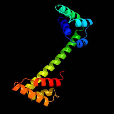
Phyre Home Retrieve Phyre Job Id
Emaill.a.kelley@imperial.ac.uk
DescriptionP0A8X0
DateThu Jan 5 11:08:53 GMT 2012
Unique Job IDdd335e3edbd2802c

Summary 

Top model
Image coloured by rainbow N → C terminus
Model (left) based on template d2p0ta1
Top template information
Fold:PSPTO4464-like
Superfamily:PSPTO4464-like
Family:PSPTO4464-like
Confidence and coverage
Confidence:100.0% Coverage: 80%
147 residues ( 80% of your sequence) have been modelled with 100.0% confidence by the single highest scoring template.
3D viewing
Interactive 3D view in Jmol

Sequence analysis 

Secondary structure and disorder prediction 

   1........10.........20.........30.........40.........50.........60
Sequence  MTKQPEDWLDDVPGDDIEDEDDEIIWVSKSEIKRDAEELKRLGAEIVDLGKNALDKIPLD
Secondary structure 


SS confidence 


Disorder  ???????????????????????
????


?


Disorder confidence 


   .........70.........80.........90.........100.........110.........120
Sequence  ADLRAAIELAQRIKMEGRRRQLQLIGKMLRQRDVEPIRQALDKLKNRHNQQVVLFHKLEN
Secondary structure 


SS confidence 


Disorder 


?


?????


Disorder confidence 


   .........130.........140.........150.........160.........170.........180
Sequence  LRDRLIDQGDDAIAEVLNLWPDADRQQLRTLIRNAKKEKEGNKPPKSARQIFQYLRELAE
Secondary structure 


SS confidence 


Disorder 


??????


??
Disorder confidence 


   ...
Sequence  NEG
Secondary structure 


SS confidence 


Disorder  ???
Disorder confidence 


Confidence Key
High(9)                    Low (0)
?Disordered
Alpha helix
Beta strand

Domain analysis 

Hover over an aligned region to see model and summary info

Please note, only up to the top 20 hits are modelled to reduce computer load

RankAligned region

PDB 2p0t chain A domain 1

3D model

Region: 36 - 182
Aligned: 147
Modelled: 147
Confidence: 100.0%
Identity: 41%
Fold: PSPTO4464-like
Superfamily: PSPTO4464-like
Family: PSPTO4464-like

Phyre2

PDB 2p0t chain A

3D model

Region: 36 - 182
Aligned: 147
Modelled: 147
Confidence: 100.0%
Identity: 41%
PDB header:structural genomics, unknown function
Chain: A: PDB Molecule:upf0307 protein pspto_4464;
PDBTitle: structural genomics, the crystal structure of a conserved putative2 protein from pseudomonas syringae pv. tomato str. dc3000

Phyre2

PDB 1naf chain A

3D model

Region: 85 - 183
Aligned: 90
Modelled: 99
Confidence: 60.9%
Identity: 13%
PDB header:signaling protein, membrane protein
Chain: A: PDB Molecule:adp-ribosylation factor binding protein gga1;
PDBTitle: crystal structure of the human gga1 gat domain

Phyre2

PDB 2kpq chain A

3D model

Region: 131 - 172
Aligned: 42
Modelled: 42
Confidence: 50.1%
Identity: 19%
PDB header:structural genomics, unknown function
Chain: A: PDB Molecule:uncharacterized protein;
PDBTitle: nmr structure of agrobacterium tumefaciens protein atu1219:2 northeast structural genomics consortium target att14

Phyre2

PDB 2kp6 chain A

3D model

Region: 116 - 158
Aligned: 43
Modelled: 43
Confidence: 42.9%
Identity: 26%
PDB header:structural genomics, unknown function
Chain: A: PDB Molecule:uncharacterized protein;
PDBTitle: solution nmr structure of protein cv0237 from2 chromobacterium violaceum. northeast structural genomics3 consortium (nesg) target cvt1

Phyre2

PDB 1tp6 chain A

3D model

Region: 109 - 155
Aligned: 45
Modelled: 47
Confidence: 40.8%
Identity: 20%
Fold: Cystatin-like
Superfamily: NTF2-like
Family: PA1314-like

Phyre2

PDB 3opc chain B

3D model

Region: 97 - 162
Aligned: 66
Modelled: 66
Confidence: 37.1%
Identity: 18%
PDB header:chaperone
Chain: B: PDB Molecule:uncharacterized protein;
PDBTitle: crystal structure of flgn chaperone from bordetella pertussis

Phyre2

PDB 3c1d chain A

3D model

Region: 87 - 175
Aligned: 86
Modelled: 89
Confidence: 26.2%
Identity: 13%
PDB header:recombination, dna binding protein
Chain: A: PDB Molecule:regulatory protein recx;
PDBTitle: x-ray crystal structure of recx

Phyre2

PDB 1oxz chain A

3D model

Region: 85 - 183
Aligned: 90
Modelled: 99
Confidence: 25.9%
Identity: 13%
Fold: Spectrin repeat-like
Superfamily: GAT-like domain
Family: GAT domain

Phyre2

PDB 1oxz chain A

3D model

Region: 85 - 183
Aligned: 90
Modelled: 99
Confidence: 25.9%
Identity: 13%
PDB header:membrane protein
Chain: A: PDB Molecule:adp-ribosylation factor binding protein gga1;
PDBTitle: crystal structure of the human gga1 gat domain

Phyre2

PDB 2bcw chain C

3D model

Region: 117 - 155
Aligned: 38
Modelled: 39
Confidence: 22.2%
Identity: 16%
PDB header:ribosome
Chain: C: PDB Molecule:elongation factor g;
PDBTitle: coordinates of the n-terminal domain of ribosomal protein2 l11,c-terminal domain of ribosomal protein l7/l12 and a3 portion of the g' domain of elongation factor g, as fitted4 into cryo-em map of an escherichia coli 70s*ef-5 g*gdp*fusidic acid complex

Phyre2

PDB 1yt3 chain A domain 2

3D model

Region: 118 - 142
Aligned: 22
Modelled: 25
Confidence: 20.2%
Identity: 23%
Fold: SAM domain-like
Superfamily: HRDC-like
Family: RNase D C-terminal domains

Phyre2

PDB 2oc6 chain A domain 1

3D model

Region: 131 - 155
Aligned: 25
Modelled: 25
Confidence: 18.5%
Identity: 36%
Fold: Secretion chaperone-like
Superfamily: YdhG-like
Family: YdhG-like

Phyre2

PDB 2dhy chain A

3D model

Region: 128 - 154
Aligned: 27
Modelled: 27
Confidence: 14.2%
Identity: 15%
PDB header:immune system
Chain: A: PDB Molecule:cue domain-containing protein 1;
PDBTitle: solution structure of the cue domain in the human cue2 domain containing protein 1 (cuedc1)

Phyre2

PDB 2i8d chain A domain 1

3D model

Region: 133 - 155
Aligned: 23
Modelled: 19
Confidence: 13.2%
Identity: 26%
Fold: Secretion chaperone-like
Superfamily: YdhG-like
Family: YdhG-like

Phyre2

PDB 1fs2 chain B domain 1

3D model

Region: 120 - 172
Aligned: 53
Modelled: 53
Confidence: 12.9%
Identity: 13%
Fold: Skp1 dimerisation domain-like
Superfamily: Skp1 dimerisation domain-like
Family: Skp1 dimerisation domain-like

Phyre2

PDB 1fs1 chain B domain 1

3D model

Region: 121 - 172
Aligned: 52
Modelled: 52
Confidence: 12.5%
Identity: 13%
Fold: Skp1 dimerisation domain-like
Superfamily: Skp1 dimerisation domain-like
Family: Skp1 dimerisation domain-like

Phyre2

PDB 3k6q chain B

3D model

Region: 61 - 101
Aligned: 41
Modelled: 41
Confidence: 10.5%
Identity: 15%
PDB header:ligand binding protein
Chain: B: PDB Molecule:putative ligand binding protein;
PDBTitle: crystal structure of an antitoxin part of a putative toxin/antitoxin2 system (swol_0700) from syntrophomonas wolfei subsp. wolfei at 1.80 a3 resolution

Phyre2

PDB 1nex chain A domain 1

3D model

Region: 120 - 172
Aligned: 53
Modelled: 53
Confidence: 10.1%
Identity: 11%
Fold: Skp1 dimerisation domain-like
Superfamily: Skp1 dimerisation domain-like
Family: Skp1 dimerisation domain-like

Phyre2

PDB 2ovr chain A domain 1

3D model

Region: 120 - 172
Aligned: 53
Modelled: 53
Confidence: 9.4%
Identity: 13%
Fold: Skp1 dimerisation domain-like
Superfamily: Skp1 dimerisation domain-like
Family: Skp1 dimerisation domain-like

Phyre2
1

d2p0ta1
2

c2p0tA_
3

c1nafA_
4

c2kpqA_
5

c2kp6A_
6

d1tp6a_
7

c3opcB_
8

c3c1dA_
9

d1oxza_
10

c1oxzA_
11

c2bcwC_
12

d1yt3a2
13

d2oc6a1
14

c2dhyA_
15

d2i8da1
16

d1fs2b1
17

d1fs1b1
18

c3k6qB_
19

d1nexa1
20

d2ovra1
21


22


23


24


25


26


27


28


29


30


31


32


33


34


35


36


37


38


39


40


41


42


43


44


45


Detailed template information 

#
Template Alignment Coverage3D Model Confidence
% i.d. Template Information
1d2p0ta1


100.0 41 Fold:PSPTO4464-like
Superfamily:PSPTO4464-like
Family:PSPTO4464-like
2c2p0tA_


100.0 41 PDB header:structural genomics, unknown function
Chain: A: PDB Molecule:upf0307 protein pspto_4464;
PDBTitle: structural genomics, the crystal structure of a conserved putative2 protein from pseudomonas syringae pv. tomato str. dc3000
3c1nafA_


60.9 13 PDB header:signaling protein, membrane protein
Chain: A: PDB Molecule:adp-ribosylation factor binding protein gga1;
PDBTitle: crystal structure of the human gga1 gat domain
4c2kpqA_


50.1 19 PDB header:structural genomics, unknown function
Chain: A: PDB Molecule:uncharacterized protein;
PDBTitle: nmr structure of agrobacterium tumefaciens protein atu1219:2 northeast structural genomics consortium target att14
5c2kp6A_


42.9 26 PDB header:structural genomics, unknown function
Chain: A: PDB Molecule:uncharacterized protein;
PDBTitle: solution nmr structure of protein cv0237 from2 chromobacterium violaceum. northeast structural genomics3 consortium (nesg) target cvt1
6d1tp6a_


40.8 20 Fold:Cystatin-like
Superfamily:NTF2-like
Family:PA1314-like
7c3opcB_


37.1 18 PDB header:chaperone
Chain: B: PDB Molecule:uncharacterized protein;
PDBTitle: crystal structure of flgn chaperone from bordetella pertussis
8c3c1dA_


26.2 13 PDB header:recombination, dna binding protein
Chain: A: PDB Molecule:regulatory protein recx;
PDBTitle: x-ray crystal structure of recx
9d1oxza_


25.9 13 Fold:Spectrin repeat-like
Superfamily:GAT-like domain
Family:GAT domain
10c1oxzA_


25.9 13 PDB header:membrane protein
Chain: A: PDB Molecule:adp-ribosylation factor binding protein gga1;
PDBTitle: crystal structure of the human gga1 gat domain
11c2bcwC_


22.2 16 PDB header:ribosome
Chain: C: PDB Molecule:elongation factor g;
PDBTitle: coordinates of the n-terminal domain of ribosomal protein2 l11,c-terminal domain of ribosomal protein l7/l12 and a3 portion of the g' domain of elongation factor g, as fitted4 into cryo-em map of an escherichia coli 70s*ef-5 g*gdp*fusidic acid complex
12d1yt3a2


20.2 23 Fold:SAM domain-like
Superfamily:HRDC-like
Family:RNase D C-terminal domains
13d2oc6a1


18.5 36 Fold:Secretion chaperone-like
Superfamily:YdhG-like
Family:YdhG-like
14c2dhyA_


14.2 15 PDB header:immune system
Chain: A: PDB Molecule:cue domain-containing protein 1;
PDBTitle: solution structure of the cue domain in the human cue2 domain containing protein 1 (cuedc1)
15d2i8da1


13.2 26 Fold:Secretion chaperone-like
Superfamily:YdhG-like
Family:YdhG-like
16d1fs2b1


12.9 13 Fold:Skp1 dimerisation domain-like
Superfamily:Skp1 dimerisation domain-like
Family:Skp1 dimerisation domain-like
17d1fs1b1


12.5 13 Fold:Skp1 dimerisation domain-like
Superfamily:Skp1 dimerisation domain-like
Family:Skp1 dimerisation domain-like
18c3k6qB_


10.5 15 PDB header:ligand binding protein
Chain: B: PDB Molecule:putative ligand binding protein;
PDBTitle: crystal structure of an antitoxin part of a putative toxin/antitoxin2 system (swol_0700) from syntrophomonas wolfei subsp. wolfei at 1.80 a3 resolution
19d1nexa1


10.1 11 Fold:Skp1 dimerisation domain-like
Superfamily:Skp1 dimerisation domain-like
Family:Skp1 dimerisation domain-like
20d2ovra1


9.4 13 Fold:Skp1 dimerisation domain-like
Superfamily:Skp1 dimerisation domain-like
Family:Skp1 dimerisation domain-like
21d2izva1


not modelled 9.4 12 Fold:SOCS box-like
Superfamily:SOCS box-like
Family:SOCS box-like
22c3oq9C_


not modelled 9.2 9 PDB header:apoptosis
Chain: C: PDB Molecule:tumor necrosis factor receptor superfamily member 6;
PDBTitle: structure of the fas/fadd death domain assembly
23c2kl4A_


not modelled 9.1 20 PDB header:structural genomics, unknown function
Chain: A: PDB Molecule:bh2032 protein;
PDBTitle: nmr structure of the protein nb7804a
24d1fkma2


not modelled 8.2 17 Fold:Left-handed superhelix
Superfamily:Ypt/Rab-GAP domain of gyp1p
Family:Ypt/Rab-GAP domain of gyp1p
25c3tj3C_


not modelled 7.9 15 PDB header:protein transport
Chain: C: PDB Molecule:nuclear pore complex protein nup50;
PDBTitle: structure of importin a5 bound to the n-terminus of nup50
26d1k91a_


not modelled 7.7 43 Fold:P-domain of calnexin/calreticulin
Superfamily:P-domain of calnexin/calreticulin
Family:P-domain of calnexin/calreticulin
27c2rqpA_


not modelled 7.2 22 PDB header:gene regulation
Chain: A: PDB Molecule:heterochromatin protein 1-binding protein 3;
PDBTitle: the solution structure of heterochromatin protein 1-binding2 protein 74 histone h1 like domain
28d2igsa1


not modelled 6.6 20 Fold:Lysozyme-like
Superfamily:Lysozyme-like
Family:PA2222-like
29d1in0a1


not modelled 6.6 18 Fold:Ferredoxin-like
Superfamily:YajQ-like
Family:YajQ-like
30c3dhyC_


not modelled 6.4 18 PDB header:hydrolase
Chain: C: PDB Molecule:adenosylhomocysteinase;
PDBTitle: crystal structures of mycobacterium tuberculosis s-adenosyl-l-2 homocysteine hydrolase in ternary complex with substrate and3 inhibitors
31d1g8eb_


not modelled 6.4 11 Fold:Flagellar transcriptional activator FlhD
Superfamily:Flagellar transcriptional activator FlhD
Family:Flagellar transcriptional activator FlhD
32d2es9a1


not modelled 6.0 45 Fold:YoaC-like
Superfamily:YoaC-like
Family:YoaC-like
33c3gehA_


not modelled 6.0 20 PDB header:hydrolase
Chain: A: PDB Molecule:trna modification gtpase mnme;
PDBTitle: crystal structure of mnme from nostoc in complex with gdp, folinic2 acid and zn
34d2choa2


not modelled 5.9 27 Fold:TIM beta/alpha-barrel
Superfamily:(Trans)glycosidases
Family:alpha-D-glucuronidase/Hyaluronidase catalytic domain
35c2kqrA_


not modelled 5.8 13 PDB header:ligase
Chain: A: PDB Molecule:asparaginyl-trna synthetase, cytoplasmic;
PDBTitle: solution structure of the n-terminal domain (residues 1-111) of brugia2 malayi asparaginyl-trna synthetase
36d1sm2a_


not modelled 5.8 24 Fold:Protein kinase-like (PK-like)
Superfamily:Protein kinase-like (PK-like)
Family:Protein kinases, catalytic subunit
37d2cbia2


not modelled 5.7 20 Fold:TIM beta/alpha-barrel
Superfamily:(Trans)glycosidases
Family:alpha-D-glucuronidase/Hyaluronidase catalytic domain
38d1li4a2


not modelled 5.6 12 Fold:Flavodoxin-like
Superfamily:Formate/glycerate dehydrogenase catalytic domain-like
Family:S-adenosylhomocystein hydrolase
39d1v8ba2


not modelled 5.4 23 Fold:Flavodoxin-like
Superfamily:Formate/glycerate dehydrogenase catalytic domain-like
Family:S-adenosylhomocystein hydrolase
40c2j8pA_


not modelled 5.4 17 PDB header:nuclear protein
Chain: A: PDB Molecule:cleavage stimulation factor 64 kda subunit;
PDBTitle: nmr structure of c-terminal domain of human cstf-64
41c1d4fD_


not modelled 5.4 12 PDB header:hydrolase
Chain: D: PDB Molecule:s-adenosylhomocysteine hydrolase;
PDBTitle: crystal structure of recombinant rat-liver d244e mutant s-2 adenosylhomocysteine hydrolase
42c3d5lA_


not modelled 5.4 16 PDB header:signaling protein
Chain: A: PDB Molecule:regulatory protein recx;
PDBTitle: crystal structure of regulatory protein recx
43d2edua1


not modelled 5.3 10 Fold:SAM domain-like
Superfamily:RuvA domain 2-like
Family:ComEA-like
44d2esna1


not modelled 5.1 15 Fold:DNA/RNA-binding 3-helical bundle
Superfamily:"Winged helix" DNA-binding domain
Family:LysR-like transcriptional regulators
45d1g8ea_


not modelled 5.0 11 Fold:Flagellar transcriptional activator FlhD
Superfamily:Flagellar transcriptional activator FlhD
Family:Flagellar transcriptional activator FlhD

Binding site prediction 

Due to computational demand, binding site predictions are not run for batch jobs

If you want to predict binding sites, please manually submit your model of choice to 3DLigandSite


Phyre is for academic use only

Please cite: Protein structure prediction on the web: a case study using the Phyre server
Kelley LA and Sternberg MJE. Nature Protocols 4, 363 - 371 (2009) [pdf] [Import into BibTeX]
 
If you use the binding site predictions from 3DLigandSite, please also cite:
3DLigandSite: predicting ligand-binding sites using similar structures.
Wass MN, Kelley LA and Sternberg MJ Nucleic Acids Research 38, W469-73 (2010) [PubMed]
 
© Structural Bioinformatics Group
Imperial College London
Lawrence Kelley, Benjamin Jefferys
Disclaimer
Terms and Conditions
Component software
Template detection: HHpred 1.51
Secondary structure prediction: Psi-pred 2.5
Disorder prediction: Disopred 2.4
Transmembrane prediction: Memsat_SVM
Multi-template modelling and ab initio: Poing 1.0