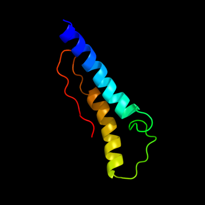
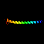
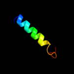
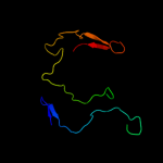
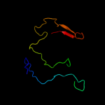
1 c2kz6A_
97.5
22
PDB header: structural genomics, unknown functionChain: A: PDB Molecule: uncharacterized protein;PDBTitle: solution structure of protein cv0426 from chromobacterium violaceum,2 northeast structural genomics consortium (nesg) target cvt2
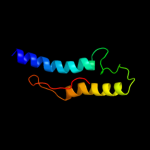
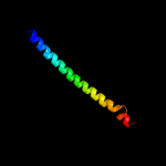
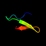
2 d1jofa_
46.0
17
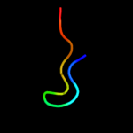
Fold: 7-bladed beta-propellerSuperfamily: 3-carboxy-cis,cis-mucoante lactonizing enzymeFamily: 3-carboxy-cis,cis-mucoante lactonizing enzyme3 c1gk6B_
39.6
20
PDB header: vimentinChain: B: PDB Molecule: vimentin;PDBTitle: human vimentin coil 2b fragment linked to gcn4 leucine2 zipper (z2b)
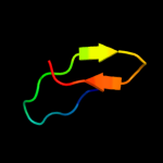
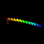
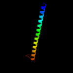
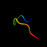
4 c2xv5A_
37.5
24
PDB header: structural proteinChain: A: PDB Molecule: lamin-a/c;PDBTitle: human lamin a coil 2b fragment
5 c1rfoC_
34.5
25
PDB header: viral proteinChain: C: PDB Molecule: whisker antigen control protein;PDBTitle: trimeric foldon of the t4 phagehead fibritin
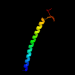
6 c2kt9A_
33.7
20
PDB header: ribosomal proteinChain: A: PDB Molecule: probable 30s ribosomal protein psrp-3;PDBTitle: solution nmr structure of probable 30s ribosomal protein2 psrp-3 (ycf65-like protein) from synechocystis sp. (strain3 pcc 6803), northeast structural genomics consortium target4 target sgr46
7 c3o0rC_
32.6
15
PDB header: immune system/oxidoreductaseChain: C: PDB Molecule: nitric oxide reductase subunit c;PDBTitle: crystal structure of nitric oxide reductase from pseudomonas2 aeruginosa in complex with antibody fragment
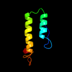
8 c1x8yA_
27.0
24
PDB header: structural proteinChain: A: PDB Molecule: lamin a/c;PDBTitle: human lamin coil 2b
9 c1v1hB_
25.7
25
PDB header: adenovirusChain: B: PDB Molecule: fibritin, fiber protein;PDBTitle: adenovirus fibre shaft sequence n-terminally fused to the2 bacteriophage t4 fibritin foldon trimerisation motif with3 a short linker
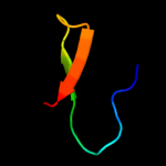
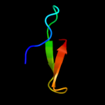
10 c2jxwA_
25.2
21
PDB header: formin binding proteinChain: A: PDB Molecule: ww domain-binding protein 4;PDBTitle: solution structure of the tandem ww domains of fbp21
11 c1k6nH_
24.3
13
PDB header: photosynthesisChain: H: PDB Molecule: photosynthetic reaction center h subunit;PDBTitle: e(l212)a,d(l213)a double mutant structure of photosynthetic reaction2 center from rhodobacter sphaeroides
12 c1gk4A_
23.7
26
PDB header: vimentinChain: A: PDB Molecule: vimentin;PDBTitle: human vimentin coil 2b fragment (cys2)
13 c1ox3A_
23.4
25
PDB header: chaperoneChain: A: PDB Molecule: fibritin;PDBTitle: crystal structure of mini-fibritin
14 c3movB_
22.3
22
PDB header: structural proteinChain: B: PDB Molecule: lamin-b1;PDBTitle: crystal structure of human lamin-b1 coil 2 segment
15 d1rzhh1
21.9
13
Fold: PRC-barrel domainSuperfamily: PRC-barrel domainFamily: Photosynthetic reaction centre, H-chain, cytoplasmic domain16 c1avyA_
21.2
25
PDB header: coiled coilChain: A: PDB Molecule: fibritin;PDBTitle: fibritin deletion mutant m (bacteriophage t4)
17 c2ov2O_
15.8
20
PDB header: protein binding/transferaseChain: O: PDB Molecule: serine/threonine-protein kinase pak 4;PDBTitle: the crystal structure of the human rac3 in complex with the crib2 domain of human p21-activated kinase 4 (pak4)
18 c2odbB_
14.1
20
PDB header: protein bindingChain: B: PDB Molecule: serine/threonine-protein kinase pak 6;PDBTitle: the crystal structure of human cdc42 in complex with the crib domain2 of human p21-activated kinase 6 (pak6)
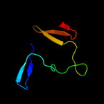
19 d2rm0w1
13.8
26
Fold: WW domain-likeSuperfamily: WW domainFamily: WW domain20 d2dk1a1
12.5
30
Fold: WW domain-likeSuperfamily: WW domainFamily: WW domain21 c2diiA_
not modelled
12.3
23
PDB header: transcriptionChain: A: PDB Molecule: tfiih basal transcription factor complex p62PDBTitle: solution structure of the bsd domain of human tfiih basal2 transcription factor complex p62 subunit
22 d2diia1
not modelled
12.2
23
Fold: BSD domain-likeSuperfamily: BSD domain-likeFamily: BSD domain23 c2v43A_
not modelled
12.1
20
PDB header: regulatorChain: A: PDB Molecule: sigma-e factor regulatory protein rseb;PDBTitle: crystal structure of rseb: a sensor for periplasmic stress2 response in e. coli
24 d1ln1a_
not modelled
11.3
17
Fold: TBP-likeSuperfamily: Bet v1-likeFamily: STAR domain25 c2dk7A_
not modelled
10.5
13
PDB header: transcriptionChain: A: PDB Molecule: transcription elongation regulator 1;PDBTitle: solution structure of ww domain in transcription elongation2 regulator 1
26 c2ghsA_
not modelled
10.5
14
PDB header: calcium-binding proteinChain: A: PDB Molecule: agr_c_1268p;PDBTitle: crystal structure of a calcium-binding protein, regucalcin2 (agr_c_1268) from agrobacterium tumefaciens str. c58 at 1.55 a3 resolution
27 d2ghsa1
not modelled
10.5
14
Fold: 6-bladed beta-propellerSuperfamily: Calcium-dependent phosphotriesteraseFamily: SGL-like28 d2oa5a1
not modelled
10.3
23
Fold: BLRF2-likeSuperfamily: BLRF2-likeFamily: BLRF2-like29 d1x4ta1
not modelled
9.3
22
Fold: Long alpha-hairpinSuperfamily: ISY1 domain-likeFamily: ISY1 N-terminal domain-like30 c1e0aB_
not modelled
8.5
50
PDB header: signalling proteinChain: B: PDB Molecule: serine/threonine-protein kinase pak-alpha;PDBTitle: cdc42 complexed with the gtpase binding domain of p212 activated kinase
31 c1w18A_
not modelled
8.4
19
PDB header: transferaseChain: A: PDB Molecule: levansucrase;PDBTitle: crystal structure of levansucrase from gluconacetobacter2 diazotrophicus
32 d1o6wa1
not modelled
8.3
26
Fold: WW domain-likeSuperfamily: WW domainFamily: WW domain33 d1ll7a2
not modelled
7.9
17
Fold: FKBP-likeSuperfamily: Chitinase insertion domainFamily: Chitinase insertion domain34 c1zr7A_
not modelled
7.3
26
PDB header: signaling proteinChain: A: PDB Molecule: huntingtin-interacting protein hypa/fbp11;PDBTitle: solution structure of the first ww domain of fbp11
35 c2c7hA_
not modelled
7.2
29
PDB header: ubiquitin-like proteinChain: A: PDB Molecule: retinoblastoma-binding protein 6, isoform 3;PDBTitle: solution nmr structure of the dwnn domain from human rbbp6
36 d1w9pa2
not modelled
6.8
25
Fold: FKBP-likeSuperfamily: Chitinase insertion domainFamily: Chitinase insertion domain37 c3nmbA_
not modelled
6.4
18
PDB header: hydrolaseChain: A: PDB Molecule: putative sugar hydrolase;PDBTitle: crystal structure of a putative sugar hydrolase (bacova_03189) from2 bacteroides ovatus at 2.40 a resolution
38 c1f3mB_
not modelled
6.4
50
PDB header: transferaseChain: B: PDB Molecule: serine/threonine-protein kinase pak-alpha;PDBTitle: crystal structure of human serine/threonine kinase pak1
39 d1pcna1
not modelled
6.3
33
Fold: Knottins (small inhibitors, toxins, lectins)Superfamily: Colipase-likeFamily: Colipase-like40 c2ysiA_
not modelled
6.3
23
PDB header: protein bindingChain: A: PDB Molecule: transcription elongation regulator 1;PDBTitle: solution structure of the first ww domain from the mouse2 transcription elongation regulator 1, transcription factor3 ca150
41 c2jz8A_
not modelled
6.2
9
PDB header: structural genomics, unknown functionChain: A: PDB Molecule: uncharacterized protein bh09830;PDBTitle: solution nmr structure of bh09830 from bartonella henselae2 modeled with one zn+2 bound. northeast structural genomics3 consortium target bnr55
42 c2dk1A_
not modelled
6.2
26
PDB header: gene regulationChain: A: PDB Molecule: ww domain-binding protein 4;PDBTitle: solution structure of ww domain in ww domain binding2 protein 4 (wbp-4)
43 c3ok8A_
not modelled
5.9
6
PDB header: protein bindingChain: A: PDB Molecule: brain-specific angiogenesis inhibitor 1-associated proteinPDBTitle: i-bar of pinkbar
44 d1wi9a_
not modelled
5.6
53
Fold: DNA/RNA-binding 3-helical bundleSuperfamily: "Winged helix" DNA-binding domainFamily: PCI domain (PINT motif)45 d2paja1
not modelled
5.6
15
Fold: Composite domain of metallo-dependent hydrolasesSuperfamily: Composite domain of metallo-dependent hydrolasesFamily: SAH/MTA deaminase-like46 d1ocya_
not modelled
5.5
14
Fold: Receptor-binding domain of short tail fibre protein gp12Superfamily: Receptor-binding domain of short tail fibre protein gp12Family: Receptor-binding domain of short tail fibre protein gp1247 c1aa0A_
not modelled
5.4
25
PDB header: attachment proteinChain: A: PDB Molecule: fibritin;PDBTitle: fibritin deletion mutant e (bacteriophage t4)
48 c2h47F_
not modelled
5.2
17
PDB header: oxidoreductase/electron transportChain: F: PDB Molecule: aromatic amine dehydrogenase;PDBTitle: crystal structure of an electron transfer complex between2 aromatic amine dephydrogenase and azurin from alcaligenes3 faecalis (form 1)
49 d1j8ra_
not modelled
5.1
31
Fold: Common fold of diphtheria toxin/transcription factors/cytochrome fSuperfamily: Bacterial adhesinsFamily: PapG adhesin receptor-binding domain