Phyre Home Retrieve Phyre Job Id
Emaill.a.kelley@imperial.ac.uk
DescriptionP45472
DateThu Jan 5 12:02:48 GMT 2012
Unique Job IDdb565b64e2b3bb38

Summary 

Top model
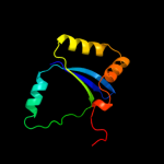
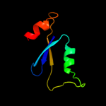
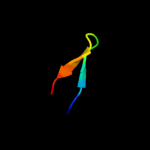
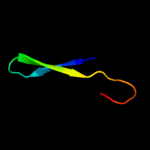
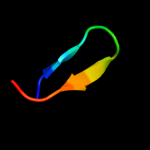
Image coloured by rainbow N → C terminus
Model (left) based on template c1zg2A_
Top template information
PDB header:structural genomics, unknown function
Chain: A: PDB Molecule:hypothetical upf0213 protein bh0048;
PDBTitle: solution nmr structure of the upf0213 protein bh0048 from2 bacillus halodurans. northeast structural genomics target3 bhr2.
Confidence and coverage
Confidence:100.0% Coverage: 93%
93 residues ( 93% of your sequence) have been modelled with 100.0% confidence by the single highest scoring template.
3D viewing
Interactive 3D view in Jmol

Sequence analysis 

Secondary structure and disorder prediction 

   1........10.........20.........30.........40.........50.........60
Sequence  MTPWFLYLIRTADNKLYTGITTDVERRYQQHQSGKGAKALRGKGELTLVFSAPVGDRSLA
Secondary structure 


SS confidence 


Disorder  ???


Disorder confidence 


   .........70.........80.........90.........100
Sequence  LRAEYRVKQLTKRQKERLVAEGAGFAELLSSLQTPEIKSD
Secondary structure 


SS confidence 


Disorder 


???????
Disorder confidence 


Confidence Key
High(9)                    Low (0)
?Disordered
Alpha helix
Beta strand

Domain analysis 

Hover over an aligned region to see model and summary info

Please note, only up to the top 20 hits are modelled to reduce computer load

RankAligned region

PDB 1zg2 chain A

3D model

Region: 3 - 95
Aligned: 93
Modelled: 93
Confidence: 100.0%
Identity: 42%
PDB header:structural genomics, unknown function
Chain: A: PDB Molecule:hypothetical upf0213 protein bh0048;
PDBTitle: solution nmr structure of the upf0213 protein bh0048 from2 bacillus halodurans. northeast structural genomics target3 bhr2.

Phyre2

PDB 1ywl chain A

3D model

Region: 2 - 81
Aligned: 80
Modelled: 80
Confidence: 100.0%
Identity: 34%
PDB header:structural genomics, unknown function
Chain: A: PDB Molecule:hypothetical upf0213 protein ef2693;
PDBTitle: solution nmr structure of the protein ef2693 from e.2 faecalis: northeast structural genomics consortium target3 efr36

Phyre2

PDB 1yd6 chain A

3D model

Region: 6 - 67
Aligned: 62
Modelled: 62
Confidence: 98.3%
Identity: 16%
PDB header:dna binding protein
Chain: A: PDB Molecule:uvrc;
PDBTitle: crystal structure of the giy-yig n-terminal endonuclease2 domain of uvrc from bacillus caldotenax

Phyre2

PDB 1yd2 chain A

3D model

Region: 6 - 67
Aligned: 62
Modelled: 62
Confidence: 97.8%
Identity: 15%
PDB header:dna binding protein
Chain: A: PDB Molecule:uvrabc system protein c;
PDBTitle: crystal structure of the giy-yig n-terminal endonuclease domain of2 uvrc from thermotoga maritima: point mutant y19f bound to the3 catalytic divalent cation

Phyre2

PDB 1mk0 chain A

3D model

Region: 1 - 67
Aligned: 66
Modelled: 67
Confidence: 97.5%
Identity: 21%
Fold: GIY-YIG endonuclease
Superfamily: GIY-YIG endonuclease
Family: GIY-YIG endonuclease

Phyre2

PDB 2wsh chain C

3D model

Region: 6 - 67
Aligned: 62
Modelled: 62
Confidence: 89.9%
Identity: 6%
PDB header:hydrolase
Chain: C: PDB Molecule:endonuclease ii;
PDBTitle: structure of bacteriophage t4 endoii e118a mutant

Phyre2

PDB 3h8t chain A

3D model

Region: 6 - 17
Aligned: 12
Modelled: 12
Confidence: 22.0%
Identity: 25%
PDB header:heme-binding protein
Chain: A: PDB Molecule:hmuy;
PDBTitle: structure of porphyromonas gingivalis heme-binding protein hmuy in2 complex with heme

Phyre2

PDB 1f0c chain B

3D model

Region: 5 - 19
Aligned: 15
Modelled: 15
Confidence: 21.0%
Identity: 53%
PDB header:viral protein
Chain: B: PDB Molecule:ice inhibitor;
PDBTitle: structure of the viral serpin crma

Phyre2

PDB 1a6q chain A domain 1

3D model

Region: 59 - 99
Aligned: 41
Modelled: 41
Confidence: 17.2%
Identity: 15%
Fold: Another 3-helical bundle
Superfamily: Protein serine/threonine phosphatase 2C, C-terminal domain
Family: Protein serine/threonine phosphatase 2C, C-terminal domain

Phyre2

PDB 2yrv chain A

3D model

Region: 7 - 18
Aligned: 12
Modelled: 12
Confidence: 11.0%
Identity: 42%
PDB header:transcription
Chain: A: PDB Molecule:at-rich interactive domain-containing protein 4a;
PDBTitle: solution structure of the rbb1nt domain of human2 rb(retinoblastoma)-binding protein 1

Phyre2

PDB 1ylx chain A domain 1

3D model

Region: 6 - 23
Aligned: 18
Modelled: 18
Confidence: 10.4%
Identity: 22%
Fold: N domain of copper amine oxidase-like
Superfamily: GK1464-like
Family: GK1464-like

Phyre2

PDB 2xk0 chain A

3D model

Region: 8 - 19
Aligned: 12
Modelled: 12
Confidence: 9.4%
Identity: 33%
PDB header:transcription
Chain: A: PDB Molecule:polycomb protein pcl;
PDBTitle: solution structure of the tudor domain from drosophila2 polycomblike (pcl)

Phyre2

PDB 2z3g chain A domain 1

3D model

Region: 8 - 20
Aligned: 13
Modelled: 13
Confidence: 9.4%
Identity: 23%
Fold: Cytidine deaminase-like
Superfamily: Cytidine deaminase-like
Family: Cytidine deaminase

Phyre2

PDB 3oj6 chain C

3D model

Region: 8 - 20
Aligned: 13
Modelled: 13
Confidence: 9.3%
Identity: 15%
PDB header:hydrolase
Chain: C: PDB Molecule:blasticidin-s deaminase;
PDBTitle: crystal structure of blasticidin s deaminase from coccidioides immitis

Phyre2

PDB 1lkt chain A

3D model

Region: 10 - 19
Aligned: 10
Modelled: 10
Confidence: 8.9%
Identity: 40%
Fold: Head-binding domain of phage P22 tailspike protein
Superfamily: Head-binding domain of phage P22 tailspike protein
Family: Head-binding domain of phage P22 tailspike protein

Phyre2

PDB 3orc chain A

3D model

Region: 6 - 27
Aligned: 22
Modelled: 22
Confidence: 8.5%
Identity: 27%
Fold: lambda repressor-like DNA-binding domains
Superfamily: lambda repressor-like DNA-binding domains
Family: Phage repressors

Phyre2

PDB 2fr5 chain A domain 1

3D model

Region: 8 - 20
Aligned: 13
Modelled: 13
Confidence: 8.3%
Identity: 23%
Fold: Cytidine deaminase-like
Superfamily: Cytidine deaminase-like
Family: Cytidine deaminase

Phyre2

PDB 3b8f chain B

3D model

Region: 9 - 20
Aligned: 12
Modelled: 12
Confidence: 8.3%
Identity: 42%
PDB header:hydrolase
Chain: B: PDB Molecule:putative blasticidin s deaminase;
PDBTitle: crystal structure of the cytidine deaminase from bacillus anthracis

Phyre2

PDB 2eqj chain A

3D model

Region: 8 - 19
Aligned: 12
Modelled: 12
Confidence: 7.8%
Identity: 42%
PDB header:transcription
Chain: A: PDB Molecule:metal-response element-binding transcription
PDBTitle: solution structure of the tudor domain of metal-response2 element-binding transcription factor 2

Phyre2

PDB 1mq0 chain A

3D model

Region: 8 - 20
Aligned: 13
Modelled: 13
Confidence: 7.8%
Identity: 15%
Fold: Cytidine deaminase-like
Superfamily: Cytidine deaminase-like
Family: Cytidine deaminase

Phyre2
1

c1zg2A_
2

c1ywlA_
3

c1yd6A_
4

c1yd2A_
5

d1mk0a_
6

c2wshC_
7

c3h8tA_
8

c1f0cB_
9

d1a6qa1
10

c2yrvA_
11

d1ylxa1
12

c2xk0A_
13

d2z3ga1
14

c3oj6C_
15

d1lkta_
16

d3orca_
17

d2fr5a1
18

c3b8fB_
19

c2eqjA_
20

d1mq0a_
21


22


23


24


25


26


27


28


29


30


31


32


33


34


35


Detailed template information 

#
Template Alignment Coverage3D Model Confidence
% i.d. Template Information
1c1zg2A_


100.0 42 PDB header:structural genomics, unknown function
Chain: A: PDB Molecule:hypothetical upf0213 protein bh0048;
PDBTitle: solution nmr structure of the upf0213 protein bh0048 from2 bacillus halodurans. northeast structural genomics target3 bhr2.
2c1ywlA_


100.0 34 PDB header:structural genomics, unknown function
Chain: A: PDB Molecule:hypothetical upf0213 protein ef2693;
PDBTitle: solution nmr structure of the protein ef2693 from e.2 faecalis: northeast structural genomics consortium target3 efr36
3c1yd6A_


98.3 16 PDB header:dna binding protein
Chain: A: PDB Molecule:uvrc;
PDBTitle: crystal structure of the giy-yig n-terminal endonuclease2 domain of uvrc from bacillus caldotenax
4c1yd2A_


97.8 15 PDB header:dna binding protein
Chain: A: PDB Molecule:uvrabc system protein c;
PDBTitle: crystal structure of the giy-yig n-terminal endonuclease domain of2 uvrc from thermotoga maritima: point mutant y19f bound to the3 catalytic divalent cation
5d1mk0a_


97.5 21 Fold:GIY-YIG endonuclease
Superfamily:GIY-YIG endonuclease
Family:GIY-YIG endonuclease
6c2wshC_


89.9 6 PDB header:hydrolase
Chain: C: PDB Molecule:endonuclease ii;
PDBTitle: structure of bacteriophage t4 endoii e118a mutant
7c3h8tA_


22.0 25 PDB header:heme-binding protein
Chain: A: PDB Molecule:hmuy;
PDBTitle: structure of porphyromonas gingivalis heme-binding protein hmuy in2 complex with heme
8c1f0cB_


21.0 53 PDB header:viral protein
Chain: B: PDB Molecule:ice inhibitor;
PDBTitle: structure of the viral serpin crma
9d1a6qa1


17.2 15 Fold:Another 3-helical bundle
Superfamily:Protein serine/threonine phosphatase 2C, C-terminal domain
Family:Protein serine/threonine phosphatase 2C, C-terminal domain
10c2yrvA_


11.0 42 PDB header:transcription
Chain: A: PDB Molecule:at-rich interactive domain-containing protein 4a;
PDBTitle: solution structure of the rbb1nt domain of human2 rb(retinoblastoma)-binding protein 1
11d1ylxa1


10.4 22 Fold:N domain of copper amine oxidase-like
Superfamily:GK1464-like
Family:GK1464-like
12c2xk0A_


9.4 33 PDB header:transcription
Chain: A: PDB Molecule:polycomb protein pcl;
PDBTitle: solution structure of the tudor domain from drosophila2 polycomblike (pcl)
13d2z3ga1


9.4 23 Fold:Cytidine deaminase-like
Superfamily:Cytidine deaminase-like
Family:Cytidine deaminase
14c3oj6C_


9.3 15 PDB header:hydrolase
Chain: C: PDB Molecule:blasticidin-s deaminase;
PDBTitle: crystal structure of blasticidin s deaminase from coccidioides immitis
15d1lkta_


8.9 40 Fold:Head-binding domain of phage P22 tailspike protein
Superfamily:Head-binding domain of phage P22 tailspike protein
Family:Head-binding domain of phage P22 tailspike protein
16d3orca_


8.5 27 Fold:lambda repressor-like DNA-binding domains
Superfamily:lambda repressor-like DNA-binding domains
Family:Phage repressors
17d2fr5a1


8.3 23 Fold:Cytidine deaminase-like
Superfamily:Cytidine deaminase-like
Family:Cytidine deaminase
18c3b8fB_


8.3 42 PDB header:hydrolase
Chain: B: PDB Molecule:putative blasticidin s deaminase;
PDBTitle: crystal structure of the cytidine deaminase from bacillus anthracis
19c2eqjA_


7.8 42 PDB header:transcription
Chain: A: PDB Molecule:metal-response element-binding transcription
PDBTitle: solution structure of the tudor domain of metal-response2 element-binding transcription factor 2
20d1mq0a_


7.8 15 Fold:Cytidine deaminase-like
Superfamily:Cytidine deaminase-like
Family:Cytidine deaminase
21d2d30a1


not modelled 7.8 38 Fold:Cytidine deaminase-like
Superfamily:Cytidine deaminase-like
Family:Cytidine deaminase
22c3r2nC_


not modelled 7.7 31 PDB header:hydrolase
Chain: C: PDB Molecule:cytidine deaminase;
PDBTitle: crystal structure of cytidine deaminase from mycobacterium leprae
23c3ijfX_


not modelled 7.6 23 PDB header:hydrolase
Chain: X: PDB Molecule:cytidine deaminase;
PDBTitle: crystal structure of cytidine deaminase from mycobacterium2 tuberculosis
24c2diiA_


not modelled 7.1 20 PDB header:transcription
Chain: A: PDB Molecule:tfiih basal transcription factor complex p62
PDBTitle: solution structure of the bsd domain of human tfiih basal2 transcription factor complex p62 subunit
25d1d0na5


not modelled 7.1 11 Fold:Gelsolin-like
Superfamily:Actin depolymerizing proteins
Family:Gelsolin-like
26d1yu0a1


not modelled 6.7 23 Fold:Pseudo beta-prism
Superfamily:Bacteriophage trimeric proteins domain
Family:Mtd domain-like
27d1uwza_


not modelled 6.5 38 Fold:Cytidine deaminase-like
Superfamily:Cytidine deaminase-like
Family:Cytidine deaminase
28d2bj7a1


not modelled 6.5 40 Fold:Ribbon-helix-helix
Superfamily:Ribbon-helix-helix
Family:CopG-like
29c2dybA_


not modelled 6.5 28 PDB header:oxidoreductase
Chain: A: PDB Molecule:neutrophil cytosol factor 4;
PDBTitle: the crystal structure of human p40(phox)
30d2diia1


not modelled 6.4 20 Fold:BSD domain-like
Superfamily:BSD domain-like
Family:BSD domain
31d2hzab1


not modelled 6.3 40 Fold:Ribbon-helix-helix
Superfamily:Ribbon-helix-helix
Family:CopG-like
32c2e5pA_


not modelled 6.3 50 PDB header:transcription
Chain: A: PDB Molecule:phd finger protein 1;
PDBTitle: solution structure of the tudor domain of phd finger2 protein 1 (phf1 protein)
33c3dmoD_


not modelled 6.0 31 PDB header:hydrolase
Chain: D: PDB Molecule:cytidine deaminase;
PDBTitle: 1.6 a crystal structure of cytidine deaminase from2 burkholderia pseudomallei
34c2vkyB_


not modelled 5.6 40 PDB header:viral protein
Chain: B: PDB Molecule:tail protein, piigcn4;
PDBTitle: headbinding domain of phage p22 tailspike c-terminally2 fused to isoleucine zipper piigcn4 (chimera i)
35d1svya_


not modelled 5.5 7 Fold:Gelsolin-like
Superfamily:Actin depolymerizing proteins
Family:Gelsolin-like

Binding site prediction 

Due to computational demand, binding site predictions are not run for batch jobs

If you want to predict binding sites, please manually submit your model of choice to 3DLigandSite


Phyre is for academic use only

Please cite: Protein structure prediction on the web: a case study using the Phyre server
Kelley LA and Sternberg MJE. Nature Protocols 4, 363 - 371 (2009) [pdf] [Import into BibTeX]
 
If you use the binding site predictions from 3DLigandSite, please also cite:
3DLigandSite: predicting ligand-binding sites using similar structures.
Wass MN, Kelley LA and Sternberg MJ Nucleic Acids Research 38, W469-73 (2010) [PubMed]
 
© Structural Bioinformatics Group
Imperial College London
Lawrence Kelley, Benjamin Jefferys
Disclaimer
Terms and Conditions
Component software
Template detection: HHpred 1.51
Secondary structure prediction: Psi-pred 2.5
Disorder prediction: Disopred 2.4
Transmembrane prediction: Memsat_SVM
Multi-template modelling and ab initio: Poing 1.0