 |
| ||||||||||||||||||||||||
| Insertion relative to template | |
| Deletion relative to template | |
| Catalytic residue from the CSA | |
| Detailed help on interpreting your alignment | |
| 30 | . | . | . | . | . | . | . | . | . | 40 | . | . | . | . | . | . | . | . | . | 50 | . | . | . | . | . | . | . | . | . | 60 | . | . | . | . | . | . | . | . | . | 70 | |||||||||||
| Predicted Secondary structure |  |  |  |  |  |  |  |  |  |  | . | . | . | . | . | . | . |  | . | . |  |  |  | ||||||||||||||||||||||||||||
| Query Sequence | E | D | K | T | A | L | T | F | R | Q | V | L | V | H | F | R | Q | K | K | Y | A | W | H | D | T | V | P | L | I | L | C | V | A | . | . | . | . | . | . | . | A | . | . | A | I | A | C | A | L | A | |
| Template Sequence | L | H | G | K | T | L | D | R | L | C | I | R | C | C | Y | C | G | G | K | L | T | K | N | E | K | H | R | H | V | L | F | N | E | P | F | C | K | T | R | A | N | I | I | R | G | R | C | Y | D | C | |
| Template Known Secondary structure |  | T | T | S | G | G | G | S |  |  | T | T | T | B |  |  |  |  |  |  |  |  |  | T | T |  |  |  | G | G | G |  |  |  | G | G | G | ||||||||||||||
| Template Predicted Secondary structure |  |  |  |  |  |  |  |  |  |  |  |  |  |  |  |  |  |  |  |  |  |  |  |  |  |  |  |  | |||||||||||||||||||||||
| 464 | . | . | . | . | . | 470 | . | . | . | . | . | . | . | . | . | 480 | . | . | . | . | . | . | . | . | . | 490 | . | . | . | . | . | . | . | . | . | 500 | . | . | . | . | . | . | . | . | . | 510 | . | . | . | ||
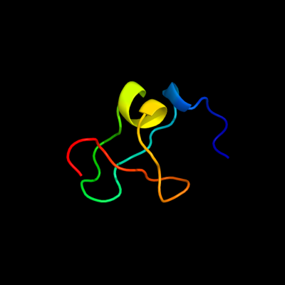
| Download: | Text version | FASTA pairwise alignment | 3D Model in PDB format |
Phyre is for academic use only
| Please cite: Protein structure prediction on the web: a case study using the Phyre server | ||||||||
| Kelley LA and Sternberg MJE. Nature Protocols 4, 363 - 371 (2009) [pdf] [Import into BibTeX] | ||||||||
|
| |||||||


