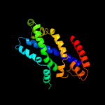
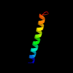
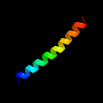
| 1 |
|
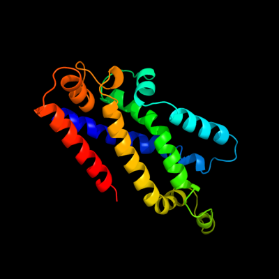
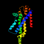
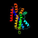
PDB 3dhw chain A domain 1
Region: 136 - 329
Aligned: 187
Modelled: 194
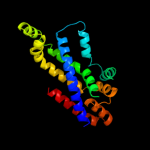
Confidence: 99.9%
Identity: 18%
Fold: MetI-like
Superfamily: MetI-like
Family: MetI-like
Phyre2
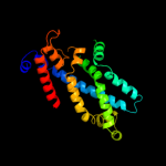
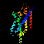
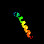
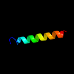
| 2 |
|
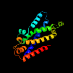
PDB 2onk chain C
Region: 134 - 331
Aligned: 192
Modelled: 198
Confidence: 99.8%
Identity: 9%
PDB header:membrane protein
Chain: C: PDB Molecule:molybdate/tungstate abc transporter, permease
PDBTitle: abc transporter modbc in complex with its binding protein2 moda
Phyre2
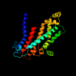
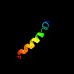
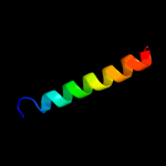
| 3 |
|
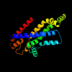
PDB 2onk chain C domain 1
Region: 134 - 331
Aligned: 192
Modelled: 198
Confidence: 99.8%
Identity: 9%
Fold: MetI-like
Superfamily: MetI-like
Family: MetI-like
Phyre2
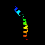
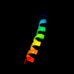
| 4 |
|
PDB 3d31 chain D
Region: 134 - 327
Aligned: 189
Modelled: 193
Confidence: 99.8%
Identity: 16%
PDB header:transport protein
Chain: D: PDB Molecule:sulfate/molybdate abc transporter, permease
PDBTitle: modbc from methanosarcina acetivorans
Phyre2
| 5 |
|
PDB 3d31 chain C domain 1
Region: 134 - 327
Aligned: 189
Modelled: 193
Confidence: 99.8%
Identity: 16%
Fold: MetI-like
Superfamily: MetI-like
Family: MetI-like
Phyre2
| 6 |
|
PDB 2r6g chain F domain 2
Region: 118 - 331
Aligned: 208
Modelled: 214
Confidence: 99.7%
Identity: 13%
Fold: MetI-like
Superfamily: MetI-like
Family: MetI-like
Phyre2
| 7 |
|
PDB 2r6g chain F
Region: 136 - 327
Aligned: 191
Modelled: 192
Confidence: 99.6%
Identity: 14%
PDB header:hydrolase/transport protein
Chain: F: PDB Molecule:maltose transport system permease protein malf;
PDBTitle: the crystal structure of the e. coli maltose transporter
Phyre2
| 8 |
|
PDB 3fh6 chain F
Region: 131 - 340
Aligned: 208
Modelled: 210
Confidence: 99.6%
Identity: 13%
PDB header:transport protein
Chain: F: PDB Molecule:maltose transport system permease protein malf;
PDBTitle: crystal structure of the resting state maltose transporter from e.2 coli
Phyre2
| 9 |
|
PDB 2r6g chain G domain 1
Region: 16 - 329
Aligned: 258
Modelled: 260
Confidence: 99.5%
Identity: 14%
Fold: MetI-like
Superfamily: MetI-like
Family: MetI-like
Phyre2
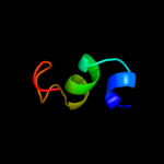
| 10 |
|
PDB 2cw1 chain A
Region: 225 - 248
Aligned: 22
Modelled: 24
Confidence: 20.0%
Identity: 45%
PDB header:de novo protein
Chain: A: PDB Molecule:sn4m;
PDBTitle: solution structure of the de novo-designed lambda cro fold2 protein
Phyre2
| 11 |
|
PDB 2jwa chain A
Region: 142 - 172
Aligned: 31
Modelled: 31
Confidence: 12.0%
Identity: 26%
PDB header:transferase
Chain: A: PDB Molecule:receptor tyrosine-protein kinase erbb-2;
PDBTitle: erbb2 transmembrane segment dimer spatial structure
Phyre2
| 12 |
|
PDB 2axt chain K domain 1
Region: 175 - 203
Aligned: 29
Modelled: 29
Confidence: 9.5%
Identity: 21%
Fold: Single transmembrane helix
Superfamily: Photosystem II reaction center protein K, PsbK
Family: PsbK-like
Phyre2
| 13 |
|
PDB 3a0b chain K
Region: 175 - 203
Aligned: 29
Modelled: 29
Confidence: 8.8%
Identity: 17%
PDB header:electron transport
Chain: K: PDB Molecule:photosystem ii reaction center protein k;
PDBTitle: crystal structure of br-substituted photosystem ii complex
Phyre2
| 14 |
|
PDB 3a0b chain K
Region: 175 - 203
Aligned: 29
Modelled: 29
Confidence: 8.8%
Identity: 17%
PDB header:electron transport
Chain: K: PDB Molecule:photosystem ii reaction center protein k;
PDBTitle: crystal structure of br-substituted photosystem ii complex
Phyre2
| 15 |
|
PDB 1wz4 chain A
Region: 332 - 340
Aligned: 9
Modelled: 9
Confidence: 7.8%
Identity: 22%
PDB header:gene regulation
Chain: A: PDB Molecule:major surface antigen;
PDBTitle: solution conformation of adr subtype hbv pre-s2 epitope
Phyre2
| 16 |
|
PDB 3hd7 chain A
Region: 6 - 37
Aligned: 32
Modelled: 32
Confidence: 7.7%
Identity: 3%
PDB header:exocytosis
Chain: A: PDB Molecule:vesicle-associated membrane protein 2;
PDBTitle: helical extension of the neuronal snare complex into the membrane,2 spacegroup c 1 2 1
Phyre2
| 17 |
|
PDB 2ka2 chain B
Region: 144 - 168
Aligned: 25
Modelled: 25
Confidence: 6.7%
Identity: 20%
PDB header:membrane protein
Chain: B: PDB Molecule:bcl2/adenovirus e1b 19 kda protein-interacting
PDBTitle: solution nmr structure of bnip3 transmembrane peptide dimer2 in detergent micelles with his173-ser172 intermonomer3 hydrogen bond restraints
Phyre2
| 18 |
|
PDB 2ka1 chain A
Region: 144 - 168
Aligned: 25
Modelled: 25
Confidence: 6.7%
Identity: 20%
PDB header:membrane protein
Chain: A: PDB Molecule:bcl2/adenovirus e1b 19 kda protein-interacting
PDBTitle: solution nmr structure of bnip3 transmembrane peptide dimer2 in detergent micelles
Phyre2
| 19 |
|
PDB 2ka2 chain A
Region: 144 - 168
Aligned: 25
Modelled: 25
Confidence: 6.7%
Identity: 20%
PDB header:membrane protein
Chain: A: PDB Molecule:bcl2/adenovirus e1b 19 kda protein-interacting
PDBTitle: solution nmr structure of bnip3 transmembrane peptide dimer2 in detergent micelles with his173-ser172 intermonomer3 hydrogen bond restraints
Phyre2
| 20 |
|
PDB 2ka1 chain B
Region: 144 - 168
Aligned: 25
Modelled: 25
Confidence: 6.7%
Identity: 20%
PDB header:membrane protein
Chain: B: PDB Molecule:bcl2/adenovirus e1b 19 kda protein-interacting
PDBTitle: solution nmr structure of bnip3 transmembrane peptide dimer2 in detergent micelles
Phyre2
| 21 |
|
| 22 |
|
| 23 |
|