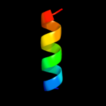
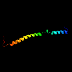
1 c1dipA_
86.2
38
PDB header: acetylationChain: A: PDB Molecule: delta-sleep-inducing peptide immunoreactivePDBTitle: the solution structure of porcine delta-sleep-inducing2 peptide immunoreactive peptide, nmr, 10 structures
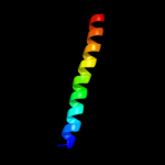
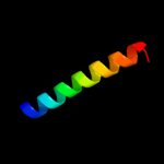
2 c3p8cE_
75.5
31
PDB header: protein bindingChain: E: PDB Molecule: probable protein brick1;PDBTitle: structure and control of the actin regulatory wave complex
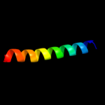
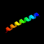
3 c3gw6F_
71.2
31
PDB header: chaperoneChain: F: PDB Molecule: endo-n-acetylneuraminidase;PDBTitle: intramolecular chaperone
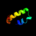
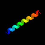
4 c1cosA_
56.7
30
PDB header: alpha-helical bundleChain: A: PDB Molecule: coiled serine;PDBTitle: crystal structure of a synthetic triple-stranded alpha-2 helical bundle
5 c1cosB_
55.6
29
PDB header: alpha-helical bundleChain: B: PDB Molecule: coiled serine;PDBTitle: crystal structure of a synthetic triple-stranded alpha-2 helical bundle
6 c3pp5A_
55.4
29
PDB header: structural proteinChain: A: PDB Molecule: brk1;PDBTitle: high-resolution structure of the trimeric scar/wave complex precursor2 brk1
7 c1coiA_
54.0
27
PDB header: alpha-helical bundleChain: A: PDB Molecule: coil-vald;PDBTitle: designed trimeric coiled coil-vald
8 c1qceB_
53.4
19
PDB header: viral proteinChain: B: PDB Molecule: protein (gp41);PDBTitle: solution nmr structure of ectodomain of siv gp41,2 restrained regularized mean structure plus 29 simulated3 annealing structures
9 c1cosC_
52.8
30
PDB header: alpha-helical bundleChain: C: PDB Molecule: coiled serine;PDBTitle: crystal structure of a synthetic triple-stranded alpha-2 helical bundle
10 c2l7kA_
52.2
13
PDB header: structural genomics, unknown functionChain: A: PDB Molecule: uncharacterized protein;PDBTitle: solution nmr structure of protein cd1104.2 from clostridium difficile,2 northeast structural genomics consortium target cfr130
11 c3p8cD_
46.8
16
PDB header: protein bindingChain: D: PDB Molecule: wiskott-aldrich syndrome protein family member 1;PDBTitle: structure and control of the actin regulatory wave complex
12 c1aq5C_
40.7
28
PDB header: coiled-coilChain: C: PDB Molecule: cartilage matrix protein;PDBTitle: high-resolution solution nmr structure of the trimeric coiled-coil2 domain of chicken cartilage matrix protein, 20 structures
13 c2jgoB_
38.0
26
PDB header: de novo proteinChain: B: PDB Molecule: coil ser l9c;PDBTitle: stucture of the arsenated de novo designed peptide coil ser2 l9c
14 c2jgoA_
38.0
26
PDB header: de novo proteinChain: A: PDB Molecule: coil ser l9c;PDBTitle: stucture of the arsenated de novo designed peptide coil ser2 l9c
15 c3ljmC_
38.0
26
PDB header: de novo proteinChain: C: PDB Molecule: coil ser l9c;PDBTitle: structure of de novo designed apo peptide coil ser l9c
16 c3ljmA_
38.0
26
PDB header: de novo proteinChain: A: PDB Molecule: coil ser l9c;PDBTitle: structure of de novo designed apo peptide coil ser l9c
17 c2ergA_
37.9
27
PDB header: transcription activator/dnaChain: A: PDB Molecule: regulatory protein leu3;PDBTitle: crystal structure of leu3 dna-binding domain with a single2 h50c mutation complexed with a 15mer dna duplex
18 c2jgoC_
36.6
25
PDB header: de novo proteinChain: C: PDB Molecule: coil ser l9c;PDBTitle: stucture of the arsenated de novo designed peptide coil ser2 l9c
19 c3ljmB_
36.6
25
PDB header: de novo proteinChain: B: PDB Molecule: coil ser l9c;PDBTitle: structure of de novo designed apo peptide coil ser l9c
20 c3h5fC_
34.0
27
PDB header: de novo proteinChain: C: PDB Molecule: coil ser l16l-pen;PDBTitle: switching the chirality of the metal environment alters the2 coordination mode in designed peptides.
21 c3h5fB_
not modelled
34.0
27
PDB header: de novo proteinChain: B: PDB Molecule: coil ser l16l-pen;PDBTitle: switching the chirality of the metal environment alters the2 coordination mode in designed peptides.
22 c3h5gA_
not modelled
34.0
27
PDB header: de novo proteinChain: A: PDB Molecule: coil ser l16d-pen;PDBTitle: switching the chirality of the metal environment alters the2 coordination mode in designed peptides.
23 c3h5gB_
not modelled
34.0
27
PDB header: de novo proteinChain: B: PDB Molecule: coil ser l16d-pen;PDBTitle: switching the chirality of the metal environment alters the2 coordination mode in designed peptides.
24 c3h5gC_
not modelled
34.0
27
PDB header: de novo proteinChain: C: PDB Molecule: coil ser l16d-pen;PDBTitle: switching the chirality of the metal environment alters the2 coordination mode in designed peptides.
25 c3h5fA_
not modelled
34.0
27
PDB header: de novo proteinChain: A: PDB Molecule: coil ser l16l-pen;PDBTitle: switching the chirality of the metal environment alters the2 coordination mode in designed peptides.
26 c1d7mA_
not modelled
33.5
39
PDB header: contractile proteinChain: A: PDB Molecule: cortexillin i;PDBTitle: coiled-coil dimerization domain from cortexillin i
27 d1grja1
not modelled
33.1
29
Fold: Long alpha-hairpinSuperfamily: GreA transcript cleavage protein, N-terminal domainFamily: GreA transcript cleavage protein, N-terminal domain28 c1g6uB_
not modelled
31.7
36
PDB header: de novo proteinChain: B: PDB Molecule: domain swapped dimer;PDBTitle: crystal structure of a domain swapped dimer
29 c3p7kA_
not modelled
31.4
24
PDB header: viral proteinChain: A: PDB Molecule: gp41 peptide;PDBTitle: gp41 peptide
30 c3iynR_
not modelled
30.3
16
PDB header: virusChain: R: PDB Molecule: hexon-associated protein;PDBTitle: 3.6-angstrom cryoem structure of human adenovirus type 5
31 d2f23a1
not modelled
29.4
29
Fold: Long alpha-hairpinSuperfamily: GreA transcript cleavage protein, N-terminal domainFamily: GreA transcript cleavage protein, N-terminal domain32 d1ohua_
not modelled
28.9
24
Fold: Toxins' membrane translocation domainsSuperfamily: Bcl-2 inhibitors of programmed cell deathFamily: Bcl-2 inhibitors of programmed cell death33 c1favA_
not modelled
28.4
22
PDB header: viral proteinChain: A: PDB Molecule: hiv-1 envelope protein chimera;PDBTitle: the structure of an hiv-1 specific cell entry inhibitor in complex2 with the hiv-1 gp41 trimeric core
34 c3m06F_
not modelled
27.1
14
PDB header: protein bindingChain: F: PDB Molecule: tnf receptor-associated factor 2;PDBTitle: crystal structure of traf2
35 c2kscA_
not modelled
25.8
13
PDB header: unknown functionChain: A: PDB Molecule: cyanoglobin;PDBTitle: solution structure of synechococcus sp. pcc 7002 hemoglobin
36 c2pnvA_
not modelled
25.5
22
PDB header: membrane proteinChain: A: PDB Molecule: small conductance calcium-activated potassiumPDBTitle: crystal structure of the leucine zipper domain of small-2 conductance ca2+-activated k+ (skca) channel from rattus3 norvegicus
37 c3a7pB_
not modelled
24.2
24
PDB header: protein transportChain: B: PDB Molecule: autophagy protein 16;PDBTitle: the crystal structure of saccharomyces cerevisiae atg16
38 c1u0iA_
not modelled
23.7
35
PDB header: de novo proteinChain: A: PDB Molecule: iaal-e3;PDBTitle: iaal-e3/k3 heterodimer
39 c2zfcB_
not modelled
23.3
14
PDB header: viral proteinChain: B: PDB Molecule: hiv-1 gp41;PDBTitle: x-ray crystal structure of an engineered n-terminal hiv-12 gp41 trimer with enhanced stability and potency
40 c3cvfA_
not modelled
19.4
21
PDB header: signaling proteinChain: A: PDB Molecule: homer protein homolog 3;PDBTitle: crystal structure of the carboxy terminus of homer3
41 c3efgA_
not modelled
19.1
37
PDB header: structural genomics, unknown functionChain: A: PDB Molecule: protein slyx homolog;PDBTitle: structure of slyx protein from xanthomonas campestris pv. campestris2 str. atcc 33913
42 c2vrsC_
not modelled
18.9
20
PDB header: viral proteinChain: C: PDB Molecule: sigma-c capsid protein;PDBTitle: structure of avian reovirus sigma c 117-326, c2 crystal form
43 c3rylB_
not modelled
18.6
31
PDB header: protein bindingChain: B: PDB Molecule: protein vpa1370;PDBTitle: dimerization domain of vibrio parahemolyticus vopl
44 c1grjA_
not modelled
17.6
29
PDB header: transcription regulationChain: A: PDB Molecule: grea protein;PDBTitle: grea transcript cleavage factor from escherichia coli
45 c3bpqC_
not modelled
16.7
29
PDB header: toxinChain: C: PDB Molecule: antitoxin relb3;PDBTitle: crystal structure of relb-rele antitoxin-toxin complex from2 methanococcus jannaschii
46 c3hnwB_
not modelled
16.3
9
PDB header: structural genomics, unknown functionChain: B: PDB Molecule: uncharacterized protein;PDBTitle: crystal structure of a basic coiled-coil protein of unknown function2 from eubacterium eligens atcc 27750
47 d1zy3a1
not modelled
16.0
20
Fold: Toxins' membrane translocation domainsSuperfamily: Bcl-2 inhibitors of programmed cell deathFamily: Bcl-2 inhibitors of programmed cell death48 c2yggA_
not modelled
15.8
40
PDB header: metal binding protein/transport proteinChain: A: PDB Molecule: sodium/hydrogen exchanger 1;PDBTitle: complex of cambr and cam
49 c2kp8A_
not modelled
15.7
27
PDB header: unknown functionChain: A: PDB Molecule: model peptide;PDBTitle: ligand bound to a model peptide that mimics the open2 fusogenic form
50 d2oa5a1
not modelled
15.5
20
Fold: BLRF2-likeSuperfamily: BLRF2-likeFamily: BLRF2-like51 c3pbjF_
not modelled
15.4
39
PDB header: de novo proteinChain: F: PDB Molecule: coil ser l9l-pen l23h;PDBTitle: hydrolytic catalysis and structural stabilization in a designed2 metalloprotein
52 c3pbjB_
not modelled
15.4
39
PDB header: de novo proteinChain: B: PDB Molecule: coil ser l9l-pen l23h;PDBTitle: hydrolytic catalysis and structural stabilization in a designed2 metalloprotein
53 c3pbjD_
not modelled
15.3
39
PDB header: de novo proteinChain: D: PDB Molecule: coil ser l9l-pen l23h;PDBTitle: hydrolytic catalysis and structural stabilization in a designed2 metalloprotein
54 c3pbjC_
not modelled
15.3
39
PDB header: de novo proteinChain: C: PDB Molecule: coil ser l9l-pen l23h;PDBTitle: hydrolytic catalysis and structural stabilization in a designed2 metalloprotein
55 c3pbjA_
not modelled
15.3
39
PDB header: de novo proteinChain: A: PDB Molecule: coil ser l9l-pen l23h;PDBTitle: hydrolytic catalysis and structural stabilization in a designed2 metalloprotein
56 c3c4mA_
not modelled
15.0
16
PDB header: membrane proteinChain: A: PDB Molecule: fusion protein of maltose-binding periplasmic protein andPDBTitle: structure of human parathyroid hormone in complex with the2 extracellular domain of its g-protein-coupled receptor (pth1r)
57 c3dbzB_
not modelled
15.0
15
PDB header: sugar binding proteinChain: B: PDB Molecule: pulmonary surfactant-associated protein d;PDBTitle: human surfactant protein d
58 d1q1va_
not modelled
14.5
27
Fold: Another 3-helical bundleSuperfamily: DEK C-terminal domainFamily: DEK C-terminal domain59 d1ivsa1
not modelled
14.4
11
Fold: Long alpha-hairpinSuperfamily: tRNA-binding armFamily: Valyl-tRNA synthetase (ValRS) C-terminal domain60 c2o2fA_
not modelled
14.2
16
PDB header: apoptosisChain: A: PDB Molecule: apoptosis regulator bcl-2;PDBTitle: solution structure of the anti-apoptotic protein bcl-2 in2 complex with an acyl-sulfonamide-based ligand
61 c2l5gA_
not modelled
13.9
33
PDB header: transcription regulatorChain: A: PDB Molecule: g protein pathway suppressor 2;PDBTitle: co-ordinates and 1h, 13c and 15n chemical shift assignments for the2 complex of gps2 53-90 and smrt 167-207
62 c1envA_
not modelled
13.9
23
PDB header: viral proteinChain: A: PDB Molecule: hiv-1 envelope protein chimera consisting of a fragment ofPDBTitle: atomic structure of the ectodomain from hiv-1 gp41
63 c2xa0A_
not modelled
13.6
16
PDB header: apoptosisChain: A: PDB Molecule: apoptosis regulator bcl-2;PDBTitle: crystal structure of bcl-2 in complex with a bax bh32 peptide
64 c1go4F_
not modelled
13.3
27
PDB header: cell cycleChain: F: PDB Molecule: mad1 (mitotic arrest deficient)-like 1;PDBTitle: crystal structure of mad1-mad2 reveals a conserved mad22 binding motif in mad1 and cdc20.
65 c3pbjE_
not modelled
13.3
28
PDB header: de novo proteinChain: E: PDB Molecule: coil ser l9l-pen l23h;PDBTitle: hydrolytic catalysis and structural stabilization in a designed2 metalloprotein
66 c1by0A_
not modelled
13.1
24
PDB header: rna binding proteinChain: A: PDB Molecule: protein (hepatitis delta antigen);PDBTitle: n-terminal leucine-repeat region of hepatitis delta antigen
67 c2x6pB_
not modelled
12.5
26
PDB header: de novo proteinChain: B: PDB Molecule: coil ser l19c;PDBTitle: crystal structure of coil ser l19c
68 c2x6pA_
not modelled
12.5
26
PDB header: de novo proteinChain: A: PDB Molecule: coil ser l19c;PDBTitle: crystal structure of coil ser l19c
69 d2p6va1
not modelled
12.4
19
Fold: TAFH domain-likeSuperfamily: TAFH domain-likeFamily: TAFH domain-like70 d1cxzb_
not modelled
12.0
21
Fold: Long alpha-hairpinSuperfamily: HR1 repeatFamily: HR1 repeat71 c1x8yA_
not modelled
11.9
31
PDB header: structural proteinChain: A: PDB Molecule: lamin a/c;PDBTitle: human lamin coil 2b
72 c2x6pC_
not modelled
11.8
25
PDB header: de novo proteinChain: C: PDB Molecule: coil ser l19c;PDBTitle: crystal structure of coil ser l19c
73 d1k4ta1
not modelled
11.7
20
Fold: Long alpha-hairpinSuperfamily: Eukaryotic DNA topoisomerase I, dispensable insert domainFamily: Eukaryotic DNA topoisomerase I, dispensable insert domain74 d1urfa_
not modelled
11.6
13
Fold: Long alpha-hairpinSuperfamily: HR1 repeatFamily: HR1 repeat75 d2rfra1
not modelled
11.2
38
Fold: Cystatin-likeSuperfamily: NTF2-likeFamily: BaiE/LinA-like76 d1pq1a_
not modelled
11.2
17
Fold: Toxins' membrane translocation domainsSuperfamily: Bcl-2 inhibitors of programmed cell deathFamily: Bcl-2 inhibitors of programmed cell death77 d2oa4a1
not modelled
10.9
36
Fold: DNA/RNA-binding 3-helical bundleSuperfamily: TrpR-likeFamily: SPO1678-like78 c2akfB_
not modelled
10.6
34
PDB header: protein bindingChain: B: PDB Molecule: coronin-1a;PDBTitle: crystal structure of the coiled-coil domain of coronin 1
79 c2akfA_
not modelled
10.6
34
PDB header: protein bindingChain: A: PDB Molecule: coronin-1a;PDBTitle: crystal structure of the coiled-coil domain of coronin 1
80 c2akfC_
not modelled
10.6
34
PDB header: protein bindingChain: C: PDB Molecule: coronin-1a;PDBTitle: crystal structure of the coiled-coil domain of coronin 1
81 c3gvmA_
not modelled
10.5
8
PDB header: viral proteinChain: A: PDB Molecule: putative uncharacterized protein sag1039;PDBTitle: structure of the homodimeric wxg-100 family protein from streptococcus2 agalactiae
82 c2elfA_
not modelled
9.7
29
PDB header: translationChain: A: PDB Molecule: protein translation elongation factor 1a;PDBTitle: crystal structure of the selb-like elongation factor ef-pyl2 from methanosarcina mazei
83 c2p4vA_
not modelled
9.6
11
PDB header: transcriptionChain: A: PDB Molecule: transcription elongation factor greb;PDBTitle: crystal structure of the transcript cleavage factor, greb2 at 2.6a resolution
84 c3exmA_
not modelled
9.4
16
PDB header: hydrolaseChain: A: PDB Molecule: phosphatase sc4828;PDBTitle: crystal structure of the phosphatase sc4828 with the non-hydrolyzable2 nucleotide gpcp
85 c1kzzA_
not modelled
9.3
25
PDB header: signaling proteinChain: A: PDB Molecule: tnf receptor associated factor 3;PDBTitle: downstream regulator tank binds to the cd40 recognition2 site on traf3
86 c3myrB_
not modelled
9.2
13
PDB header: oxidoreductaseChain: B: PDB Molecule: nickel-dependent hydrogenase large subunit;PDBTitle: crystal structure of [nife] hydrogenase from allochromatium vinosum in2 its ni-a state
87 c2qnlA_
not modelled
9.1
36
PDB header: signaling proteinChain: A: PDB Molecule: uncharacterized protein;PDBTitle: crystal structure of a putative dna damage-inducible protein2 (chu_0679) from cytophaga hutchinsonii atcc 33406 at 1.50 a3 resolution
88 c2jrtA_
not modelled
9.1
29
PDB header: structural genomics, unknown functionChain: A: PDB Molecule: uncharacterized protein;PDBTitle: nmr solution structure of the protein coded by gene2 rhos4_12090 of rhodobacter sphaeroides. northeast3 structural genomics target rhr5
89 c2vofA_
not modelled
9.1
16
PDB header: apoptosisChain: A: PDB Molecule: bcl-2-related protein a1;PDBTitle: structure of mouse a1 bound to the puma bh3-domain
90 c2a5yA_
not modelled
9.0
24
PDB header: apoptosisChain: A: PDB Molecule: apoptosis regulator ced-9;PDBTitle: structure of a ced-4/ced-9 complex
91 c3m0dC_
not modelled
8.9
22
PDB header: signaling proteinChain: C: PDB Molecule: tnf receptor-associated factor 1;PDBTitle: crystal structure of the traf1:traf2:ciap2 complex
92 c1jekA_
not modelled
8.8
16
PDB header: viral proteinChain: A: PDB Molecule: env polyprotein;PDBTitle: visna tm core structure
93 d2it9a1
not modelled
8.6
12
Fold: ssDNA-binding transcriptional regulator domainSuperfamily: ssDNA-binding transcriptional regulator domainFamily: PMN2A0962/syc2379c-like94 d1pzra_
not modelled
8.5
30
Fold: HLH-likeSuperfamily: Docking domain B of the erythromycin polyketide synthase (DEBS)Family: Docking domain B of the erythromycin polyketide synthase (DEBS)95 c1jocA_
not modelled
8.5
15
PDB header: membrane proteinChain: A: PDB Molecule: early endosomal autoantigen 1;PDBTitle: eea1 homodimer of c-terminal fyve domain bound to inositol2 1,3-diphosphate
96 c1p9iA_
not modelled
8.0
19
PDB header: unknown functionChain: A: PDB Molecule: cortexillin i/gcn4 hybrid peptide;PDBTitle: coiled-coil x-ray structure at 1.17 a resolution
97 c3ci9B_
not modelled
7.8
18
PDB header: transcriptionChain: B: PDB Molecule: heat shock factor-binding protein 1;PDBTitle: crystal structure of the human hsbp1
98 c2yv6A_
not modelled
7.7
14
PDB header: apoptosisChain: A: PDB Molecule: bcl-2 homologous antagonist/killer;PDBTitle: crystal structure of human bcl-2 family protein bak
99 c1pyiA_
not modelled
7.6
18
PDB header: transcription/dnaChain: A: PDB Molecule: protein (pyrimidine pathway regulator 1);PDBTitle: crystal structure of a ppr1-dna complex: dna recognition by2 proteins containing a zn2cys6 binuclear cluster