Phyre Home Retrieve Phyre Job Id
Emaill.a.kelley@imperial.ac.uk
DescriptionP0AG96
DateThu Jan 5 11:28:31 GMT 2012
Unique Job IDc6bcf1b1399ef897

Summary 

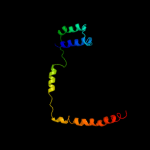
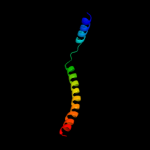
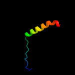
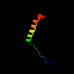
Top model
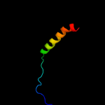
Image coloured by rainbow N → C terminus
Model (left) based on template c2akhZ_
Top template information
PDB header:protein transport
Chain: Z: PDB Molecule:preprotein translocase sece subunit;
PDBTitle: normal mode-based flexible fitted coordinates of a non-2 translocating secyeg protein-conducting channel into the3 cryo-em map of a secyeg-nascent chain-70s ribosome complex4 from e. coli
Confidence and coverage
Confidence:100.0% Coverage: 87%
111 residues ( 87% of your sequence) have been modelled with 100.0% confidence by the single highest scoring template.
3D viewing
Interactive 3D view in Jmol

Sequence analysis 

Secondary structure and disorder prediction 

   1........10.........20.........30.........40.........50.........60
Sequence  MSANTEAQGSGRGLEAMKWVVVVALLLVAIVGNYLYRDIMLPLRALAVVILIAAAGGVAL
Secondary structure 


SS confidence 


Disorder  ?????????????


?
Disorder confidence 


   .........70.........80.........90.........100.........110.........120
Sequence  LTTKGKATVAFAREARTEVRKVIWPTRQETLHTTLIVAAVTAVMSLILWGLDGILVRLVS
Secondary structure 


SS confidence 


Disorder 

????


Disorder confidence 


   .......
Sequence  FITGLRF
Secondary structure 


SS confidence 


Disorder 


????
Disorder confidence 


Confidence Key
High(9)                    Low (0)
?Disordered
Alpha helix
Beta strand

Domain analysis 

Hover over an aligned region to see model and summary info

Please note, only up to the top 20 hits are modelled to reduce computer load

RankAligned region

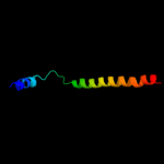
PDB 2akh chain Z

3D model

Region: 17 - 127
Aligned: 111
Modelled: 111
Confidence: 100.0%
Identity: 100%
PDB header:protein transport
Chain: Z: PDB Molecule:preprotein translocase sece subunit;
PDBTitle: normal mode-based flexible fitted coordinates of a non-2 translocating secyeg protein-conducting channel into the3 cryo-em map of a secyeg-nascent chain-70s ribosome complex4 from e. coli

Phyre2

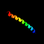
PDB 3din chain D

3D model

Region: 74 - 124
Aligned: 51
Modelled: 51
Confidence: 99.7%
Identity: 35%
PDB header:membrane protein, protein transport
Chain: D: PDB Molecule:preprotein translocase subunit sece;
PDBTitle: crystal structure of the protein-translocation complex formed by the2 secy channel and the seca atpase

Phyre2

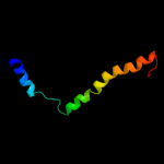
PDB 2zjs chain E

3D model

Region: 75 - 120
Aligned: 46
Modelled: 46
Confidence: 98.9%
Identity: 30%
PDB header:protein transport/immune system
Chain: E: PDB Molecule:preprotein translocase sece subunit;
PDBTitle: crystal structure of secye translocon from thermus thermophilus with a2 fab fragment

Phyre2

PDB 3dl8 chain D

3D model

Region: 92 - 126
Aligned: 35
Modelled: 35
Confidence: 97.2%
Identity: 31%
PDB header:protein transport
Chain: D: PDB Molecule:sece;
PDBTitle: structure of the complex of aquifex aeolicus secyeg and2 bacillus subtilis seca

Phyre2

PDB 2wwb chain B

3D model

Region: 68 - 124
Aligned: 57
Modelled: 57
Confidence: 84.5%
Identity: 21%
PDB header:ribosome
Chain: B: PDB Molecule:protein transport protein sec61 subunit gamma;
PDBTitle: cryo-em structure of the mammalian sec61 complex bound to the2 actively translating wheat germ 80s ribosome

Phyre2

PDB 1rhz chain B

3D model

Region: 66 - 122
Aligned: 57
Modelled: 57
Confidence: 83.0%
Identity: 16%
Fold: Single transmembrane helix
Superfamily: Preprotein translocase SecE subunit
Family: Preprotein translocase SecE subunit

Phyre2

PDB 2r6g chain F domain 1

3D model

Region: 15 - 72
Aligned: 56
Modelled: 58
Confidence: 81.3%
Identity: 20%
Fold: MalF N-terminal region-like
Superfamily: MalF N-terminal region-like
Family: MalF N-terminal region-like

Phyre2

PDB 2ww9 chain B

3D model

Region: 68 - 122
Aligned: 55
Modelled: 55
Confidence: 75.6%
Identity: 20%
PDB header:ribosome
Chain: B: PDB Molecule:protein transport protein sss1;
PDBTitle: cryo-em structure of the active yeast ssh1 complex bound to the2 yeast 80s ribosome

Phyre2

PDB 1rh5 chain B

3D model

Region: 70 - 122
Aligned: 53
Modelled: 53
Confidence: 45.5%
Identity: 17%
Fold: Single transmembrane helix
Superfamily: Preprotein translocase SecE subunit
Family: Preprotein translocase SecE subunit

Phyre2

PDB 3mp7 chain B

3D model

Region: 70 - 115
Aligned: 46
Modelled: 46
Confidence: 38.9%
Identity: 17%
PDB header:protein transport
Chain: B: PDB Molecule:preprotein translocase subunit sece;
PDBTitle: lateral opening of a translocon upon entry of protein suggests the2 mechanism of insertion into membranes

Phyre2

PDB 1s5l chain L

3D model

Region: 1 - 36
Aligned: 36
Modelled: 36
Confidence: 34.4%
Identity: 17%
PDB header:photosynthesis
Chain: L: PDB Molecule:photosystem ii reaction center l protein;
PDBTitle: architecture of the photosynthetic oxygen evolving center

Phyre2

PDB 1s5l chain L

3D model

Region: 1 - 36
Aligned: 36
Modelled: 36
Confidence: 34.4%
Identity: 17%
PDB header:photosynthesis
Chain: L: PDB Molecule:photosystem ii reaction center l protein;
PDBTitle: architecture of the photosynthetic oxygen evolving center

Phyre2

PDB 2axt chain L

3D model

Region: 1 - 36
Aligned: 36
Modelled: 36
Confidence: 34.4%
Identity: 17%
PDB header:electron transport
Chain: L: PDB Molecule:photosystem ii reaction center l protein;
PDBTitle: crystal structure of photosystem ii from thermosynechococcus elongatus

Phyre2

PDB 2axt chain L

3D model

Region: 1 - 36
Aligned: 36
Modelled: 36
Confidence: 34.4%
Identity: 17%
PDB header:electron transport
Chain: L: PDB Molecule:photosystem ii reaction center l protein;
PDBTitle: crystal structure of photosystem ii from thermosynechococcus elongatus

Phyre2

PDB 3a0h chain L

3D model

Region: 1 - 36
Aligned: 36
Modelled: 36
Confidence: 34.4%
Identity: 17%
PDB header:electron transport
Chain: L: PDB Molecule:photosystem ii reaction center protein l;
PDBTitle: crystal structure of i-substituted photosystem ii complex

Phyre2

PDB 3a0h chain L

3D model

Region: 1 - 36
Aligned: 36
Modelled: 36
Confidence: 34.4%
Identity: 17%
PDB header:electron transport
Chain: L: PDB Molecule:photosystem ii reaction center protein l;
PDBTitle: crystal structure of i-substituted photosystem ii complex

Phyre2

PDB 2axt chain L domain 1

3D model

Region: 1 - 36
Aligned: 36
Modelled: 36
Confidence: 34.4%
Identity: 17%
Fold: Single transmembrane helix
Superfamily: Photosystem II reaction center protein L, PsbL
Family: PsbL-like

Phyre2

PDB 3a0b chain L

3D model

Region: 1 - 36
Aligned: 36
Modelled: 36
Confidence: 34.4%
Identity: 17%
PDB header:electron transport
Chain: L: PDB Molecule:photosystem ii reaction center protein l;
PDBTitle: crystal structure of br-substituted photosystem ii complex

Phyre2

PDB 3a0b chain L

3D model

Region: 1 - 36
Aligned: 36
Modelled: 36
Confidence: 34.4%
Identity: 17%
PDB header:electron transport
Chain: L: PDB Molecule:photosystem ii reaction center protein l;
PDBTitle: crystal structure of br-substituted photosystem ii complex

Phyre2

PDB 3arc chain L

3D model

Region: 1 - 36
Aligned: 36
Modelled: 36
Confidence: 34.4%
Identity: 17%
PDB header:electron transport, photosynthesis
Chain: L: PDB Molecule:photosystem ii reaction center protein l;
PDBTitle: crystal structure of oxygen-evolving photosystem ii at 1.9 angstrom2 resolution

Phyre2
1

c2akhZ_
2

c3dinD_
3

c2zjsE_
4

c3dl8D_
5

c2wwbB_
6

d1rhzb_
7

d2r6gf1
8

c2ww9B_
9

d1rh5b_
10

c3mp7B_
11

c1s5ll_
12

c1s5lL_
13

c2axtl_
14

c2axtL_
15

c3a0hl_
16

c3a0hL_
17

d2axtl1
18

c3a0bL_
19

c3a0bl_
20

c3arcL_
21


22


23


24


25


26


27


28


29


30


31


32


33


34


35


36


37


38


39


Detailed template information 

#
Template Alignment Coverage3D Model Confidence
% i.d. Template Information
1c2akhZ_


100.0 100 PDB header:protein transport
Chain: Z: PDB Molecule:preprotein translocase sece subunit;
PDBTitle: normal mode-based flexible fitted coordinates of a non-2 translocating secyeg protein-conducting channel into the3 cryo-em map of a secyeg-nascent chain-70s ribosome complex4 from e. coli
2c3dinD_


99.7 35 PDB header:membrane protein, protein transport
Chain: D: PDB Molecule:preprotein translocase subunit sece;
PDBTitle: crystal structure of the protein-translocation complex formed by the2 secy channel and the seca atpase
3c2zjsE_


98.9 30 PDB header:protein transport/immune system
Chain: E: PDB Molecule:preprotein translocase sece subunit;
PDBTitle: crystal structure of secye translocon from thermus thermophilus with a2 fab fragment
4c3dl8D_


97.2 31 PDB header:protein transport
Chain: D: PDB Molecule:sece;
PDBTitle: structure of the complex of aquifex aeolicus secyeg and2 bacillus subtilis seca
5c2wwbB_


84.5 21 PDB header:ribosome
Chain: B: PDB Molecule:protein transport protein sec61 subunit gamma;
PDBTitle: cryo-em structure of the mammalian sec61 complex bound to the2 actively translating wheat germ 80s ribosome
6d1rhzb_


83.0 16 Fold:Single transmembrane helix
Superfamily:Preprotein translocase SecE subunit
Family:Preprotein translocase SecE subunit
7d2r6gf1


81.3 20 Fold:MalF N-terminal region-like
Superfamily:MalF N-terminal region-like
Family:MalF N-terminal region-like
8c2ww9B_


75.6 20 PDB header:ribosome
Chain: B: PDB Molecule:protein transport protein sss1;
PDBTitle: cryo-em structure of the active yeast ssh1 complex bound to the2 yeast 80s ribosome
9d1rh5b_


45.5 17 Fold:Single transmembrane helix
Superfamily:Preprotein translocase SecE subunit
Family:Preprotein translocase SecE subunit
10c3mp7B_


38.9 17 PDB header:protein transport
Chain: B: PDB Molecule:preprotein translocase subunit sece;
PDBTitle: lateral opening of a translocon upon entry of protein suggests the2 mechanism of insertion into membranes
11c1s5ll_


34.4 17 PDB header:photosynthesis
Chain: L: PDB Molecule:photosystem ii reaction center l protein;
PDBTitle: architecture of the photosynthetic oxygen evolving center
12c1s5lL_


34.4 17 PDB header:photosynthesis
Chain: L: PDB Molecule:photosystem ii reaction center l protein;
PDBTitle: architecture of the photosynthetic oxygen evolving center
13c2axtl_


34.4 17 PDB header:electron transport
Chain: L: PDB Molecule:photosystem ii reaction center l protein;
PDBTitle: crystal structure of photosystem ii from thermosynechococcus elongatus
14c2axtL_


34.4 17 PDB header:electron transport
Chain: L: PDB Molecule:photosystem ii reaction center l protein;
PDBTitle: crystal structure of photosystem ii from thermosynechococcus elongatus
15c3a0hl_


34.4 17 PDB header:electron transport
Chain: L: PDB Molecule:photosystem ii reaction center protein l;
PDBTitle: crystal structure of i-substituted photosystem ii complex
16c3a0hL_


34.4 17 PDB header:electron transport
Chain: L: PDB Molecule:photosystem ii reaction center protein l;
PDBTitle: crystal structure of i-substituted photosystem ii complex
17d2axtl1


34.4 17 Fold:Single transmembrane helix
Superfamily:Photosystem II reaction center protein L, PsbL
Family:PsbL-like
18c3a0bL_


34.4 17 PDB header:electron transport
Chain: L: PDB Molecule:photosystem ii reaction center protein l;
PDBTitle: crystal structure of br-substituted photosystem ii complex
19c3a0bl_


34.4 17 PDB header:electron transport
Chain: L: PDB Molecule:photosystem ii reaction center protein l;
PDBTitle: crystal structure of br-substituted photosystem ii complex
20c3arcL_


34.4 17 PDB header:electron transport, photosynthesis
Chain: L: PDB Molecule:photosystem ii reaction center protein l;
PDBTitle: crystal structure of oxygen-evolving photosystem ii at 1.9 angstrom2 resolution
21c3bz1L_


not modelled 34.4 17 PDB header:electron transport
Chain: L: PDB Molecule:photosystem ii reaction center protein l;
PDBTitle: crystal structure of cyanobacterial photosystem ii (part 12 of 2). this file contains first monomer of psii dimer
22c3prrL_


not modelled 34.4 17 PDB header:photosynthesis
Chain: L: PDB Molecule:photosystem ii reaction center protein l;
PDBTitle: crystal structure of cyanobacterial photosystem ii in complex with2 terbutryn (part 2 of 2). this file contains second monomer of psii3 dimer
23c3kziL_


not modelled 34.4 17 PDB header:electron transport
Chain: L: PDB Molecule:photosystem ii reaction center protein l;
PDBTitle: crystal structure of monomeric form of cyanobacterial photosystem ii
24c3prqL_


not modelled 34.4 17 PDB header:photosynthesis
Chain: L: PDB Molecule:photosystem ii reaction center protein l;
PDBTitle: crystal structure of cyanobacterial photosystem ii in complex with2 terbutryn (part 1 of 2). this file contains first monomer of psii3 dimer
25c3bz2L_


not modelled 34.4 17 PDB header:electron transport
Chain: L: PDB Molecule:photosystem ii reaction center protein l;
PDBTitle: crystal structure of cyanobacterial photosystem ii (part 22 of 2). this file contains second monomer of psii dimer
26c3arcl_


not modelled 26.0 17 PDB header:electron transport, photosynthesis
Chain: L: PDB Molecule:photosystem ii reaction center protein l;
PDBTitle: crystal structure of oxygen-evolving photosystem ii at 1.9 angstrom2 resolution
27c1yq3C_


not modelled 24.1 12 PDB header:oxidoreductase
Chain: C: PDB Molecule:succinate dehydrogenase cytochrome b, large subunit;
PDBTitle: avian respiratory complex ii with oxaloacetate and ubiquinone
28c2ehbD_


not modelled 11.7 18 PDB header:signalling protein/transferase
Chain: D: PDB Molecule:cbl-interacting serine/threonine-protein kinase 24;
PDBTitle: the structure of the c-terminal domain of the protein kinase atsos22 bound to the calcium sensor atsos3
29d1jy1a2


not modelled 11.1 15 Fold:Phospholipase D/nuclease
Superfamily:Phospholipase D/nuclease
Family:Tyrosyl-DNA phosphodiesterase TDP1
30c3fwbB_


not modelled 9.9 9 PDB header:cell cycle, transcription
Chain: B: PDB Molecule:nuclear mrna export protein sac3;
PDBTitle: sac3:sus1:cdc31 complex
31c2okqB_


not modelled 7.5 13 PDB header:structural genomics, unknown function
Chain: B: PDB Molecule:hypothetical protein ybaa;
PDBTitle: crystal structure of unknown conserved ybaa protein from2 shigella flexneri
32c3sq3C_


not modelled 7.3 38 PDB header:hydrolase
Chain: C: PDB Molecule:tyrosyl-dna phosphodiesterase 1;
PDBTitle: crystal structure analysis of the yeast tyrosyl-dna phosphodiesterase2 h182a mutant
33c2qvoA_


not modelled 6.7 17 PDB header:structural genomics, unknown function
Chain: A: PDB Molecule:uncharacterized protein af_1382;
PDBTitle: crystal structure of af1382 from archaeoglobus fulgidus
34d1q32a2


not modelled 6.5 42 Fold:Phospholipase D/nuclease
Superfamily:Phospholipase D/nuclease
Family:Tyrosyl-DNA phosphodiesterase TDP1
35d2okqa1


not modelled 6.0 13 Fold:Ferredoxin-like
Superfamily:Dimeric alpha+beta barrel
Family:YbaA-like
36c1usdA_


not modelled 5.8 36 PDB header:signaling protein
Chain: A: PDB Molecule:vasodilator-stimulated phosphoprotein;
PDBTitle: human vasp tetramerisation domain l352m
37c2wmhA_


not modelled 5.8 29 PDB header:hydrolase
Chain: A: PDB Molecule:fucolectin-related protein;
PDBTitle: crystal structure of the catalytic module of a family 982 glycoside hydrolase from streptococcus pneumoniae tigr4 in3 complex with the h-disaccharide blood group antigen.
38d3buxb2


not modelled 5.8 19 Fold:N-cbl like
Superfamily:N-terminal domain of cbl (N-cbl)
Family:N-terminal domain of cbl (N-cbl)
39c1nopB_


not modelled 5.7 17 PDB header:hydrolase/dna
Chain: B: PDB Molecule:tyrosyl-dna phosphodiesterase 1;
PDBTitle: crystal structure of human tyrosyl-dna phosphodiesterase2 (tdp1) in complex with vanadate, dna and a human3 topoisomerase i-derived peptide

Binding site prediction 

Due to computational demand, binding site predictions are not run for batch jobs

If you want to predict binding sites, please manually submit your model of choice to 3DLigandSite


Phyre is for academic use only

Please cite: Protein structure prediction on the web: a case study using the Phyre server
Kelley LA and Sternberg MJE. Nature Protocols 4, 363 - 371 (2009) [pdf] [Import into BibTeX]
 
If you use the binding site predictions from 3DLigandSite, please also cite:
3DLigandSite: predicting ligand-binding sites using similar structures.
Wass MN, Kelley LA and Sternberg MJ Nucleic Acids Research 38, W469-73 (2010) [PubMed]
 
© Structural Bioinformatics Group
Imperial College London
Lawrence Kelley, Benjamin Jefferys
Disclaimer
Terms and Conditions
Component software
Template detection: HHpred 1.51
Secondary structure prediction: Psi-pred 2.5
Disorder prediction: Disopred 2.4
Transmembrane prediction: Memsat_SVM
Multi-template modelling and ab initio: Poing 1.0