Phyre Home Retrieve Phyre Job Id
Emaill.a.kelley@imperial.ac.uk
DescriptionP75826
DateThu Jan 5 12:14:42 GMT 2012
Unique Job IDc3d70911400a2792

Summary 

Top model
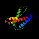
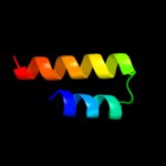
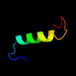
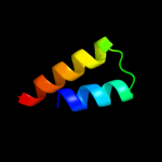
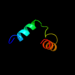
Image coloured by rainbow N → C terminus
Model (left) based on template c1zcdA_
Top template information
PDB header:membrane protein
Chain: A: PDB Molecule:na(+)/h(+) antiporter 1;
PDBTitle: crystal structure of the na+/h+ antiporter nhaa
Confidence and coverage
Confidence: 73.7% Coverage: 38%
114 residues ( 38% of your sequence) have been modelled with 73.7% confidence by the single highest scoring template.
You may wish to submit your sequence to Phyrealarm. This will automatically scan your sequence every week for new potential templates as they appear in the Phyre2 library.
Please note: You must be registered and logged in to use Phyrealarm.
3D viewing
Interactive 3D view in Jmol

Sequence analysis 

Secondary structure and disorder prediction 

   1........10.........20.........30.........40.........50.........60
Sequence  MFSGLLIILVPLIVGYLIPLRQQAALKVINQLLSWMVYLILFFMGISLAFLDNLASNLLA
Secondary structure 


SS confidence 


Disorder  ????


?


??


Disorder confidence 


   .........70.........80.........90.........100.........110.........120
Sequence  ILHYSAVSITVILLCNIAALMWLERGLPWRNHHQQEKLPSRIAMALESLKLCGVVVIGFA
Secondary structure 


SS confidence 


Disorder 


??????????????????
?


Disorder confidence 


   .........130.........140.........150.........160.........170.........180
Sequence  IGLSGLAFLQHATEASEYTLILLLFLVGIQLRNNGMTLKQIVLNRRGMIVAVVVVVSSLI
Secondary structure 


SS confidence 


Disorder 


?


??
?????


??


Disorder confidence 


   .........190.........200.........210.........220.........230.........240
Sequence  GGLINAFILDLPINTALAMASGFGWYSLSGILLTESFGPVIGSAAFFNDLARELIAIMLI
Secondary structure 


SS confidence 


Disorder 


Disorder confidence 


   .........250.........260.........270.........280.........290.........
Sequence  PGLIRRSRSTALGLCGATSMDFTLPVLQRTGGLDMVPAAIVHGFILSLLVPILIAFFSA
Secondary structure 


SS confidence 


Disorder 


?

??


???
Disorder confidence 


Confidence Key
High(9)                    Low (0)
?Disordered
Alpha helix
Beta strand

Domain analysis 

Hover over an aligned region to see model and summary info

Please note, only up to the top 20 hits are modelled to reduce computer load

RankAligned region

PDB 1zcd chain A

3D model

Region: 128 - 243
Aligned: 114
Modelled: 116
Confidence: 73.7%
Identity: 11%
PDB header:membrane protein
Chain: A: PDB Molecule:na(+)/h(+) antiporter 1;
PDBTitle: crystal structure of the na+/h+ antiporter nhaa

Phyre2

PDB 3gra chain A

3D model

Region: 253 - 276
Aligned: 24
Modelled: 24
Confidence: 23.2%
Identity: 13%
PDB header:transcription regulator
Chain: A: PDB Molecule:transcriptional regulator, arac family;
PDBTitle: crystal structure of arac family transcriptional regulator from2 pseudomonas putida

Phyre2

PDB 2oar chain A

3D model

Region: 158 - 246
Aligned: 88
Modelled: 89
Confidence: 20.3%
Identity: 11%
PDB header:membrane protein
Chain: A: PDB Molecule:large-conductance mechanosensitive channel;
PDBTitle: mechanosensitive channel of large conductance (mscl)

Phyre2

PDB 2oar chain A domain 1

3D model

Region: 166 - 246
Aligned: 79
Modelled: 81
Confidence: 20.1%
Identity: 10%
Fold: Gated mechanosensitive channel
Superfamily: Gated mechanosensitive channel
Family: Gated mechanosensitive channel

Phyre2

PDB 3pl2 chain D

3D model

Region: 178 - 209
Aligned: 32
Modelled: 32
Confidence: 16.9%
Identity: 22%
PDB header:transferase
Chain: D: PDB Molecule:sugar kinase, ribokinase family;
PDBTitle: crystal structure of a 5-keto-2-deoxygluconokinase (ncgl0155, cgl0158)2 from corynebacterium glutamicum atcc 13032 kitasato at 1.89 a3 resolution

Phyre2

PDB 2f02 chain A domain 1

3D model

Region: 178 - 209
Aligned: 32
Modelled: 32
Confidence: 16.6%
Identity: 22%
Fold: Ribokinase-like
Superfamily: Ribokinase-like
Family: Ribokinase-like

Phyre2

PDB 2afb chain A domain 1

3D model

Region: 178 - 209
Aligned: 32
Modelled: 32
Confidence: 16.4%
Identity: 16%
Fold: Ribokinase-like
Superfamily: Ribokinase-like
Family: Ribokinase-like

Phyre2

PDB 1vm7 chain A

3D model

Region: 178 - 209
Aligned: 32
Modelled: 32
Confidence: 15.0%
Identity: 13%
Fold: Ribokinase-like
Superfamily: Ribokinase-like
Family: Ribokinase-like

Phyre2

PDB 1cii chain A

3D model

Region: 199 - 226
Aligned: 28
Modelled: 28
Confidence: 14.2%
Identity: 14%
PDB header:transmembrane protein
Chain: A: PDB Molecule:colicin ia;
PDBTitle: colicin ia

Phyre2

PDB 3er6 chain D

3D model

Region: 253 - 279
Aligned: 27
Modelled: 27
Confidence: 12.6%
Identity: 7%
PDB header:structural genomics, unknown function
Chain: D: PDB Molecule:putative transcriptional regulator protein;
PDBTitle: crystal structure of a putative transcriptional regulator2 protein from vibrio parahaemolyticus

Phyre2

PDB 3noo chain B

3D model

Region: 253 - 276
Aligned: 24
Modelled: 24
Confidence: 12.5%
Identity: 17%
PDB header:lyase
Chain: B: PDB Molecule:thij/pfpi family protein;
PDBTitle: crystal structure of c101a isocyanide hydratase from pseudomonas2 fluorescens

Phyre2

PDB 3b3l chain C

3D model

Region: 178 - 209
Aligned: 32
Modelled: 32
Confidence: 11.8%
Identity: 9%
PDB header:transferase
Chain: C: PDB Molecule:ketohexokinase;
PDBTitle: crystal structures of alternatively-spliced isoforms of human2 ketohexokinase

Phyre2

PDB 3iq0 chain B

3D model

Region: 178 - 209
Aligned: 32
Modelled: 32
Confidence: 9.8%
Identity: 22%
PDB header:transferase
Chain: B: PDB Molecule:putative ribokinase ii;
PDBTitle: crystal structure of a putative ribokinase ii in complex2 with atp and mg+2 from e.coli

Phyre2

PDB 2ibg chain E domain 1

3D model

Region: 197 - 205
Aligned: 9
Modelled: 9
Confidence: 9.4%
Identity: 56%
Fold: Hedgehog/DD-peptidase
Superfamily: Hedgehog/DD-peptidase
Family: Hedgehog (development protein), N-terminal signaling domain

Phyre2

PDB 2jg1 chain C

3D model

Region: 178 - 209
Aligned: 32
Modelled: 32
Confidence: 9.4%
Identity: 19%
PDB header:transferase
Chain: C: PDB Molecule:tagatose-6-phosphate kinase;
PDBTitle: structure of staphylococcus aureus d-tagatose-6-phosphate2 kinase with cofactor and substrate

Phyre2

PDB 3ot1 chain B

3D model

Region: 253 - 276
Aligned: 24
Modelled: 24
Confidence: 9.0%
Identity: 17%
PDB header:structural genomics
Chain: B: PDB Molecule:4-methyl-5(b-hydroxyethyl)-thiazole monophosphate
PDBTitle: crystal structure of vc2308 protein

Phyre2

PDB 2dkv chain A

3D model

Region: 202 - 238
Aligned: 37
Modelled: 37
Confidence: 8.9%
Identity: 22%
PDB header:hydrolase
Chain: A: PDB Molecule:chitinase;
PDBTitle: crystal structure of class i chitinase from oryza sativa l.2 japonica

Phyre2

PDB 3ewn chain A

3D model

Region: 253 - 276
Aligned: 24
Modelled: 24
Confidence: 8.6%
Identity: 17%
PDB header:structural genomics, unknown function
Chain: A: PDB Molecule:thij/pfpi family protein;
PDBTitle: crystal structure of a thij/pfpi family protein from pseudomonas2 syringae

Phyre2

PDB 3d1m chain A domain 1

3D model

Region: 197 - 205
Aligned: 9
Modelled: 9
Confidence: 8.1%
Identity: 56%
Fold: Hedgehog/DD-peptidase
Superfamily: Hedgehog/DD-peptidase
Family: Hedgehog (development protein), N-terminal signaling domain

Phyre2

PDB 2pkk chain A

3D model

Region: 178 - 209
Aligned: 32
Modelled: 32
Confidence: 7.6%
Identity: 13%
PDB header:transferase
Chain: A: PDB Molecule:adenosine kinase;
PDBTitle: crystal structure of m tuberculosis adenosine kinase complexed with 2-2 fluro adenosine

Phyre2
1

c1zcdA_
2

c3graA_
3

c2oarA_
4

d2oara1
5

c3pl2D_
6

d2f02a1
7

d2afba1
8

d1vm7a_
9

c1ciiA_
10

c3er6D_
11

c3nooB_
12

c3b3lC_
13

c3iq0B_
14

d2ibge1
15

c2jg1C_
16

c3ot1B_
17

c2dkvA_
18

c3ewnA_
19

d3d1ma1
20

c2pkkA_
21


22


23


24


25


26


Detailed template information 

#
Template Alignment Coverage3D Model Confidence
% i.d. Template Information
1c1zcdA_


73.7 11 PDB header:membrane protein
Chain: A: PDB Molecule:na(+)/h(+) antiporter 1;
PDBTitle: crystal structure of the na+/h+ antiporter nhaa
2c3graA_


23.2 13 PDB header:transcription regulator
Chain: A: PDB Molecule:transcriptional regulator, arac family;
PDBTitle: crystal structure of arac family transcriptional regulator from2 pseudomonas putida
3c2oarA_


20.3 11 PDB header:membrane protein
Chain: A: PDB Molecule:large-conductance mechanosensitive channel;
PDBTitle: mechanosensitive channel of large conductance (mscl)
4d2oara1


20.1 10 Fold:Gated mechanosensitive channel
Superfamily:Gated mechanosensitive channel
Family:Gated mechanosensitive channel
5c3pl2D_


16.9 22 PDB header:transferase
Chain: D: PDB Molecule:sugar kinase, ribokinase family;
PDBTitle: crystal structure of a 5-keto-2-deoxygluconokinase (ncgl0155, cgl0158)2 from corynebacterium glutamicum atcc 13032 kitasato at 1.89 a3 resolution
6d2f02a1


16.6 22 Fold:Ribokinase-like
Superfamily:Ribokinase-like
Family:Ribokinase-like
7d2afba1


16.4 16 Fold:Ribokinase-like
Superfamily:Ribokinase-like
Family:Ribokinase-like
8d1vm7a_


15.0 13 Fold:Ribokinase-like
Superfamily:Ribokinase-like
Family:Ribokinase-like
9c1ciiA_


14.2 14 PDB header:transmembrane protein
Chain: A: PDB Molecule:colicin ia;
PDBTitle: colicin ia
10c3er6D_


12.6 7 PDB header:structural genomics, unknown function
Chain: D: PDB Molecule:putative transcriptional regulator protein;
PDBTitle: crystal structure of a putative transcriptional regulator2 protein from vibrio parahaemolyticus
11c3nooB_


12.5 17 PDB header:lyase
Chain: B: PDB Molecule:thij/pfpi family protein;
PDBTitle: crystal structure of c101a isocyanide hydratase from pseudomonas2 fluorescens
12c3b3lC_


11.8 9 PDB header:transferase
Chain: C: PDB Molecule:ketohexokinase;
PDBTitle: crystal structures of alternatively-spliced isoforms of human2 ketohexokinase
13c3iq0B_


9.8 22 PDB header:transferase
Chain: B: PDB Molecule:putative ribokinase ii;
PDBTitle: crystal structure of a putative ribokinase ii in complex2 with atp and mg+2 from e.coli
14d2ibge1


9.4 56 Fold:Hedgehog/DD-peptidase
Superfamily:Hedgehog/DD-peptidase
Family:Hedgehog (development protein), N-terminal signaling domain
15c2jg1C_


9.4 19 PDB header:transferase
Chain: C: PDB Molecule:tagatose-6-phosphate kinase;
PDBTitle: structure of staphylococcus aureus d-tagatose-6-phosphate2 kinase with cofactor and substrate
16c3ot1B_


9.0 17 PDB header:structural genomics
Chain: B: PDB Molecule:4-methyl-5(b-hydroxyethyl)-thiazole monophosphate
PDBTitle: crystal structure of vc2308 protein
17c2dkvA_


8.9 22 PDB header:hydrolase
Chain: A: PDB Molecule:chitinase;
PDBTitle: crystal structure of class i chitinase from oryza sativa l.2 japonica
18c3ewnA_


8.6 17 PDB header:structural genomics, unknown function
Chain: A: PDB Molecule:thij/pfpi family protein;
PDBTitle: crystal structure of a thij/pfpi family protein from pseudomonas2 syringae
19d3d1ma1


8.1 56 Fold:Hedgehog/DD-peptidase
Superfamily:Hedgehog/DD-peptidase
Family:Hedgehog (development protein), N-terminal signaling domain
20c2pkkA_


7.6 13 PDB header:transferase
Chain: A: PDB Molecule:adenosine kinase;
PDBTitle: crystal structure of m tuberculosis adenosine kinase complexed with 2-2 fluro adenosine
21c3m1nB_


not modelled 7.5 56 PDB header:signaling protein
Chain: B: PDB Molecule:sonic hedgehog protein;
PDBTitle: crystal structure of human sonic hedgehog n-terminal domain
22d2ab0a1


not modelled 7.5 21 Fold:Flavodoxin-like
Superfamily:Class I glutamine amidotransferase-like
Family:DJ-1/PfpI
23d1rkda_


not modelled 7.4 26 Fold:Ribokinase-like
Superfamily:Ribokinase-like
Family:Ribokinase-like
24c3b1qD_


not modelled 6.2 26 PDB header:transferase
Chain: D: PDB Molecule:ribokinase, putative;
PDBTitle: structure of burkholderia thailandensis nucleoside kinase (bthnk) in2 complex with inosine
25c3gbuD_


not modelled 6.1 16 PDB header:transferase
Chain: D: PDB Molecule:uncharacterized sugar kinase ph1459;
PDBTitle: crystal structure of an uncharacterized sugar kinase ph1459 from2 pyrococcus horikoshii in complex with atp
26c3k11A_


not modelled 5.8 19 PDB header:hydrolase
Chain: A: PDB Molecule:putative glycosyl hydrolase;
PDBTitle: crystal structure of putative glycosyl hydrolase (np_813087.1) from2 bacteroides thetaiotaomicron vpi-5482 at 1.80 a resolution

Binding site prediction 

Due to computational demand, binding site predictions are not run for batch jobs

If you want to predict binding sites, please manually submit your model of choice to 3DLigandSite


Phyre is for academic use only

Please cite: Protein structure prediction on the web: a case study using the Phyre server
Kelley LA and Sternberg MJE. Nature Protocols 4, 363 - 371 (2009) [pdf] [Import into BibTeX]
 
If you use the binding site predictions from 3DLigandSite, please also cite:
3DLigandSite: predicting ligand-binding sites using similar structures.
Wass MN, Kelley LA and Sternberg MJ Nucleic Acids Research 38, W469-73 (2010) [PubMed]
 
© Structural Bioinformatics Group
Imperial College London
Lawrence Kelley, Benjamin Jefferys
Disclaimer
Terms and Conditions
Component software
Template detection: HHpred 1.51
Secondary structure prediction: Psi-pred 2.5
Disorder prediction: Disopred 2.4
Transmembrane prediction: Memsat_SVM
Multi-template modelling and ab initio: Poing 1.0