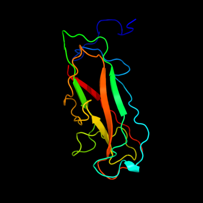
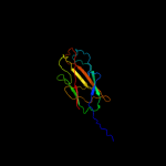
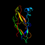
| 1 |
|
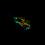
PDB 2jty chain A
Region: 23 - 178
Aligned: 155
Modelled: 156
Confidence: 99.9%
Identity: 32%
PDB header:structural protein
Chain: A: PDB Molecule:type-1 fimbrial protein, a chain;
PDBTitle: self-complemented variant of fima, the main subunit of type 1 pilus
Phyre2
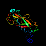
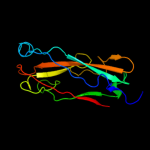
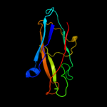
| 2 |
|
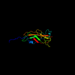
PDB 3jwn chain L
Region: 28 - 178
Aligned: 149
Modelled: 151
Confidence: 99.9%
Identity: 22%
PDB header:protein binding/cell adhesion
Chain: L: PDB Molecule:protein fimf;
PDBTitle: complex of fimc, fimf, fimg and fimh
Phyre2
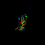
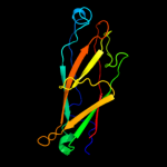
| 3 |
|
PDB 3jwn chain K
Region: 28 - 178
Aligned: 149
Modelled: 151
Confidence: 99.9%
Identity: 22%
PDB header:protein binding/cell adhesion
Chain: K: PDB Molecule:protein fimf;
PDBTitle: complex of fimc, fimf, fimg and fimh
Phyre2
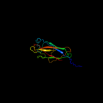
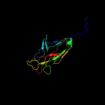
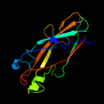
| 4 |
|
PDB 3jwn chain E
Region: 28 - 178
Aligned: 149
Modelled: 151
Confidence: 99.9%
Identity: 22%
PDB header:protein binding/cell adhesion
Chain: E: PDB Molecule:protein fimf;
PDBTitle: complex of fimc, fimf, fimg and fimh
Phyre2
| 5 |
|
PDB 3jwn chain F
Region: 28 - 178
Aligned: 149
Modelled: 151
Confidence: 99.9%
Identity: 22%
PDB header:protein binding/cell adhesion
Chain: F: PDB Molecule:protein fimf;
PDBTitle: complex of fimc, fimf, fimg and fimh
Phyre2
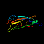
| 6 |
|
PDB 2jmr chain A
Region: 27 - 178
Aligned: 150
Modelled: 152
Confidence: 99.9%
Identity: 22%
PDB header:cell adhesion
Chain: A: PDB Molecule:fimf;
PDBTitle: nmr structure of the e. coli type 1 pilus subunit fimf
Phyre2
| 7 |
|
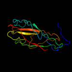
PDB 2j2z chain B domain 1
Region: 32 - 178
Aligned: 140
Modelled: 144
Confidence: 99.9%
Identity: 25%
Fold: Common fold of diphtheria toxin/transcription factors/cytochrome f
Superfamily: Bacterial adhesins
Family: Pilus subunits
Phyre2
| 8 |
|
PDB 2uy6 chain B domain 1
Region: 31 - 178
Aligned: 141
Modelled: 148
Confidence: 99.9%
Identity: 23%
Fold: Common fold of diphtheria toxin/transcription factors/cytochrome f
Superfamily: Bacterial adhesins
Family: Pilus subunits
Phyre2
| 9 |
|
PDB 1pdk chain B
Region: 38 - 178
Aligned: 138
Modelled: 139
Confidence: 99.9%
Identity: 20%
Fold: Common fold of diphtheria toxin/transcription factors/cytochrome f
Superfamily: Bacterial adhesins
Family: Pilus subunits
Phyre2
| 10 |
|
PDB 3bfw chain A
Region: 40 - 178
Aligned: 130
Modelled: 134
Confidence: 99.8%
Identity: 28%
PDB header:structural protein/structural protein
Chain: A: PDB Molecule:protein fimg;
PDBTitle: crystal structure of truncated fimg (fimgt) in complex with the donor2 strand peptide of fimf (dsf)
Phyre2
| 11 |
|
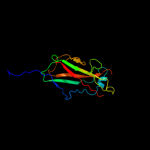
PDB 1klf chain P
Region: 31 - 178
Aligned: 131
Modelled: 148
Confidence: 99.8%
Identity: 24%
PDB header:chaperone/adhesin complex
Chain: P: PDB Molecule:fimh protein;
PDBTitle: fimh adhesin-fimc chaperone complex with d-mannose
Phyre2
| 12 |
|
PDB 1ze3 chain H domain 1
Region: 41 - 178
Aligned: 119
Modelled: 136
Confidence: 99.8%
Identity: 25%
Fold: Common fold of diphtheria toxin/transcription factors/cytochrome f
Superfamily: Bacterial adhesins
Family: Pilus subunits
Phyre2
| 13 |
|
PDB 3bwu chain F
Region: 58 - 178
Aligned: 119
Modelled: 121
Confidence: 99.8%
Identity: 22%
PDB header:chaperone, structural, membrane protein
Chain: F: PDB Molecule:protein fimf;
PDBTitle: crystal structure of the ternary complex of fimd (n-terminal domain,2 fimdn) with fimc and the n-terminally truncated pilus subunit fimf3 (fimft)
Phyre2
| 14 |
|
PDB 2w07 chain B
Region: 37 - 179
Aligned: 119
Modelled: 139
Confidence: 99.7%
Identity: 17%
PDB header:cell adhesion
Chain: B: PDB Molecule:minor pilin subunit papf;
PDBTitle: structural determinants of polymerization reactivity of the2 p pilus adaptor subunit papf
Phyre2
| 15 |
|
PDB 1n12 chain A
Region: 41 - 178
Aligned: 128
Modelled: 138
Confidence: 99.4%
Identity: 18%
Fold: Common fold of diphtheria toxin/transcription factors/cytochrome f
Superfamily: Bacterial adhesins
Family: Pilus subunits
Phyre2
| 16 |
|
PDB 2jna chain A domain 1
Region: 1 - 24
Aligned: 24
Modelled: 24
Confidence: 18.3%
Identity: 33%
Fold: Dodecin subunit-like
Superfamily: YdgH-like
Family: YdgH-like
Phyre2
| 17 |
|
PDB 2j6g chain A
Region: 169 - 179
Aligned: 11
Modelled: 11
Confidence: 6.7%
Identity: 45%
PDB header:cell adhesion
Chain: A: PDB Molecule:faeg;
PDBTitle: faeg from f4ac etec strain 5_95, produced in tobacco plant2 chloroplast
Phyre2
| 18 |
|
PDB 3mn8 chain A
Region: 75 - 155
Aligned: 80
Modelled: 81
Confidence: 6.0%
Identity: 15%
PDB header:hydrolase
Chain: A: PDB Molecule:lp15968p;
PDBTitle: structure of drosophila melanogaster carboxypeptidase d isoform 1b2 short
Phyre2
| 19 |
|
PDB 1h6t chain A domain 1
Region: 30 - 41
Aligned: 12
Modelled: 12
Confidence: 5.4%
Identity: 33%
Fold: Immunoglobulin-like beta-sandwich
Superfamily: E set domains
Family: Internalin Ig-like domain
Phyre2