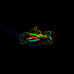
| 1 |
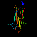
|
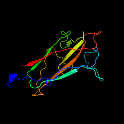
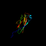
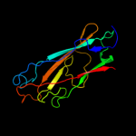
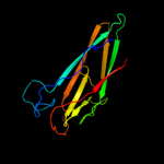
PDB 2w07 chain B
Region: 184 - 327
Aligned: 121
Modelled: 144
Confidence: 99.9%
Identity: 19%
PDB header:cell adhesion
Chain: B: PDB Molecule:minor pilin subunit papf;
PDBTitle: structural determinants of polymerization reactivity of the2 p pilus adaptor subunit papf
Phyre2
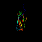
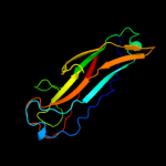
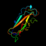
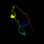
| 2 |
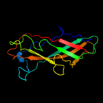
|
PDB 1klf chain P
Region: 16 - 327
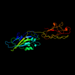
Aligned: 268
Modelled: 292
Confidence: 99.9%
Identity: 17%
PDB header:chaperone/adhesin complex
Chain: P: PDB Molecule:fimh protein;
PDBTitle: fimh adhesin-fimc chaperone complex with d-mannose
Phyre2
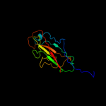
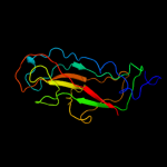
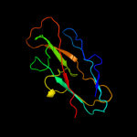
| 3 |
|
PDB 1n12 chain A
Region: 189 - 326
Aligned: 128
Modelled: 138
Confidence: 99.9%
Identity: 17%
Fold: Common fold of diphtheria toxin/transcription factors/cytochrome f
Superfamily: Bacterial adhesins
Family: Pilus subunits
Phyre2
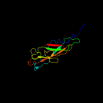
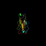
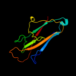
| 4 |
|
PDB 3jwn chain L
Region: 178 - 327
Aligned: 144
Modelled: 150
Confidence: 99.9%
Identity: 17%
PDB header:protein binding/cell adhesion
Chain: L: PDB Molecule:protein fimf;
PDBTitle: complex of fimc, fimf, fimg and fimh
Phyre2
| 5 |
|
PDB 3jwn chain K
Region: 178 - 327
Aligned: 144
Modelled: 150
Confidence: 99.9%
Identity: 17%
PDB header:protein binding/cell adhesion
Chain: K: PDB Molecule:protein fimf;
PDBTitle: complex of fimc, fimf, fimg and fimh
Phyre2
| 6 |
|
PDB 2j2z chain B domain 1
Region: 178 - 327
Aligned: 144
Modelled: 150
Confidence: 99.8%
Identity: 16%
Fold: Common fold of diphtheria toxin/transcription factors/cytochrome f
Superfamily: Bacterial adhesins
Family: Pilus subunits
Phyre2
| 7 |
|
PDB 3jwn chain F
Region: 178 - 327
Aligned: 144
Modelled: 150
Confidence: 99.8%
Identity: 17%
PDB header:protein binding/cell adhesion
Chain: F: PDB Molecule:protein fimf;
PDBTitle: complex of fimc, fimf, fimg and fimh
Phyre2
| 8 |
|
PDB 3jwn chain E
Region: 178 - 327
Aligned: 144
Modelled: 150
Confidence: 99.8%
Identity: 17%
PDB header:protein binding/cell adhesion
Chain: E: PDB Molecule:protein fimf;
PDBTitle: complex of fimc, fimf, fimg and fimh
Phyre2
| 9 |
|
PDB 3bfw chain A
Region: 188 - 326
Aligned: 131
Modelled: 139
Confidence: 99.8%
Identity: 14%
PDB header:structural protein/structural protein
Chain: A: PDB Molecule:protein fimg;
PDBTitle: crystal structure of truncated fimg (fimgt) in complex with the donor2 strand peptide of fimf (dsf)
Phyre2
| 10 |
|
PDB 2jmr chain A
Region: 178 - 327
Aligned: 144
Modelled: 150
Confidence: 99.8%
Identity: 18%
PDB header:cell adhesion
Chain: A: PDB Molecule:fimf;
PDBTitle: nmr structure of the e. coli type 1 pilus subunit fimf
Phyre2
| 11 |
|
PDB 2uy6 chain B domain 1
Region: 178 - 327
Aligned: 147
Modelled: 150
Confidence: 99.8%
Identity: 18%
Fold: Common fold of diphtheria toxin/transcription factors/cytochrome f
Superfamily: Bacterial adhesins
Family: Pilus subunits
Phyre2
| 12 |
|
PDB 1pdk chain B
Region: 186 - 327
Aligned: 140
Modelled: 142
Confidence: 99.8%
Identity: 11%
Fold: Common fold of diphtheria toxin/transcription factors/cytochrome f
Superfamily: Bacterial adhesins
Family: Pilus subunits
Phyre2
| 13 |
|
PDB 2jty chain A
Region: 179 - 327
Aligned: 142
Modelled: 149
Confidence: 99.8%
Identity: 20%
PDB header:structural protein
Chain: A: PDB Molecule:type-1 fimbrial protein, a chain;
PDBTitle: self-complemented variant of fima, the main subunit of type 1 pilus
Phyre2
| 14 |
|
PDB 1ze3 chain H domain 1
Region: 190 - 327
Aligned: 115
Modelled: 122
Confidence: 99.6%
Identity: 21%
Fold: Common fold of diphtheria toxin/transcription factors/cytochrome f
Superfamily: Bacterial adhesins
Family: Pilus subunits
Phyre2
| 15 |
|
PDB 3bwu chain F
Region: 205 - 327
Aligned: 118
Modelled: 123
Confidence: 99.6%
Identity: 14%
PDB header:chaperone, structural, membrane protein
Chain: F: PDB Molecule:protein fimf;
PDBTitle: crystal structure of the ternary complex of fimd (n-terminal domain,2 fimdn) with fimc and the n-terminally truncated pilus subunit fimf3 (fimft)
Phyre2
| 16 |
|
PDB 2wmp chain B
Region: 191 - 326
Aligned: 118
Modelled: 136
Confidence: 84.1%
Identity: 18%
PDB header:chaperone
Chain: B: PDB Molecule:papg protein;
PDBTitle: structure of the e. coli chaperone papd in complex with the pilin2 domain of the papgii adhesin
Phyre2
| 17 |
|
PDB 1p5v chain B
Region: 276 - 327
Aligned: 50
Modelled: 52
Confidence: 8.1%
Identity: 20%
Fold: Common fold of diphtheria toxin/transcription factors/cytochrome f
Superfamily: Bacterial adhesins
Family: Pilus subunits
Phyre2