Phyre Home Retrieve Phyre Job Id
Emaill.a.kelley@imperial.ac.uk
DescriptionP51024
DateThu Jan 5 12:04:50 GMT 2012
Unique Job IDc1a5f782e8e94219

Summary 

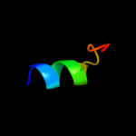
Top model
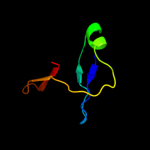
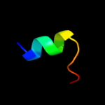
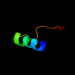
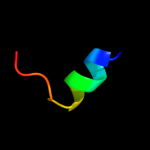
Image coloured by rainbow N → C terminus
Model (left) based on template c2l0cA_
Top template information
PDB header:structural genomics, unknown function
Chain: A: PDB Molecule:putative membrane protein;
PDBTitle: solution nmr structure of protein sty4237 (residues 36-120) from2 salmonella enterica, northeast structural genomics consortium target3 slr115
Confidence and coverage
Confidence: 28.2% Coverage: 23%
41 residues ( 23% of your sequence) have been modelled with 28.2% confidence by the single highest scoring template.
You may wish to submit your sequence to Phyrealarm. This will automatically scan your sequence every week for new potential templates as they appear in the Phyre2 library.
Please note: You must be registered and logged in to use Phyrealarm.
3D viewing
Interactive 3D view in Jmol

Sequence analysis 

Secondary structure and disorder prediction 

   1........10.........20.........30.........40.........50.........60
Sequence  MAKLTLQEQLLKAGLVTSKKAAKVERTAKKSRVQAREARAAVEENKKAQLERDKQLSEQQ
Secondary structure 


SS confidence 


Disorder  ?????


?


???????????


Disorder confidence 


   .........70.........80.........90.........100.........110.........120
Sequence  KQAALAKEYKAQVKQLIEMNRITIANGDIGFNFTDGNLIKKIFVDKLTQAQLINGRLAIA
Secondary structure 


SS confidence 


Disorder 


?


??????


Disorder confidence 


   .........130.........140.........150.........160.........170.........
Sequence  RLLVDNNSEGEYAIIPASVADKIAQRDASSIVLHSALSAEEQDEDDPYADFKVPDDLMW
Secondary structure 


SS confidence 


Disorder 


?
???


?????????


????
Disorder confidence 


Confidence Key
High(9)                    Low (0)
?Disordered
Alpha helix
Beta strand

Domain analysis 

Hover over an aligned region to see model and summary info

Please note, only up to the top 20 hits are modelled to reduce computer load

RankAligned region

PDB 2l0c chain A

3D model

Region: 88 - 134
Aligned: 41
Modelled: 47
Confidence: 28.2%
Identity: 17%
PDB header:structural genomics, unknown function
Chain: A: PDB Molecule:putative membrane protein;
PDBTitle: solution nmr structure of protein sty4237 (residues 36-120) from2 salmonella enterica, northeast structural genomics consortium target3 slr115

Phyre2

PDB 3ep1 chain B

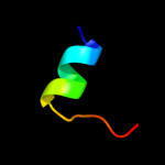
3D model

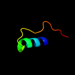
Region: 72 - 97
Aligned: 26
Modelled: 26
Confidence: 20.9%
Identity: 23%
PDB header:immune system
Chain: B: PDB Molecule:pgrp-hd - peptidoglycan recognition protein
PDBTitle: structure of the pgrp-hd from alvinella pompejana

Phyre2

PDB 3ky8 chain B

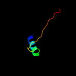
3D model

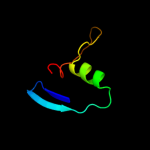
Region: 5 - 18
Aligned: 14
Modelled: 14
Confidence: 18.0%
Identity: 21%
PDB header:biosynthetic protein
Chain: B: PDB Molecule:putative riboflavin biosynthesis protein;
PDBTitle: crystal structure of putative riboflavin biosynthesis protein2 (yp_001092907.1) from shewanella sp. pv-4 at 2.12 a resolution

Phyre2

PDB 3jtw chain B

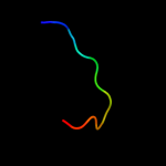
3D model

Region: 5 - 18
Aligned: 14
Modelled: 14
Confidence: 17.7%
Identity: 21%
PDB header:oxidoreductase
Chain: B: PDB Molecule:dihydrofolate reductase;
PDBTitle: crystal structure of putative dihydrofolate reductase (yp_805003.1)2 from pediococcus pentosaceus atcc 25745 at 1.90 a resolution

Phyre2

PDB 2gd9 chain A

3D model

Region: 5 - 18
Aligned: 14
Modelled: 14
Confidence: 15.9%
Identity: 29%
PDB header:oxidoreductase
Chain: A: PDB Molecule:hypothetical protein yyap;
PDBTitle: crystal structure of a putative dihydrofolate reductase (bsu40760,2 yyap) from bacillus subtilis at 2.30 a resolution

Phyre2

PDB 3kgy chain A

3D model

Region: 5 - 18
Aligned: 14
Modelled: 14
Confidence: 15.1%
Identity: 36%
PDB header:oxidoreductase
Chain: A: PDB Molecule:bifunctional deaminase-reductase domain protein;
PDBTitle: crystal structure of putative dihydrofolate reductase (yp_001636057.1)2 from chloroflexus aurantiacus j-10-fl at 1.50 a resolution

Phyre2

PDB 2xw7 chain A

3D model

Region: 5 - 18
Aligned: 14
Modelled: 14
Confidence: 14.2%
Identity: 21%
PDB header:oxidoreductase
Chain: A: PDB Molecule:dihydrofolate reductase;
PDBTitle: structure of mycobacterium smegmatis putative reductase ms0308

Phyre2

PDB 2xz8 chain A

3D model

Region: 71 - 97
Aligned: 27
Modelled: 27
Confidence: 13.4%
Identity: 22%
PDB header:immune system
Chain: A: PDB Molecule:peptidoglycan-recognition protein lf;
PDBTitle: crystal structure of the lfw ectodomain of the2 peptidoglycan recognition protein lf

Phyre2

PDB 1sxr chain A

3D model

Region: 71 - 97
Aligned: 27
Modelled: 27
Confidence: 10.3%
Identity: 22%
Fold: N-acetylmuramoyl-L-alanine amidase-like
Superfamily: N-acetylmuramoyl-L-alanine amidase-like
Family: N-acetylmuramoyl-L-alanine amidase-like

Phyre2

PDB 1d1g chain A

3D model

Region: 5 - 18
Aligned: 14
Modelled: 14
Confidence: 10.1%
Identity: 29%
Fold: Dihydrofolate reductase-like
Superfamily: Dihydrofolate reductase-like
Family: Dihydrofolate reductases

Phyre2

PDB 3hls chain E

3D model

Region: 132 - 143
Aligned: 12
Modelled: 12
Confidence: 9.9%
Identity: 42%
PDB header:signaling protein
Chain: E: PDB Molecule:guanylate cyclase soluble subunit beta-1;
PDBTitle: crystal structure of the signaling helix coiled-coil doimain2 of the beta-1 subunit of the soluble guanylyl cyclase

Phyre2

PDB 2b3z chain A domain 1

3D model

Region: 5 - 18
Aligned: 14
Modelled: 14
Confidence: 9.5%
Identity: 14%
Fold: Dihydrofolate reductase-like
Superfamily: Dihydrofolate reductase-like
Family: RibD C-terminal domain-like

Phyre2

PDB 2d28 chain C domain 1

3D model

Region: 1 - 31
Aligned: 31
Modelled: 31
Confidence: 9.3%
Identity: 13%
Fold: Alpha-lytic protease prodomain-like
Superfamily: EspE N-terminal domain-like
Family: GSPII protein E N-terminal domain-like

Phyre2

PDB 2v5i chain A

3D model

Region: 88 - 99
Aligned: 12
Modelled: 12
Confidence: 9.0%
Identity: 58%
PDB header:viral protein
Chain: A: PDB Molecule:salmonella typhimurium db7155 bacteriophage det7
PDBTitle: structure of the receptor-binding protein of bacteriophage2 det7: a podoviral tailspike in a myovirus

Phyre2

PDB 3iyj chain E

3D model

Region: 157 - 171
Aligned: 15
Modelled: 15
Confidence: 8.9%
Identity: 40%
PDB header:virus
Chain: E: PDB Molecule:major capsid protein l1;
PDBTitle: bovine papillomavirus type 1 outer capsid

Phyre2

PDB 2azn chain A domain 1

3D model

Region: 5 - 18
Aligned: 14
Modelled: 14
Confidence: 8.7%
Identity: 43%
Fold: Dihydrofolate reductase-like
Superfamily: Dihydrofolate reductase-like
Family: RibD C-terminal domain-like

Phyre2

PDB 1s2j chain A

3D model

Region: 71 - 97
Aligned: 27
Modelled: 27
Confidence: 8.3%
Identity: 22%
PDB header:hydrolase
Chain: A: PDB Molecule:peptidoglycan recognition protein sa cg11709-pa;
PDBTitle: crystal structure of the drosophila pattern-recognition2 receptor pgrp-sa

Phyre2

PDB 3op0 chain B

3D model

Region: 89 - 136
Aligned: 36
Modelled: 48
Confidence: 8.2%
Identity: 25%
PDB header:signaling protein/signaling protein regu
Chain: B: PDB Molecule:signal transduction protein cbl-c;
PDBTitle: crystal structure of cbl-c (cbl-3) tkb domain in complex with egfr2 py1069 peptide

Phyre2

PDB 2p4g chain A

3D model

Region: 5 - 18
Aligned: 14
Modelled: 14
Confidence: 8.2%
Identity: 29%
PDB header:oxidoreductase
Chain: A: PDB Molecule:hypothetical protein;
PDBTitle: crystal structure of a pyrimidine reductase-like protein (dip1392)2 from corynebacterium diphtheriae nctc at 2.30 a resolution

Phyre2

PDB 1hk9 chain A

3D model

Region: 5 - 13
Aligned: 9
Modelled: 9
Confidence: 8.0%
Identity: 44%
Fold: Sm-like fold
Superfamily: Sm-like ribonucleoproteins
Family: Pleiotropic translational regulator Hfq

Phyre2
1

c2l0cA_
2

c3ep1B_
3

c3ky8B_
4

c3jtwB_
5

c2gd9A_
6

c3kgyA_
7

c2xw7A_
8

c2xz8A_
9

d1sxra_
10

d1d1ga_
11

c3hlsE_
12

d2b3za1
13

d2d28c1
14

c2v5iA_
15

c3iyjE_
16

d2azna1
17

c1s2jA_
18

c3op0B_
19

c2p4gA_
20

d1hk9a_
21


22


23


24


25


26


27


28


29


30


31


32


33


34


Detailed template information 

#
Template Alignment Coverage3D Model Confidence
% i.d. Template Information
1c2l0cA_


28.2 17 PDB header:structural genomics, unknown function
Chain: A: PDB Molecule:putative membrane protein;
PDBTitle: solution nmr structure of protein sty4237 (residues 36-120) from2 salmonella enterica, northeast structural genomics consortium target3 slr115
2c3ep1B_


20.9 23 PDB header:immune system
Chain: B: PDB Molecule:pgrp-hd - peptidoglycan recognition protein
PDBTitle: structure of the pgrp-hd from alvinella pompejana
3c3ky8B_


18.0 21 PDB header:biosynthetic protein
Chain: B: PDB Molecule:putative riboflavin biosynthesis protein;
PDBTitle: crystal structure of putative riboflavin biosynthesis protein2 (yp_001092907.1) from shewanella sp. pv-4 at 2.12 a resolution
4c3jtwB_


17.7 21 PDB header:oxidoreductase
Chain: B: PDB Molecule:dihydrofolate reductase;
PDBTitle: crystal structure of putative dihydrofolate reductase (yp_805003.1)2 from pediococcus pentosaceus atcc 25745 at 1.90 a resolution
5c2gd9A_


15.9 29 PDB header:oxidoreductase
Chain: A: PDB Molecule:hypothetical protein yyap;
PDBTitle: crystal structure of a putative dihydrofolate reductase (bsu40760,2 yyap) from bacillus subtilis at 2.30 a resolution
6c3kgyA_


15.1 36 PDB header:oxidoreductase
Chain: A: PDB Molecule:bifunctional deaminase-reductase domain protein;
PDBTitle: crystal structure of putative dihydrofolate reductase (yp_001636057.1)2 from chloroflexus aurantiacus j-10-fl at 1.50 a resolution
7c2xw7A_


14.2 21 PDB header:oxidoreductase
Chain: A: PDB Molecule:dihydrofolate reductase;
PDBTitle: structure of mycobacterium smegmatis putative reductase ms0308
8c2xz8A_


13.4 22 PDB header:immune system
Chain: A: PDB Molecule:peptidoglycan-recognition protein lf;
PDBTitle: crystal structure of the lfw ectodomain of the2 peptidoglycan recognition protein lf
9d1sxra_


10.3 22 Fold:N-acetylmuramoyl-L-alanine amidase-like
Superfamily:N-acetylmuramoyl-L-alanine amidase-like
Family:N-acetylmuramoyl-L-alanine amidase-like
10d1d1ga_


10.1 29 Fold:Dihydrofolate reductase-like
Superfamily:Dihydrofolate reductase-like
Family:Dihydrofolate reductases
11c3hlsE_


9.9 42 PDB header:signaling protein
Chain: E: PDB Molecule:guanylate cyclase soluble subunit beta-1;
PDBTitle: crystal structure of the signaling helix coiled-coil doimain2 of the beta-1 subunit of the soluble guanylyl cyclase
12d2b3za1


9.5 14 Fold:Dihydrofolate reductase-like
Superfamily:Dihydrofolate reductase-like
Family:RibD C-terminal domain-like
13d2d28c1


9.3 13 Fold:Alpha-lytic protease prodomain-like
Superfamily:EspE N-terminal domain-like
Family:GSPII protein E N-terminal domain-like
14c2v5iA_


9.0 58 PDB header:viral protein
Chain: A: PDB Molecule:salmonella typhimurium db7155 bacteriophage det7
PDBTitle: structure of the receptor-binding protein of bacteriophage2 det7: a podoviral tailspike in a myovirus
15c3iyjE_


8.9 40 PDB header:virus
Chain: E: PDB Molecule:major capsid protein l1;
PDBTitle: bovine papillomavirus type 1 outer capsid
16d2azna1


8.7 43 Fold:Dihydrofolate reductase-like
Superfamily:Dihydrofolate reductase-like
Family:RibD C-terminal domain-like
17c1s2jA_


8.3 22 PDB header:hydrolase
Chain: A: PDB Molecule:peptidoglycan recognition protein sa cg11709-pa;
PDBTitle: crystal structure of the drosophila pattern-recognition2 receptor pgrp-sa
18c3op0B_


8.2 25 PDB header:signaling protein/signaling protein regu
Chain: B: PDB Molecule:signal transduction protein cbl-c;
PDBTitle: crystal structure of cbl-c (cbl-3) tkb domain in complex with egfr2 py1069 peptide
19c2p4gA_


8.2 29 PDB header:oxidoreductase
Chain: A: PDB Molecule:hypothetical protein;
PDBTitle: crystal structure of a pyrimidine reductase-like protein (dip1392)2 from corynebacterium diphtheriae nctc at 2.30 a resolution
20d1hk9a_


8.0 44 Fold:Sm-like fold
Superfamily:Sm-like ribonucleoproteins
Family:Pleiotropic translational regulator Hfq
21c2o7pA_


not modelled 8.0 57 PDB header:hydrolase, oxidoreductase
Chain: A: PDB Molecule:riboflavin biosynthesis protein ribd;
PDBTitle: the crystal structure of ribd from escherichia coli in complex with2 the oxidised nadp+ cofactor in the active site of the reductase3 domain
22d1vr0a1


not modelled 7.8 17 Fold:ComB-like
Superfamily:ComB-like
Family:ComB-like
23c2zxeG_


not modelled 7.8 33 PDB header:hydrolase/transport protein
Chain: G: PDB Molecule:phospholemman-like protein;
PDBTitle: crystal structure of the sodium - potassium pump in the e2.2k+.pi2 state
24c2zy3A_


not modelled 7.7 16 PDB header:lyase
Chain: A: PDB Molecule:l-aspartate beta-decarboxylase;
PDBTitle: dodecameric l-aspartate beta-decarboxylase
25d2p4pa1


not modelled 7.1 21 Fold:FAD-binding/transporter-associated domain-like
Superfamily:FAD-binding/transporter-associated domain-like
Family:CorC/HlyC domain-like
26c3ol0C_


not modelled 7.1 18 PDB header:de novo protein
Chain: C: PDB Molecule:de novo designed monomer trefoil-fold sub-domain which
PDBTitle: crystal structure of monofoil-4p homo-trimer: de novo designed monomer2 trefoil-fold sub-domain which forms homo-trimer assembly
27d1zhva1


not modelled 6.9 48 Fold:Ferredoxin-like
Superfamily:ACT-like
Family:Atu0741-like
28c2d5nB_


not modelled 6.1 14 PDB header:hydrolase, oxidoreductase
Chain: B: PDB Molecule:riboflavin biosynthesis protein ribd;
PDBTitle: crystal structure of a bifunctional deaminase and reductase2 involved in riboflavin biosynthesis
29d1z7ga1


not modelled 5.7 21 Fold:PRTase-like
Superfamily:PRTase-like
Family:Phosphoribosyltransferases (PRTases)
30d1u1sa1


not modelled 5.5 33 Fold:Sm-like fold
Superfamily:Sm-like ribonucleoproteins
Family:Pleiotropic translational regulator Hfq
31d1dzla_


not modelled 5.4 13 Fold:Nucleoplasmin-like/VP (viral coat and capsid proteins)
Superfamily:Group I dsDNA viruses
Family:Papovaviridae-like VP
32c1dzlA_


not modelled 5.4 13 PDB header:virus
Chain: A: PDB Molecule:late major capsid protein l1;
PDBTitle: l1 protein of human papillomavirus 16
33d1tzpa_


not modelled 5.2 25 Fold:Hedgehog/DD-peptidase
Superfamily:Hedgehog/DD-peptidase
Family:MepA-like
34c3dsoA_


not modelled 5.2 25 PDB header:metal binding protein
Chain: A: PDB Molecule:putative uncharacterized protein copk;
PDBTitle: crystal structure of cu(i) bound copper resistance protein copk

Binding site prediction 

Due to computational demand, binding site predictions are not run for batch jobs

If you want to predict binding sites, please manually submit your model of choice to 3DLigandSite


Phyre is for academic use only

Please cite: Protein structure prediction on the web: a case study using the Phyre server
Kelley LA and Sternberg MJE. Nature Protocols 4, 363 - 371 (2009) [pdf] [Import into BibTeX]
 
If you use the binding site predictions from 3DLigandSite, please also cite:
3DLigandSite: predicting ligand-binding sites using similar structures.
Wass MN, Kelley LA and Sternberg MJ Nucleic Acids Research 38, W469-73 (2010) [PubMed]
 
© Structural Bioinformatics Group
Imperial College London
Lawrence Kelley, Benjamin Jefferys
Disclaimer
Terms and Conditions
Component software
Template detection: HHpred 1.51
Secondary structure prediction: Psi-pred 2.5
Disorder prediction: Disopred 2.4
Transmembrane prediction: Memsat_SVM
Multi-template modelling and ab initio: Poing 1.0