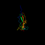
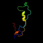
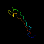
| 1 |
|
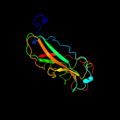
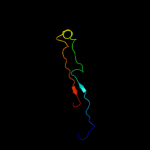
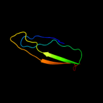
PDB 2jty chain A
Region: 17 - 237
Aligned: 154
Modelled: 158
Confidence: 97.8%
Identity: 15%
PDB header:structural protein
Chain: A: PDB Molecule:type-1 fimbrial protein, a chain;
PDBTitle: self-complemented variant of fima, the main subunit of type 1 pilus
Phyre2
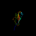
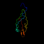
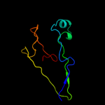
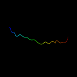
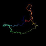
| 2 |
|
PDB 3jwn chain K
Region: 24 - 185
Aligned: 137
Modelled: 138
Confidence: 97.4%
Identity: 12%
PDB header:protein binding/cell adhesion
Chain: K: PDB Molecule:protein fimf;
PDBTitle: complex of fimc, fimf, fimg and fimh
Phyre2
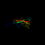
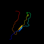
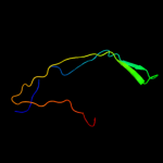
| 3 |
|
PDB 3jwn chain L
Region: 24 - 185
Aligned: 137
Modelled: 138
Confidence: 97.4%
Identity: 12%
PDB header:protein binding/cell adhesion
Chain: L: PDB Molecule:protein fimf;
PDBTitle: complex of fimc, fimf, fimg and fimh
Phyre2
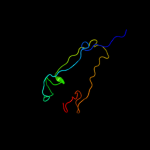
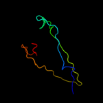
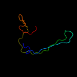
| 4 |
|
PDB 3jwn chain E
Region: 24 - 185
Aligned: 137
Modelled: 138
Confidence: 97.3%
Identity: 12%
PDB header:protein binding/cell adhesion
Chain: E: PDB Molecule:protein fimf;
PDBTitle: complex of fimc, fimf, fimg and fimh
Phyre2
| 5 |
|
PDB 3jwn chain F
Region: 24 - 185
Aligned: 137
Modelled: 138
Confidence: 97.2%
Identity: 12%
PDB header:protein binding/cell adhesion
Chain: F: PDB Molecule:protein fimf;
PDBTitle: complex of fimc, fimf, fimg and fimh
Phyre2
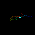
| 6 |
|
PDB 2w07 chain B
Region: 32 - 106
Aligned: 70
Modelled: 75
Confidence: 96.3%
Identity: 16%
PDB header:cell adhesion
Chain: B: PDB Molecule:minor pilin subunit papf;
PDBTitle: structural determinants of polymerization reactivity of the2 p pilus adaptor subunit papf
Phyre2
| 7 |
|
PDB 1n12 chain A
Region: 35 - 88
Aligned: 48
Modelled: 54
Confidence: 94.9%
Identity: 17%
Fold: Common fold of diphtheria toxin/transcription factors/cytochrome f
Superfamily: Bacterial adhesins
Family: Pilus subunits
Phyre2
| 8 |
|
PDB 2jmr chain A
Region: 25 - 133
Aligned: 95
Modelled: 109
Confidence: 94.7%
Identity: 8%
PDB header:cell adhesion
Chain: A: PDB Molecule:fimf;
PDBTitle: nmr structure of the e. coli type 1 pilus subunit fimf
Phyre2
| 9 |
|
PDB 2uy6 chain B domain 1
Region: 25 - 80
Aligned: 54
Modelled: 56
Confidence: 94.1%
Identity: 15%
Fold: Common fold of diphtheria toxin/transcription factors/cytochrome f
Superfamily: Bacterial adhesins
Family: Pilus subunits
Phyre2
| 10 |
|
PDB 3bfw chain A
Region: 35 - 87
Aligned: 51
Modelled: 53
Confidence: 93.7%
Identity: 16%
PDB header:structural protein/structural protein
Chain: A: PDB Molecule:protein fimg;
PDBTitle: crystal structure of truncated fimg (fimgt) in complex with the donor2 strand peptide of fimf (dsf)
Phyre2
| 11 |
|
PDB 2j2z chain B domain 1
Region: 26 - 79
Aligned: 52
Modelled: 54
Confidence: 93.6%
Identity: 13%
Fold: Common fold of diphtheria toxin/transcription factors/cytochrome f
Superfamily: Bacterial adhesins
Family: Pilus subunits
Phyre2
| 12 |
|
PDB 1ze3 chain H domain 1
Region: 35 - 134
Aligned: 74
Modelled: 87
Confidence: 93.1%
Identity: 19%
Fold: Common fold of diphtheria toxin/transcription factors/cytochrome f
Superfamily: Bacterial adhesins
Family: Pilus subunits
Phyre2
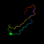
| 13 |
|
PDB 1klf chain P
Region: 31 - 93
Aligned: 50
Modelled: 63
Confidence: 92.0%
Identity: 16%
PDB header:chaperone/adhesin complex
Chain: P: PDB Molecule:fimh protein;
PDBTitle: fimh adhesin-fimc chaperone complex with d-mannose
Phyre2
| 14 |
|
PDB 2wmp chain B
Region: 35 - 126
Aligned: 64
Modelled: 71
Confidence: 91.0%
Identity: 23%
PDB header:chaperone
Chain: B: PDB Molecule:papg protein;
PDBTitle: structure of the e. coli chaperone papd in complex with the pilin2 domain of the papgii adhesin
Phyre2
| 15 |
|
PDB 1pdk chain B
Region: 32 - 79
Aligned: 47
Modelled: 48
Confidence: 88.4%
Identity: 13%
Fold: Common fold of diphtheria toxin/transcription factors/cytochrome f
Superfamily: Bacterial adhesins
Family: Pilus subunits
Phyre2
| 16 |
|
PDB 2jna chain A domain 1
Region: 1 - 22
Aligned: 22
Modelled: 22
Confidence: 16.5%
Identity: 27%
Fold: Dodecin subunit-like
Superfamily: YdgH-like
Family: YdgH-like
Phyre2
| 17 |
|
PDB 3qbt chain H
Region: 37 - 93
Aligned: 47
Modelled: 57
Confidence: 12.7%
Identity: 15%
PDB header:protein transport/hydrolase
Chain: H: PDB Molecule:inositol polyphosphate 5-phosphatase ocrl-1;
PDBTitle: crystal structure of ocrl1 540-678 in complex with rab8a:gppnhp
Phyre2
| 18 |
|
PDB 2tbv chain C
Region: 42 - 106
Aligned: 65
Modelled: 65
Confidence: 9.6%
Identity: 11%
Fold: Nucleoplasmin-like/VP (viral coat and capsid proteins)
Superfamily: Positive stranded ssRNA viruses
Family: Tombusviridae-like VP
Phyre2
| 19 |
|
PDB 3klq chain B
Region: 34 - 92
Aligned: 59
Modelled: 59
Confidence: 7.4%
Identity: 7%
PDB header:cell adhesion
Chain: B: PDB Molecule:putative pilus anchoring protein;
PDBTitle: crystal structure of the minor pilin fctb from streptococcus pyogenes2 90/306s
Phyre2
| 20 |
|
PDB 2tbv chain B
Region: 42 - 106
Aligned: 65
Modelled: 65
Confidence: 6.9%
Identity: 11%
PDB header:virus
Chain: B: PDB Molecule:tomato bushy stunt virus;
PDBTitle: structure of tomato bushy stunt virus. v. coat protein2 sequence determination and its structural implications
Phyre2
| 21 |
|