| 1 |
|
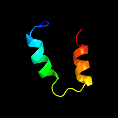
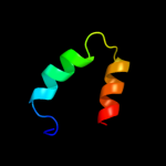
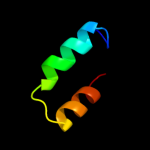
PDB 1m9f chain D
Region: 22 - 53
Aligned: 32
Modelled: 32
Confidence: 35.7%
Identity: 25%
Fold: Retrovirus capsid protein, N-terminal core domain
Superfamily: Retrovirus capsid protein, N-terminal core domain
Family: Retrovirus capsid protein, N-terminal core domain
Phyre2
| 2 |
|
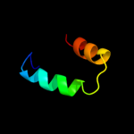
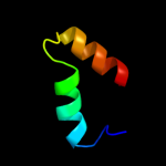
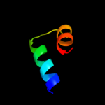
PDB 1m9d chain C
Region: 22 - 53
Aligned: 32
Modelled: 32
Confidence: 25.3%
Identity: 25%
Fold: Retrovirus capsid protein, N-terminal core domain
Superfamily: Retrovirus capsid protein, N-terminal core domain
Family: Retrovirus capsid protein, N-terminal core domain
Phyre2
| 3 |
|
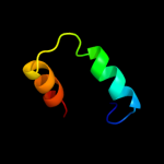
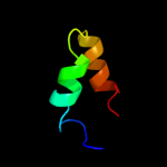
PDB 2wlv chain A
Region: 22 - 53
Aligned: 32
Modelled: 32
Confidence: 24.4%
Identity: 31%
PDB header:virus protein
Chain: A: PDB Molecule:gag polyprotein;
PDBTitle: structure of the n-terminal capsid domain of hiv-2
Phyre2
| 4 |
|
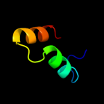
PDB 2pxr chain C domain 1
Region: 22 - 53
Aligned: 32
Modelled: 32
Confidence: 20.6%
Identity: 25%
Fold: Retrovirus capsid protein, N-terminal core domain
Superfamily: Retrovirus capsid protein, N-terminal core domain
Family: Retrovirus capsid protein, N-terminal core domain
Phyre2
| 5 |
|
PDB 2eia chain A domain 2
Region: 22 - 53
Aligned: 32
Modelled: 32
Confidence: 20.2%
Identity: 19%
Fold: Retrovirus capsid protein, N-terminal core domain
Superfamily: Retrovirus capsid protein, N-terminal core domain
Family: Retrovirus capsid protein, N-terminal core domain
Phyre2
| 6 |
|
PDB 1l6n chain A
Region: 22 - 53
Aligned: 32
Modelled: 32
Confidence: 17.0%
Identity: 25%
PDB header:viral protein
Chain: A: PDB Molecule:gag polyprotein;
PDBTitle: structure of the n-terminal 283-residue fragment of the hiv-2 1 gag polyprotein
Phyre2
| 7 |
|
PDB 2xgv chain A
Region: 22 - 53
Aligned: 32
Modelled: 32
Confidence: 7.2%
Identity: 22%
PDB header:viral protein
Chain: A: PDB Molecule:psiv capsid n-terminal domain;
PDBTitle: structure of the n-terminal domain of capsid protein from2 rabbit endogenous lentivirus (relik)
Phyre2
| 8 |
|
PDB 1q08 chain A
Region: 13 - 37
Aligned: 25
Modelled: 25
Confidence: 6.7%
Identity: 32%
Fold: Putative DNA-binding domain
Superfamily: Putative DNA-binding domain
Family: DNA-binding N-terminal domain of transcription activators
Phyre2