| 1 |
|
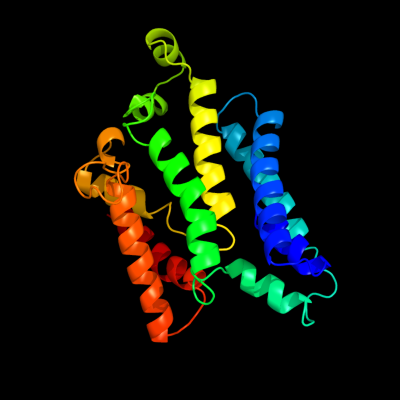
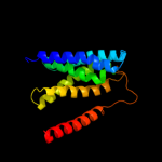
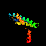
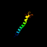
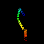
PDB 2nq2 chain A
Region: 74 - 304
Aligned: 216
Modelled: 231
Confidence: 97.5%
Identity: 17%
PDB header:metal transport
Chain: A: PDB Molecule:hypothetical abc transporter permease protein
PDBTitle: an inward-facing conformation of a putative metal-chelate2 type abc transporter.
Phyre2
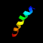
| 2 |
|
PDB 1l7v chain A
Region: 2 - 304
Aligned: 287
Modelled: 290
Confidence: 96.8%
Identity: 16%
Fold: ABC transporter involved in vitamin B12 uptake, BtuC
Superfamily: ABC transporter involved in vitamin B12 uptake, BtuC
Family: ABC transporter involved in vitamin B12 uptake, BtuC
Phyre2
| 3 |
|
PDB 1u7g chain A
Region: 6 - 181
Aligned: 176
Modelled: 176
Confidence: 35.4%
Identity: 11%
Fold: Ammonium transporter
Superfamily: Ammonium transporter
Family: Ammonium transporter
Phyre2
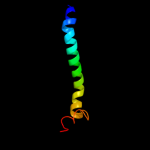
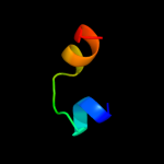
| 4 |
|
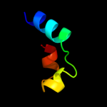
PDB 2jp3 chain A
Region: 287 - 332
Aligned: 46
Modelled: 46
Confidence: 30.6%
Identity: 17%
PDB header:transcription
Chain: A: PDB Molecule:fxyd domain-containing ion transport regulator 4;
PDBTitle: solution structure of the human fxyd4 (chif) protein in sds2 micelles
Phyre2
| 5 |
|
PDB 2b2h chain A
Region: 6 - 206
Aligned: 194
Modelled: 201
Confidence: 25.6%
Identity: 12%
PDB header:transport protein
Chain: A: PDB Molecule:ammonium transporter;
PDBTitle: ammonium transporter amt-1 from a. fulgidus (as)
Phyre2
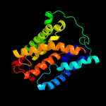
| 6 |
|
PDB 3k3g chain A
Region: 72 - 340
Aligned: 230
Modelled: 230
Confidence: 25.2%
Identity: 13%
PDB header:transport protein
Chain: A: PDB Molecule:urea transporter;
PDBTitle: crystal structure of the urea transporter from desulfovibrio vulgaris2 bound to 1,3-dimethylurea
Phyre2
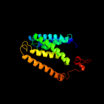
| 7 |
|
PDB 3pxp chain A
Region: 177 - 209
Aligned: 33
Modelled: 32
Confidence: 20.8%
Identity: 21%
PDB header:transcription regulator
Chain: A: PDB Molecule:helix-turn-helix domain protein;
PDBTitle: crystal structure of a pas and dna binding domain containing protein2 (caur_2278) from chloroflexus aurantiacus j-10-fl at 2.30 a3 resolution
Phyre2
| 8 |
|
PDB 3b9y chain A
Region: 11 - 116
Aligned: 106
Modelled: 106
Confidence: 17.0%
Identity: 11%
PDB header:transport protein
Chain: A: PDB Molecule:ammonium transporter family rh-like protein;
PDBTitle: crystal structure of the nitrosomonas europaea rh protein
Phyre2
| 9 |
|
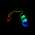
PDB 2e74 chain G domain 1
Region: 212 - 235
Aligned: 24
Modelled: 24
Confidence: 15.3%
Identity: 21%
Fold: Single transmembrane helix
Superfamily: PetG subunit of the cytochrome b6f complex
Family: PetG subunit of the cytochrome b6f complex
Phyre2
| 10 |
|
PDB 1y9q chain A
Region: 179 - 209
Aligned: 31
Modelled: 26
Confidence: 11.4%
Identity: 13%
PDB header:transcription regulator
Chain: A: PDB Molecule:transcriptional regulator, hth_3 family;
PDBTitle: crystal structure of hth_3 family transcriptional regulator2 from vibrio cholerae
Phyre2
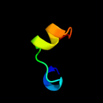
| 11 |
|
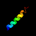
PDB 2jo1 chain A
Region: 287 - 334
Aligned: 48
Modelled: 48
Confidence: 8.9%
Identity: 17%
PDB header:hydrolase regulator
Chain: A: PDB Molecule:phospholemman;
PDBTitle: structure of the na,k-atpase regulatory protein fxyd1 in2 micelles
Phyre2
| 12 |
|
PDB 1y6u chain A
Region: 193 - 210
Aligned: 18
Modelled: 18
Confidence: 8.0%
Identity: 11%
PDB header:dna binding protein
Chain: A: PDB Molecule:excisionase from transposon tn916;
PDBTitle: the structure of the excisionase (xis) protein from2 conjugative transposon tn916 provides insights into the3 regulation of heterobivalent tyrosine recombinases
Phyre2
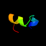
| 13 |
|
PDB 4a18 chain U
Region: 185 - 203
Aligned: 19
Modelled: 19
Confidence: 8.0%
Identity: 21%
PDB header:ribosome
Chain: U: PDB Molecule:rpl13;
PDBTitle: t.thermophila 60s ribosomal subunit in complex with initiation2 factor 6. this file contains 26s rrna and proteins of molecule 1
Phyre2
| 14 |
|
PDB 3u5e chain L
Region: 185 - 203
Aligned: 19
Modelled: 19
Confidence: 7.0%
Identity: 26%
PDB header:ribosome
Chain: L: PDB Molecule:60s ribosomal protein l13-a;
PDBTitle: the structure of the eukaryotic ribosome at 3.0 resolution
Phyre2
| 15 |
|
PDB 2w8a chain C
Region: 262 - 332
Aligned: 71
Modelled: 71
Confidence: 6.2%
Identity: 15%
PDB header:membrane protein
Chain: C: PDB Molecule:glycine betaine transporter betp;
PDBTitle: crystal structure of the sodium-coupled glycine betaine2 symporter betp from corynebacterium glutamicum with bound3 substrate
Phyre2
| 16 |
|
PDB 1vf5 chain G
Region: 212 - 231
Aligned: 20
Modelled: 20
Confidence: 5.6%
Identity: 25%
PDB header:photosynthesis
Chain: G: PDB Molecule:protein pet g;
PDBTitle: crystal structure of cytochrome b6f complex from m.laminosus
Phyre2
| 17 |
|
PDB 1vf5 chain G
Region: 212 - 231
Aligned: 20
Modelled: 20
Confidence: 5.6%
Identity: 25%
Fold: Single transmembrane helix
Superfamily: PetG subunit of the cytochrome b6f complex
Family: PetG subunit of the cytochrome b6f complex
Phyre2