| Secondary structure and disorder prediction | |
| | |
1 | . | . | . | . | . | . | . | . | 10 | . | . | . | . | . | . | . | . | . | 20 | . | . | . | . | . | . | . | . | . | 30 | . | . | . | . | . | . | . | . | . | 40 | . | . | . | . | . | . | . | . | . |
| Sequence | |
M | L | E | L | L | K | S | L | V | F | A | V | I | M | V | P | V | V | M | A | I | I | L | G | L | I | Y | G | L | G | E | V | F | N | I | F | S | G | V | G | K | K | D | Q | P | G | Q | N | H |
| Secondary structure | |
|  | | | | | | | | | | | | | | | | | | | | | | | | | | | | | | | | | | | | | |
|
|
|
|
|
|
|
|
|
|
| SS confidence | |
|
|
|
|
|
|
|
|
|
|
|
|
|
|
|
|
|
|
|
|
|
|
|
|
|
|
|
|
|
|
|
|
|
|
|
|
|
|
|
|
|
|
|
|
|
|
|
|
|
| Disorder | |
? |
|
|
|
|
|
|
|
|
|
|
|
|
|
|
|
|
|
|
|
|
|
|
|
|
|
|
|
|
|
|
|
|
|
|
|
|
| ? | ? | ? | ? | ? | ? | ? | ? | ? | ? | ? |
| Disorder confidence | |
|
|
|
|
|
|
|
|
|
|
|
|
|
|
|
|
|
|
|
|
|
|
|
|
|
|
|
|
|
|
|
|
|
|
|
|
|
|
|
|
|
|
|
|
|
|
|
|
|
| |
| Confidence Key |
| High(9) | |
|
|
|
|
|
|
|
|
|
Low (0) |
| ? | Disordered |
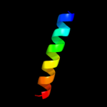
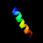
| Alpha helix |
  | Beta strand |
Hover over an aligned region to see model and summary info
Please note, only up to the top 20 hits are modelled to reduce computer load
|
| 1 |
|
PDB 2ajm chain A
Region: 22 - 34
Aligned: 13
Modelled: 13
Confidence: 23.7%
Identity: 46%
PDB header:membrane protein
Chain: A: PDB Molecule:nonstructural protein 5a;
PDBTitle: nmr structure of the in-plane membrane anchor domain [1-28]2 of the monotopic nonstructural protein 5a (ns5a) from the3 bovine viral diarrhea virus (bvdv)
Phyre2
| 2 |
|
PDB 3din chain D
Region: 3 - 36
Aligned: 34
Modelled: 34
Confidence: 19.6%
Identity: 21%
PDB header:membrane protein, protein transport
Chain: D: PDB Molecule:preprotein translocase subunit sece;
PDBTitle: crystal structure of the protein-translocation complex formed by the2 secy channel and the seca atpase
Phyre2
| 3 |
|
PDB 1jb0 chain I
Region: 12 - 29
Aligned: 18
Modelled: 18
Confidence: 7.9%
Identity: 33%
Fold: Single transmembrane helix
Superfamily: Subunit VIII of photosystem I reaction centre, PsaI
Family: Subunit VIII of photosystem I reaction centre, PsaI
Phyre2
| 4 |
|
PDB 3g79 chain A
Region: 38 - 49
Aligned: 12
Modelled: 12
Confidence: 7.3%
Identity: 25%
PDB header:oxidoreductase
Chain: A: PDB Molecule:ndp-n-acetyl-d-galactosaminuronic acid dehydrogenase;
PDBTitle: crystal structure of ndp-n-acetyl-d-galactosaminuronic acid2 dehydrogenase from methanosarcina mazei go1
Phyre2
| 5 |
|
PDB 1afo chain B
Region: 16 - 37
Aligned: 22
Modelled: 22
Confidence: 6.4%
Identity: 27%
PDB header:integral membrane protein
Chain: B: PDB Molecule:glycophorin a;
PDBTitle: dimeric transmembrane domain of human glycophorin a, nmr,2 20 structures
Phyre2
| 6 |
|
PDB 2kv5 chain A
Region: 5 - 22
Aligned: 18
Modelled: 18
Confidence: 6.0%
Identity: 50%
PDB header:toxin
Chain: A: PDB Molecule:putative uncharacterized protein rnai;
PDBTitle: solution structure of the par toxin fst in dpc micelles
Phyre2
|
| Detailed template information | |
Due to computational demand, binding site predictions are not run for batch jobs
If you want to predict binding sites, please manually submit your model of choice to 3DLigandSite
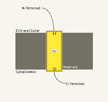
| Transmembrane helix prediction | |
Transmembrane helices have been predicted in your sequence to adopt the topology shown below
Phyre is for academic use only
| Please cite: Protein structure prediction on
the web: a case study using the Phyre server |
| Kelley LA and Sternberg MJE. Nature Protocols
4, 363 - 371 (2009) [pdf] [Import into BibTeX] |
| |
| If you use the binding site
predictions from 3DLigandSite, please also cite: |
| 3DLigandSite: predicting ligand-binding sites using similar structures. |
| Wass MN, Kelley LA and Sternberg
MJ Nucleic Acids Research 38, W469-73 (2010) [PubMed] |
| |
|
|
|
|