1 c3rpjA_
100.0
48
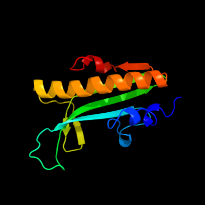
PDB header: transcription regulatorChain: A: PDB Molecule: curlin genes transcriptional regulator;PDBTitle: structure of a curlin genes transcriptional regulator protein from2 proteus mirabilis hi4320.
2 d2bhua1
67.4
20
Fold: Immunoglobulin-like beta-sandwichSuperfamily: E set domainsFamily: E-set domains of sugar-utilizing enzymes3 c2b9bA_
62.2
44
PDB header: viral proteinChain: A: PDB Molecule: fusion glycoprotein f0;PDBTitle: structure of the parainfluenza virus 5 f protein in its metastable,2 pre-fusion conformation
4 d2a1va1
49.9
18
Fold: Secretion chaperone-likeSuperfamily: YjbR-likeFamily: YjbR-like5 d3duea1
45.9
23
Fold: BLIP-likeSuperfamily: BT0923-likeFamily: BT0923-like6 d2fkia1
32.8
22
Fold: Secretion chaperone-likeSuperfamily: YjbR-likeFamily: YjbR-like7 c3nrlB_
22.2
25
PDB header: structural genomics, unknown functionChain: B: PDB Molecule: uncharacterized protein rumgna_01417;PDBTitle: crystal structure of protein rumgna_01417 from ruminococcus gnavus,2 northeast structural genomics consortium target ugr76
8 d1axib1
18.0
28
Fold: Immunoglobulin-like beta-sandwichSuperfamily: Fibronectin type IIIFamily: Fibronectin type III9 d3elga1
17.7
15
Fold: BLIP-likeSuperfamily: BT0923-likeFamily: BT0923-like10 c1agqB_
16.4
31
PDB header: growth factorChain: B: PDB Molecule: glial cell-derived neurotrophic factor;PDBTitle: glial cell-derived neurotrophic factor from rat
11 d1b63a1
16.0
21
Fold: Ribosomal protein S5 domain 2-likeSuperfamily: Ribosomal protein S5 domain 2-likeFamily: DNA gyrase/MutL, second domain12 d2p02a2
15.1
35
Fold: S-adenosylmethionine synthetaseSuperfamily: S-adenosylmethionine synthetaseFamily: S-adenosylmethionine synthetase13 d1mxaa2
14.3
30
Fold: S-adenosylmethionine synthetaseSuperfamily: S-adenosylmethionine synthetaseFamily: S-adenosylmethionine synthetase14 d1qm4a2
13.0
30
Fold: S-adenosylmethionine synthetaseSuperfamily: S-adenosylmethionine synthetaseFamily: S-adenosylmethionine synthetase15 c2gyrB_
11.9
13
PDB header: hormone/growth factorChain: B: PDB Molecule: neurotrophic factor artemin, isoform 3;PDBTitle: crystal structure of human artemin
16 d1zyma1
10.4
19
Fold: SAM domain-likeSuperfamily: Enzyme I of the PEP:sugar phosphotransferase system HPr-binding (sub)domainFamily: Enzyme I of the PEP:sugar phosphotransferase system HPr-binding (sub)domain17 c2w82C_
9.7
28
PDB header: replication inhibitorChain: C: PDB Molecule: orf18;PDBTitle: the structure of arda
18 c1lu0A_
9.6
86
PDB header: hydrolase inhibitorChain: A: PDB Molecule: trypsin inhibitor i;PDBTitle: atomic resolution structure of squash trypsin inhibitor: unexpected2 metal coordination
19 d1lu0a_
9.6
86
Fold: Knottins (small inhibitors, toxins, lectins)Superfamily: Plant inhibitors of proteinases and amylasesFamily: Plant inhibitors of proteinases and amylases20 c3ctiA_
9.6
86
PDB header: proteinase inhibitor (trypsin)Chain: A: PDB Molecule: trypsin inhibitor;PDBTitle: relaxation matrix refinement of the solution structure of2 squash trypsin inhibitor
21 c2ctiA_
not modelled
9.6
86
PDB header: proteinase inhibitor (trypsin)Chain: A: PDB Molecule: trypsin inhibitor;PDBTitle: determination of the complete three-dimensional structure2 of the trypsin inhibitor from squash seeds in aqueous3 solution by nuclear magnetic resonance and a combination4 of distance geometry and dynamical simulated annealing
22 c1ppeI_
not modelled
9.6
86
PDB header: hydrolase(serine proteinase)Chain: I: PDB Molecule: trypsin inhibitor cmti-i;PDBTitle: the refined 2.0 angstroms x-ray crystal structure of the2 complex formed between bovine beta-trypsin and cmti-i, a3 trypsin inhibitor from squash seeds (cucurbita maxima):4 topological similarity of the squash seed inhibitors with5 the carboxypeptidase a inhibitor from potatoes
23 c1ctiA_
not modelled
9.6
86
PDB header: proteinase inhibitor (trypsin)Chain: A: PDB Molecule: trypsin inhibitor;PDBTitle: determination of the complete three-dimensional structure2 of the trypsin inhibitor from squash seeds in aqueous3 solution by nuclear magnetic resonance and a combination4 of distance geometry and dynamical simulated annealing
24 c2staI_
not modelled
9.6
86
PDB header: hydrolase/hydrolase inhibitorChain: I: PDB Molecule: protein (trypsin inhibitor);PDBTitle: anionic salmon trypsin in complex with squash seed2 inhibitor (cucurbita maxima trypsin inhibitor i)
25 d2id1a1
not modelled
9.5
25
Fold: NucleotidyltransferaseSuperfamily: NucleotidyltransferaseFamily: Iojap/YbeB-like26 c1lu0B_
not modelled
9.5
86
PDB header: hydrolase inhibitorChain: B: PDB Molecule: trypsin inhibitor i;PDBTitle: atomic resolution structure of squash trypsin inhibitor: unexpected2 metal coordination
27 c2v1vA_
not modelled
9.5
86
PDB header: hydrolase inhibitorChain: A: PDB Molecule: trypsin inhibitor 1;PDBTitle: 3d structure of the m8l mutant of squash trypsin inhibitor2 cmti-i
28 c2a7oA_
not modelled
9.2
50
PDB header: transcriptionChain: A: PDB Molecule: huntingtin interacting protein b;PDBTitle: solution structure of the hset2/hypb sri domain
29 d2c4ba2
not modelled
9.2
86
Fold: Knottins (small inhibitors, toxins, lectins)Superfamily: Plant inhibitors of proteinases and amylasesFamily: Plant inhibitors of proteinases and amylases30 c2vpzG_
not modelled
9.2
50
PDB header: oxidoreductaseChain: G: PDB Molecule: hypothetical membrane spanning protein;PDBTitle: polysulfide reductase native structure
31 d1h9ii_
not modelled
9.1
60
Fold: Knottins (small inhibitors, toxins, lectins)Superfamily: Plant inhibitors of proteinases and amylasesFamily: Plant inhibitors of proteinases and amylases32 d1h9hi_
not modelled
8.8
86
Fold: Knottins (small inhibitors, toxins, lectins)Superfamily: Plant inhibitors of proteinases and amylasesFamily: Plant inhibitors of proteinases and amylases33 c1mcvI_
not modelled
8.8
86
PDB header: hydrolaseChain: I: PDB Molecule: hei-toe i;PDBTitle: crystal structure analysis of a hybrid squash inhibitor in2 complex with porcine pancreatic elastase
34 d1mcvi_
not modelled
8.8
86
Fold: Knottins (small inhibitors, toxins, lectins)Superfamily: Plant inhibitors of proteinases and amylasesFamily: Plant inhibitors of proteinases and amylases35 c2it7A_
not modelled
8.7
86
PDB header: plant proteinChain: A: PDB Molecule: trypsin inhibitor 2;PDBTitle: solution structure of the squash trypsin inhibitor eeti-ii
36 d2it7a1
not modelled
8.7
86
Fold: Knottins (small inhibitors, toxins, lectins)Superfamily: Plant inhibitors of proteinases and amylasesFamily: Plant inhibitors of proteinases and amylases37 c2letA_
not modelled
8.5
86
PDB header: proteinase inhibitor(trypsin)Chain: A: PDB Molecule: trypsin inhibitor ii;PDBTitle: an 1h nmr determination of the three dimensional structures2 of mirror image forms of a leu-5 variant of the trypsin3 inhibitor ecballium elaterium (eeti-ii)
38 d1rpya_
not modelled
8.1
19
Fold: SH2-likeSuperfamily: SH2 domainFamily: SH2 domain39 c3m07A_
not modelled
8.0
11
PDB header: unknown functionChain: A: PDB Molecule: putative alpha amylase;PDBTitle: 1.4 angstrom resolution crystal structure of putative alpha2 amylase from salmonella typhimurium.
40 c3fg7A_
not modelled
7.6
16
PDB header: structural proteinChain: A: PDB Molecule: villin-1;PDBTitle: the crystal structure of villin domain 6
41 d1xa6a2
not modelled
7.6
18
Fold: SH2-likeSuperfamily: SH2 domainFamily: SH2 domain42 d2evra2
not modelled
7.3
26
Fold: Cysteine proteinasesSuperfamily: Cysteine proteinasesFamily: NlpC/P6043 c1ik9C_
not modelled
7.3
38
PDB header: gene regulation/ligaseChain: C: PDB Molecule: dna ligase iv;PDBTitle: crystal structure of a xrcc4-dna ligase iv complex
44 d2o62a1
not modelled
7.2
23
Fold: LipocalinsSuperfamily: LipocalinsFamily: All1756-like45 d2i1sa1
not modelled
6.9
27
Fold: MM3350-likeSuperfamily: MM3350-likeFamily: MM3350-like46 d1exbe_
not modelled
6.6
21
Fold: POZ domainSuperfamily: POZ domainFamily: Tetramerization domain of potassium channels47 d2btci_
not modelled
6.5
71
Fold: Knottins (small inhibitors, toxins, lectins)Superfamily: Plant inhibitors of proteinases and amylasesFamily: Plant inhibitors of proteinases and amylases48 c2btcI_
not modelled
6.5
71
PDB header: hydrolase/hydrolase inhibitorChain: I: PDB Molecule: protein (trypsin inhibitor);PDBTitle: bovine trypsin in complex with squash seed inhibitor2 (cucurbita pepo trypsin inhibitor ii)
49 c2stbI_
not modelled
6.5
71
PDB header: hydrolase/hydrolase inhibitorChain: I: PDB Molecule: protein (trypsin inhibitor);PDBTitle: anionic salmon trypsin in complex with squash seed2 inhibitor (cucurbita pepo trypsin inhibitor ii)
50 c3h9xB_
not modelled
6.3
13
PDB header: structural genomics, unknown functionChain: B: PDB Molecule: uncharacterized protein pspto_3016;PDBTitle: crystal structure of the pspto_3016 protein from2 pseudomonas syringae, northeast structural genomics3 consortium target psr293
51 c2ysxA_
not modelled
6.2
15
PDB header: signaling proteinChain: A: PDB Molecule: signaling inositol polyphosphate phosphatasePDBTitle: solution structure of the human ship sh2 domain
52 c2yh5A_
not modelled
6.1
12
PDB header: lipid binding proteinChain: A: PDB Molecule: dapx protein;PDBTitle: structure of the c-terminal domain of bamc
53 c2xznZ_
not modelled
6.1
11
PDB header: ribosomeChain: Z: PDB Molecule: rps21e;PDBTitle: crystal structure of the eukaryotic 40s ribosomal2 subunit in complex with initiation factor 1. this file3 contains the 40s subunit and initiation factor for4 molecule 2
54 d1t6aa_
not modelled
6.0
12
Fold: TBP-likeSuperfamily: Rbstp2229 proteinFamily: Rbstp2229 protein55 d2eyva1
not modelled
6.0
19
Fold: SH2-likeSuperfamily: SH2 domainFamily: SH2 domain56 d1eysl_
not modelled
5.9
67
Fold: Bacterial photosystem II reaction centre, L and M subunitsSuperfamily: Bacterial photosystem II reaction centre, L and M subunitsFamily: Bacterial photosystem II reaction centre, L and M subunits57 c2kkuA_
not modelled
5.8
38
PDB header: structural genomics, unknown functionChain: A: PDB Molecule: uncharacterized protein;PDBTitle: solution structure of protein af2351 from archaeoglobus2 fulgidus. northeast structural genomics consortium target3 att9/ontario center for structural proteomics target af2351
58 d2bcqa3
not modelled
5.5
25
Fold: NucleotidyltransferaseSuperfamily: NucleotidyltransferaseFamily: DNA polymerase beta-like59 c2nvgA_
not modelled
5.5
13
PDB header: oxidoreductaseChain: A: PDB Molecule: ubiquinol-cytochrome c reductase iron-sulfur subunit;PDBTitle: soluble domain of rieske iron sulfur protein.
60 c2qv5A_
not modelled
5.5
23
PDB header: structural genomics, unknown functionChain: A: PDB Molecule: uncharacterized protein atu2773;PDBTitle: crystal structure of uncharacterized protein atu2773 from2 agrobacterium tumefaciens c58
61 c3rv2B_
not modelled
5.4
43
PDB header: transferaseChain: B: PDB Molecule: s-adenosylmethionine synthase;PDBTitle: crystal structure of s-adenosylmethionine synthetase from2 mycobacterium marinum