| Secondary structure and disorder prediction | |
 |
| | |
1 | . | . | . | . | . | . | . | . | 10 | . | . | . | . | . | . | . | . | . | 20 | . | . | . | . | . | . | . | . | . | 30 | . | . | . | . | . | . | . | . | . | 40 | . | . | . | . | . | . | . | . | . | 50 | . | . | . | . | . | . | . | . | . | 60 |
| Sequence | |
M | F | V | P | F | L | I | M | L | R | E | G | L | E | A | A | L | I | V | S | L | I | A | S | Y | L | K | R | T | Q | R | G | M | X | D | C | V | M | W | I | G | V | L | L | A | A | A | L | C | L | G | L | G | I | F | I | N | E | T | T |
| Secondary structure | |
|  | | | | | | | | | | | | | | | | | | | | | | | | | | |
|
|
| | | | | | | | | | | | | | | | | | | | | | | | | | | | | |
| SS confidence | |
|
|
|
|
|
|
|
|
|
|
|
|
|
|
|
|
|
|
|
|
|
|
|
|
|
|
|
|
|
|
|
|
|
|
|
|
|
|
|
|
|
|
|
|
|
|
|
|
|
|
|
|
|
|
|
|
|
|
|
|
| Disorder | |
? |
|
|
|
|
|
|
|
|
|
|
|
|
|
|
|
|
|
|
|
|
|
|
|
|
|
|
|
|
|
|
|
|
|
|
|
|
|
|
|
|
|
|
|
|
|
|
|
|
|
|
|
|
|
|
|
|
|
|
|
| Disorder confidence | |
|
|
|
|
|
|
|
|
|
|
|
|
|
|
|
|
|
|
|
|
|
|
|
|
|
|
|
|
|
|
|
|
|
|
|
|
|
|
|
|
|
|
|
|
|
|
|
|
|
|
|
|
|
|
|
|
|
|
|
|
| |
| | |
. | . | . | . | . | . | . | . | . | 70 | . | . | . | . | . | . | . | . | . | 80 | . | . | . | . | . | . | . | . | . | 90 | . | . | . | . | . | . | . | . | . | 100 | . | . | . | . | . | . | . | . | . | 110 | . | . | . | . | . | . | . | . | . | 120 |
| Sequence | |
G | E | F | P | Q | K | E | Q | E | L | F | E | G | I | V | A | V | I | A | V | V | I | L | T | W | M | V | F | W | M | R | K | V | S | R | N | V | K | V | Q | L | E | Q | A | V | D | S | A | L | Q | R | G | N | H | H | G | W | A | L | V |
| Secondary structure | |
|
|
|
|
|
| | | | | | | | | | | | | | | | | | | | | | | | | | |
| | | | | | | | | | | | | | | | |
|
|
|
| | | | | | | |
| SS confidence | |
|
|
|
|
|
|
|
|
|
|
|
|
|
|
|
|
|
|
|
|
|
|
|
|
|
|
|
|
|
|
|
|
|
|
|
|
|
|
|
|
|
|
|
|
|
|
|
|
|
|
|
|
|
|
|
|
|
|
|
|
| Disorder | |
? |
|
|
|
|
|
|
|
|
|
|
|
|
|
|
|
|
|
|
|
|
|
|
|
|
|
|
|
|
|
|
|
|
|
|
|
|
|
|
|
|
|
|
|
|
|
|
| ? | ? | ? | ? | ? | ? |
|
|
|
|
|
|
| Disorder confidence | |
|
|
|
|
|
|
|
|
|
|
|
|
|
|
|
|
|
|
|
|
|
|
|
|
|
|
|
|
|
|
|
|
|
|
|
|
|
|
|
|
|
|
|
|
|
|
|
|
|
|
|
|
|
|
|
|
|
|
|
|
| |
| | |
. | . | . | . | . | . | . | . | . | 130 | . | . | . | . | . | . | . | . | . | 140 | . | . | . | . | . | . | . | . | . | 150 | . | . | . | . | . | . | . | . | . | 160 | . | . | . | . | . | . | . | . | . | 170 | . | . | . | . | . | . | . | . | . | 180 |
| Sequence | |
M | M | V | F | F | A | V | A | R | E | G | L | E | S | V | F | F | L | L | A | A | F | Q | Q | D | V | G | I | W | P | P | L | G | A | M | L | G | L | A | T | A | V | V | L | G | F | L | L | Y | W | G | G | I | R | L | N | L | G | A | F |
| Secondary structure | |
| | | | | | | | | | | | | | | | | | | | | |
|
|
|
|
|
|
| | | | | | | | | | | | | | | | | | | | | | | | | |
|
| | | | |
| SS confidence | |
|
|
|
|
|
|
|
|
|
|
|
|
|
|
|
|
|
|
|
|
|
|
|
|
|
|
|
|
|
|
|
|
|
|
|
|
|
|
|
|
|
|
|
|
|
|
|
|
|
|
|
|
|
|
|
|
|
|
|
|
| Disorder | |
|
|
|
|
|
|
|
|
|
|
|
|
|
|
|
|
|
|
|
|
|
| ? |
| ? | ? | ? | ? |
|
|
|
|
|
|
|
|
|
|
|
|
|
|
|
|
|
|
|
|
|
|
|
|
|
| ? |
|
|
|
|
|
| Disorder confidence | |
|
|
|
|
|
|
|
|
|
|
|
|
|
|
|
|
|
|
|
|
|
|
|
|
|
|
|
|
|
|
|
|
|
|
|
|
|
|
|
|
|
|
|
|
|
|
|
|
|
|
|
|
|
|
|
|
|
|
|
|
| |
| | |
. | . | . | . | . | . | . | . | . | 190 | . | . | . | . | . | . | . | . | . | 200 | . | . | . | . | . | . | . | . | . | 210 | . | . | . | . | . | . | . | . | . | 220 | . | . | . | . | . | . | . | . | . | 230 | . | . | . | . | . | . | . | . | . | 240 |
| Sequence | |
F | K | W | T | S | L | F | I | L | F | V | A | A | G | L | A | A | G | A | I | R | A | F | H | E | A | G | L | W | N | H | F | Q | E | I | A | F | D | M | S | A | V | L | S | T | H | S | L | F | G | T | L | M | E | G | I | F | G | Y | Q |
| Secondary structure | |
| | | | | | | | | | | | | | | | | | | | | | | | |
|
|
|
|
|
|
|
|
|
|
|
|
| | | |
|
|
|
|
|
|
| | | | | | | | | |
|
|
|
| SS confidence | |
|
|
|
|
|
|
|
|
|
|
|
|
|
|
|
|
|
|
|
|
|
|
|
|
|
|
|
|
|
|
|
|
|
|
|
|
|
|
|
|
|
|
|
|
|
|
|
|
|
|
|
|
|
|
|
|
|
|
|
|
| Disorder | |
|
|
|
|
|
|
|
|
|
|
|
|
|
|
|
|
|
|
|
|
|
|
|
|
|
|
|
|
|
|
|
|
|
|
|
|
|
|
|
|
|
|
|
|
|
|
|
|
|
|
|
|
|
|
|
|
|
|
|
|
| Disorder confidence | |
|
|
|
|
|
|
|
|
|
|
|
|
|
|
|
|
|
|
|
|
|
|
|
|
|
|
|
|
|
|
|
|
|
|
|
|
|
|
|
|
|
|
|
|
|
|
|
|
|
|
|
|
|
|
|
|
|
|
|
|
| |
| | |
. | . | . | . | . | . | . | . | . | 250 | . | . | . | . | . | . | . | . | . | 260 | . | . | . | . | . | . | . | . | . | 270 | . | . | . | . | . | . |
| Sequence | |
E | A | P | S | V | S | E | V | A | V | W | F | I | Y | L | I | P | A | L | V | A | F | A | L | P | P | R | A | G | A | T | A | S | R | S | A |
| Secondary structure | |
|
|
|
| | | | | | | | | | | | | | | | | | | |
|
|
|
|
|
|
|
|
|
|
|
|
|
| SS confidence | |
|
|
|
|
|
|
|
|
|
|
|
|
|
|
|
|
|
|
|
|
|
|
|
|
|
|
|
|
|
|
|
|
|
|
|
|
| Disorder | |
|
|
|
|
|
|
|
|
|
|
|
|
|
|
|
|
|
|
|
|
|
|
| ? | ? | ? | ? | ? | ? | ? | ? | ? | ? | ? | ? | ? |
| Disorder confidence | |
|
|
|
|
|
|
|
|
|
|
|
|
|
|
|
|
|
|
|
|
|
|
|
|
|
|
|
|
|
|
|
|
|
|
|
|
| |
| Confidence Key |
| High(9) | |
|
|
|
|
|
|
|
|
|
Low (0) |
| ? | Disordered |
| Alpha helix |
  | Beta strand |
Hover over an aligned region to see model and summary info
Please note, only up to the top 20 hits are modelled to reduce computer load
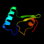
|
| 1 |
|
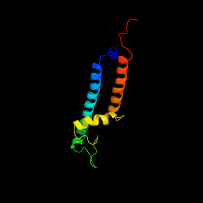
PDB 3jyc chain A
Region: 168 - 274
Aligned: 107
Modelled: 107
Confidence: 14.4%
Identity: 13%
PDB header:metal transport
Chain: A: PDB Molecule:inward-rectifier k+ channel kir2.2;
PDBTitle: crystal structure of the eukaryotic strong inward-rectifier2 k+ channel kir2.2 at 3.1 angstrom resolution
Phyre2
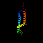
| 2 |
|
PDB 2knc chain A
Region: 150 - 172
Aligned: 23
Modelled: 23
Confidence: 10.9%
Identity: 22%
PDB header:cell adhesion
Chain: A: PDB Molecule:integrin alpha-iib;
PDBTitle: platelet integrin alfaiib-beta3 transmembrane-cytoplasmic2 heterocomplex
Phyre2
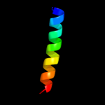
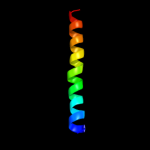
| 3 |
|
PDB 2rdd chain B
Region: 245 - 275
Aligned: 31
Modelled: 31
Confidence: 10.0%
Identity: 6%
PDB header:membrane protein/transport protein
Chain: B: PDB Molecule:upf0092 membrane protein yajc;
PDBTitle: x-ray crystal structure of acrb in complex with a novel2 transmembrane helix.
Phyre2
| 4 |
|
PDB 2rmi chain A
Region: 9 - 15
Aligned: 7
Modelled: 7
Confidence: 7.4%
Identity: 86%
PDB header:neuropeptide
Chain: A: PDB Molecule:astressin;
PDBTitle: 3d nmr structure of astressin
Phyre2
| 5 |
|
PDB 1cii chain A
Region: 176 - 262
Aligned: 82
Modelled: 87
Confidence: 6.8%
Identity: 10%
PDB header:transmembrane protein
Chain: A: PDB Molecule:colicin ia;
PDBTitle: colicin ia
Phyre2
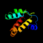
| 6 |
|
PDB 1d0n chain A domain 5
Region: 89 - 146
Aligned: 46
Modelled: 58
Confidence: 6.7%
Identity: 17%
Fold: Gelsolin-like
Superfamily: Actin depolymerizing proteins
Family: Gelsolin-like
Phyre2
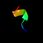
| 7 |
|
PDB 2axt chain Z domain 1
Region: 16 - 55
Aligned: 40
Modelled: 40
Confidence: 6.4%
Identity: 18%
Fold: Transmembrane helix hairpin
Superfamily: PsbZ-like
Family: PsbZ-like
Phyre2
|
| Detailed template information | |
Due to computational demand, binding site predictions are not run for batch jobs
If you want to predict binding sites, please manually submit your model of choice to 3DLigandSite
Phyre is for academic use only
| Please cite: Protein structure prediction on
the web: a case study using the Phyre server |
| Kelley LA and Sternberg MJE. Nature Protocols
4, 363 - 371 (2009) [pdf] [Import into BibTeX] |
| |
| If you use the binding site
predictions from 3DLigandSite, please also cite: |
| 3DLigandSite: predicting ligand-binding sites using similar structures. |
| Wass MN, Kelley LA and Sternberg
MJ Nucleic Acids Research 38, W469-73 (2010) [PubMed] |
| |
|
|
|
|