| 1 |
|
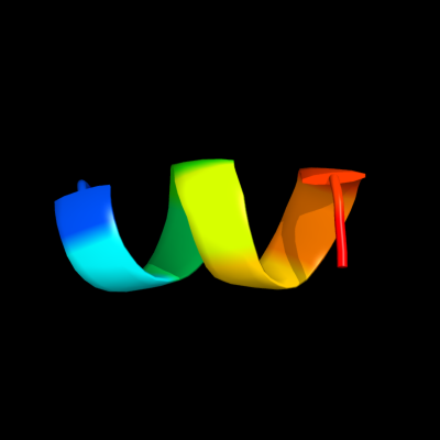
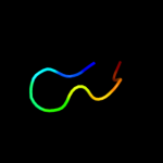
PDB 3owt chain C
Region: 164 - 173
Aligned: 10
Modelled: 10
Confidence: 30.5%
Identity: 60%
PDB header:protein binding
Chain: C: PDB Molecule:regulatory protein sir3;
PDBTitle: crystal structure of s. cerevisiae rap1-sir3 complex
Phyre2
| 2 |
|
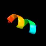
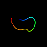
PDB 3s32 chain A
Region: 67 - 81
Aligned: 15
Modelled: 15
Confidence: 15.1%
Identity: 47%
PDB header:transcription
Chain: A: PDB Molecule:set1/ash2 histone methyltransferase complex subunit ash2;
PDBTitle: crystal structure of ash2l n-terminal domain
Phyre2
| 3 |
|
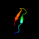
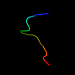
PDB 1wig chain A domain 2
Region: 73 - 80
Aligned: 8
Modelled: 8
Confidence: 7.6%
Identity: 63%
Fold: Glucocorticoid receptor-like (DNA-binding domain)
Superfamily: Glucocorticoid receptor-like (DNA-binding domain)
Family: LIM domain
Phyre2
| 4 |
|
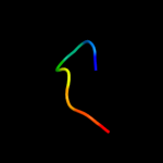
PDB 1g47 chain A domain 1
Region: 72 - 81
Aligned: 10
Modelled: 10
Confidence: 7.5%
Identity: 40%
Fold: Glucocorticoid receptor-like (DNA-binding domain)
Superfamily: Glucocorticoid receptor-like (DNA-binding domain)
Family: LIM domain
Phyre2
| 5 |
|
PDB 1zfo chain A
Region: 72 - 80
Aligned: 9
Modelled: 9
Confidence: 6.8%
Identity: 56%
Fold: Glucocorticoid receptor-like (DNA-binding domain)
Superfamily: Glucocorticoid receptor-like (DNA-binding domain)
Family: LASP-1
Phyre2
| 6 |
|
PDB 2dlo chain A domain 2
Region: 73 - 85
Aligned: 13
Modelled: 13
Confidence: 5.7%
Identity: 23%
Fold: Glucocorticoid receptor-like (DNA-binding domain)
Superfamily: Glucocorticoid receptor-like (DNA-binding domain)
Family: LIM domain
Phyre2
| 7 |
|
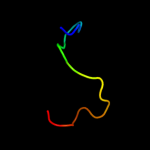
PDB 1zup chain A domain 1
Region: 51 - 96
Aligned: 46
Modelled: 46
Confidence: 5.7%
Identity: 7%
Fold: Ribonuclease H-like motif
Superfamily: Ribonuclease H-like
Family: TM1739-like
Phyre2
| 8 |
|
PDB 1es6 chain A domain 2
Region: 63 - 83
Aligned: 21
Modelled: 21
Confidence: 5.5%
Identity: 29%
Fold: EV matrix protein
Superfamily: EV matrix protein
Family: EV matrix protein
Phyre2
| 9 |
|
PDB 2cup chain A domain 2
Region: 73 - 80
Aligned: 8
Modelled: 8
Confidence: 5.4%
Identity: 38%
Fold: Glucocorticoid receptor-like (DNA-binding domain)
Superfamily: Glucocorticoid receptor-like (DNA-binding domain)
Family: LIM domain
Phyre2