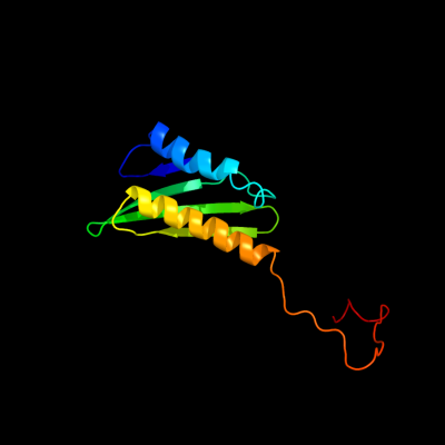
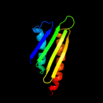
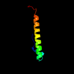
1 d1l4sa_
100.0
100
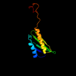
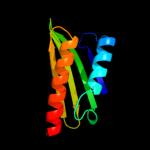
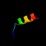
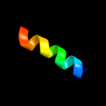
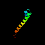
Fold: Ribosome binding protein Y (YfiA homologue)Superfamily: Ribosome binding protein Y (YfiA homologue)Family: Ribosome binding protein Y (YfiA homologue)2 d1imua_
100.0
64
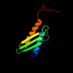
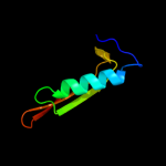
Fold: Ribosome binding protein Y (YfiA homologue)Superfamily: Ribosome binding protein Y (YfiA homologue)Family: Ribosome binding protein Y (YfiA homologue)3 c2rqlA_
100.0
38
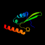
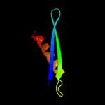
PDB header: translationChain: A: PDB Molecule: probable sigma-54 modulation protein;PDBTitle: solution structure of the e. coli ribosome hibernation2 promoting factor hpf
4 c3tqmD_
99.9
26
PDB header: protein bindingChain: D: PDB Molecule: ribosome-associated factor y;PDBTitle: structure of an ribosomal subunit interface protein from coxiella2 burnetii
5 d2ywqa1
99.9
27
Fold: Ribosome binding protein Y (YfiA homologue)Superfamily: Ribosome binding protein Y (YfiA homologue)Family: Ribosome binding protein Y (YfiA homologue)6 d1uila_
25.1
27
Fold: dsRBD-likeSuperfamily: dsRNA-binding domain-likeFamily: Double-stranded RNA-binding domain (dsRBD)7 c3kxeD_
23.0
6
PDB header: protein bindingChain: D: PDB Molecule: antitoxin protein pard-1;PDBTitle: a conserved mode of protein recognition and binding in a2 pard-pare toxin-antitoxin complex
8 c1by0A_
16.6
44
PDB header: rna binding proteinChain: A: PDB Molecule: protein (hepatitis delta antigen);PDBTitle: n-terminal leucine-repeat region of hepatitis delta antigen
9 c1i7nA_
15.7
12
PDB header: neuropeptideChain: A: PDB Molecule: synapsin ii;PDBTitle: crystal structure analysis of the c domain of synapsin ii2 from rat brain
10 c2kxoA_
13.7
24
PDB header: cell cycleChain: A: PDB Molecule: cell division topological specificity factor;PDBTitle: solution nmr structure of the cell division regulator mine protein2 from neisseria gonorrhoeae
11 d1o6da_
12.7
10
Fold: alpha/beta knotSuperfamily: alpha/beta knotFamily: YbeA-like12 c3f6hA_
12.5
12
PDB header: transferaseChain: A: PDB Molecule: alpha-isopropylmalate synthase;PDBTitle: crystal structure of the regulatory domain of licms in2 complexed with isoleucine - type iii
13 c2yh5A_
12.1
12
PDB header: lipid binding proteinChain: A: PDB Molecule: dapx protein;PDBTitle: structure of the c-terminal domain of bamc
14 d2e9xd1
12.1
33
Fold: GINS helical bundle-likeSuperfamily: GINS helical bundle-likeFamily: SLD5 N-terminal domain-like15 d1lr0a_
10.2
7
Fold: TolA/TonB C-terminal domainSuperfamily: TolA/TonB C-terminal domainFamily: TolA16 c1hk8A_
10.2
17
PDB header: oxidoreductaseChain: A: PDB Molecule: anaerobic ribonucleotide-triphosphate reductase;PDBTitle: structural basis for allosteric substrate specificity2 regulation in class iii ribonucleotide reductases:3 nrdd in complex with dgtp
17 d1hk8a_
10.2
17
Fold: PFL-like glycyl radical enzymesSuperfamily: PFL-like glycyl radical enzymesFamily: Class III anaerobic ribonucleotide reductase NRDD subunit18 d2i52a1
9.5
13
Fold: MK0786-likeSuperfamily: MK0786-likeFamily: MK0786-like19 c3rd6A_
9.0
12
PDB header: structural genomics, unknown functionChain: A: PDB Molecule: mll3558 protein;PDBTitle: crystal structure of mll3558 protein from rhizobium loti. northeast2 structural genomics consortium target id mlr403
20 c3r9jD_
8.9
11
PDB header: cell cycle,hydrolase/cell cycleChain: D: PDB Molecule: cell division topological specificity factor;PDBTitle: 4.3a resolution structure of a mind-mine(i24n) protein complex
21 c1pk8D_
not modelled
8.3
10
PDB header: membrane proteinChain: D: PDB Molecule: rat synapsin i;PDBTitle: crystal structure of rat synapsin i c domain complexed to2 ca.atp
22 c3q2oB_
not modelled
7.9
10
PDB header: lyaseChain: B: PDB Molecule: phosphoribosylaminoimidazole carboxylase, atpase subunit;PDBTitle: crystal structure of purk: n5-carboxyaminoimidazole ribonucleotide2 synthetase
23 c1hf9B_
not modelled
7.8
13
PDB header: atpase inhibitorChain: B: PDB Molecule: atpase inhibitor (mitochondrial);PDBTitle: c-terminal coiled-coil domain from bovine if1
24 c3zqbB_
not modelled
7.3
19
PDB header: cell invasionChain: B: PDB Molecule: protein prgi, cell invasion protein sipd;PDBTitle: prgi-sipd from salmonella typhimurium
25 d1sr9a3
not modelled
6.9
13
Fold: 2-isopropylmalate synthase LeuA, allosteric (dimerisation) domainSuperfamily: 2-isopropylmalate synthase LeuA, allosteric (dimerisation) domainFamily: 2-isopropylmalate synthase LeuA, allosteric (dimerisation) domain26 c1mx0D_
not modelled
6.5
17
PDB header: isomeraseChain: D: PDB Molecule: type ii dna topoisomerase vi subunit b;PDBTitle: structure of topoisomerase subunit
27 d1kr4a_
not modelled
6.5
13
Fold: Ferredoxin-likeSuperfamily: GlnB-likeFamily: Divalent ion tolerance proteins CutA (CutA1)28 c2kpoA_
not modelled
6.3
5
PDB header: de novo proteinChain: A: PDB Molecule: rossmann 2x2 fold protein;PDBTitle: solution nmr structure of de novo designed rossmann 2x2 fold protein,2 northeast structural genomics consortium target or16
29 d2r85a2
not modelled
6.0
6
Fold: ATP-graspSuperfamily: Glutathione synthetase ATP-binding domain-likeFamily: PurP ATP-binding domain-like30 d1ghha_
not modelled
6.0
10
Fold: DNA damage-inducible protein DinISuperfamily: DNA damage-inducible protein DinIFamily: DNA damage-inducible protein DinI31 c2l9pA_
not modelled
5.7
17
PDB header: structural genomics, unknown functionChain: A: PDB Molecule: uncharacterized protein;PDBTitle: solution nmr structure of q5hli9 from staphylococcus epidermidis,2 northeast structural genomics consortium target ser147
32 d1gsoa3
not modelled
5.6
9
Fold: ATP-graspSuperfamily: Glutathione synthetase ATP-binding domain-likeFamily: BC ATP-binding domain-like33 c2l5gB_
not modelled
5.3
29
PDB header: transcription regulatorChain: B: PDB Molecule: putative uncharacterized protein ncor2;PDBTitle: co-ordinates and 1h, 13c and 15n chemical shift assignments for the2 complex of gps2 53-90 and smrt 167-207
34 c2q9qF_
not modelled
5.2
33
PDB header: replicationChain: F: PDB Molecule: gins complex subunit 4;PDBTitle: the crystal structure of full length human gins complex
35 d1vkza1
not modelled
5.2
19
Fold: Barrel-sandwich hybridSuperfamily: Rudiment single hybrid motifFamily: BC C-terminal domain-like