1 c3c1qA_
99.9
51
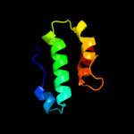
PDB header: transport proteinChain: A: PDB Molecule: general secretion pathway protein f;PDBTitle: the three-dimensional structure of the cytoplasmic domains of epsf2 from the type 2 secretion system of vibrio cholerae
2 c2whnA_
99.9
23
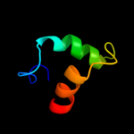
PDB header: protein transportChain: A: PDB Molecule: pilus assembly protein pilc;PDBTitle: n-terminal domain from the pilc type iv pilus biogenesis2 protein
3 c3c8mA_
42.1
18
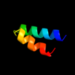
PDB header: oxidoreductaseChain: A: PDB Molecule: homoserine dehydrogenase;PDBTitle: crystal structure of homoserine dehydrogenase from thermoplasma2 volcanium
4 d3cuma1
38.1
8
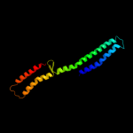
Fold: 6-phosphogluconate dehydrogenase C-terminal domain-likeSuperfamily: 6-phosphogluconate dehydrogenase C-terminal domain-likeFamily: Hydroxyisobutyrate and 6-phosphogluconate dehydrogenase domain5 c3i3aC_
36.5
8
PDB header: transferaseChain: C: PDB Molecule: acyl-[acyl-carrier-protein]--udp-n-PDBTitle: structural basis for the sugar nucleotide and acyl chain2 selectivity of leptospira interrogans lpxa
6 c3ingA_
34.4
27
PDB header: oxidoreductaseChain: A: PDB Molecule: homoserine dehydrogenase;PDBTitle: crystal structure of homoserine dehydrogenase (np_394635.1) from2 thermoplasma acidophilum at 1.95 a resolution
7 c2ejwB_
34.2
16
PDB header: oxidoreductaseChain: B: PDB Molecule: homoserine dehydrogenase;PDBTitle: homoserine dehydrogenase from thermus thermophilus hb8
8 c2d1lA_
33.4
10
PDB header: protein bindingChain: A: PDB Molecule: metastasis suppressor protein 1;PDBTitle: structure of f-actin binding domain imd of mim (missing in metastasis)
9 c3ok8A_
31.0
15
PDB header: protein bindingChain: A: PDB Molecule: brain-specific angiogenesis inhibitor 1-associated proteinPDBTitle: i-bar of pinkbar
10 d1y2oa1
29.7
15
Fold: BAR/IMD domain-likeSuperfamily: BAR/IMD domain-likeFamily: IMD domain11 c3iugA_
20.0
17
PDB header: splicingChain: A: PDB Molecule: rho/cdc42/rac gtpase-activating protein rics;PDBTitle: crystal structure of the rhogap domain of rics
12 d2jf2a1
17.0
11
Fold: Single-stranded left-handed beta-helixSuperfamily: Trimeric LpxA-like enzymesFamily: UDP N-acetylglucosamine acyltransferase13 c3k4iC_
15.1
25
PDB header: structural genomics, unknown functionChain: C: PDB Molecule: uncharacterized protein;PDBTitle: crystal structure of uncharacterized protein pspto_3204 from2 pseudomonas syringae pv. tomato str. dc3000
14 c3r1fO_
14.5
15
PDB header: transcriptionChain: O: PDB Molecule: esx-1 secretion-associated regulator espr;PDBTitle: crystal structure of a key regulator of virulence in mycobacterium2 tuberculosis
15 c2c5qE_
14.1
19
PDB header: structural genomics,unknown functionChain: E: PDB Molecule: rraa-like protein yer010c;PDBTitle: crystal structure of yeast yer010cp
16 c2bbjB_
13.0
17
PDB header: metal transport/membrane proteinChain: B: PDB Molecule: divalent cation transport-related protein;PDBTitle: crystal structure of the cora mg2+ transporter
17 d1xa6a1
12.8
13
Fold: GTPase activation domain, GAPSuperfamily: GTPase activation domain, GAPFamily: BCR-homology GTPase activation domain (BH-domain)18 c3kuqA_
12.8
15
PDB header: hydrolase activatorChain: A: PDB Molecule: rho gtpase-activating protein 7;PDBTitle: crystal structure of the dlc1 rhogap domain
19 c3byiA_
12.7
19
PDB header: signaling proteinChain: A: PDB Molecule: rho gtpase activating protein 15;PDBTitle: crystal structure of human rho gtpase activating protein 15 (arhgap15)
20 d1vq3a_
11.9
13
Fold: PurS-likeSuperfamily: PurS-likeFamily: PurS subunit of FGAM synthetase21 d2ezla_
not modelled
11.4
14
Fold: DNA/RNA-binding 3-helical bundleSuperfamily: Homeodomain-likeFamily: Recombinase DNA-binding domain22 c2f4qA_
not modelled
10.7
18
PDB header: isomeraseChain: A: PDB Molecule: type i topoisomerase, putative;PDBTitle: crystal structure of deinococcus radiodurans topoisomerase ib
23 c3ibqA_
not modelled
10.7
22
PDB header: transferaseChain: A: PDB Molecule: pyridoxal kinase;PDBTitle: crystal structure of pyridoxal kinase from lactobacillus2 plantarum in complex with atp
24 c3r0sA_
not modelled
10.5
16
PDB header: transferaseChain: A: PDB Molecule: acyl-[acyl-carrier-protein]--udp-n-acetylglucosamine o-PDBTitle: udp-n-acetylglucosamine acyltransferase from campylobacter jejuni
25 c3do5A_
not modelled
10.5
18
PDB header: oxidoreductaseChain: A: PDB Molecule: homoserine dehydrogenase;PDBTitle: crystal structure of putative homoserine dehydrogenase (np_069768.1)2 from archaeoglobus fulgidus at 2.20 a resolution
26 c2hx1D_
not modelled
10.4
11
PDB header: hydrolaseChain: D: PDB Molecule: predicted sugar phosphatases of the hadPDBTitle: crystal structure of possible sugar phosphatase, had2 superfamily (zp_00311070.1) from cytophaga hutchinsonii3 atcc 33406 at 2.10 a resolution
27 d1j2za_
not modelled
9.3
12
Fold: Single-stranded left-handed beta-helixSuperfamily: Trimeric LpxA-like enzymesFamily: UDP N-acetylglucosamine acyltransferase28 d1gtda_
not modelled
9.3
4
Fold: PurS-likeSuperfamily: PurS-likeFamily: PurS subunit of FGAM synthetase29 c2zw2B_
not modelled
9.2
13
PDB header: ligaseChain: B: PDB Molecule: putative uncharacterized protein sts178;PDBTitle: crystal structure of formylglycinamide ribonucleotide amidotransferase2 iii from sulfolobus tokodaii (stpurs)
30 c1xa6A_
not modelled
8.9
13
PDB header: signaling proteinChain: A: PDB Molecule: beta2-chimaerin;PDBTitle: crystal structure of the human beta2-chimaerin
31 c3neuA_
not modelled
8.8
15
PDB header: structural genomics, unknown functionChain: A: PDB Molecule: lin1836 protein;PDBTitle: the crystal structure of a functionally-unknown protein lin1836 from2 listeria innocua clip11262
32 c2pmzN_
not modelled
8.4
13
PDB header: translation, transferaseChain: N: PDB Molecule: dna-directed rna polymerase subunit n;PDBTitle: archaeal rna polymerase from sulfolobus solfataricus
33 d1hara_
not modelled
8.3
5
Fold: DNA/RNA polymerasesSuperfamily: DNA/RNA polymerasesFamily: Reverse transcriptase34 d2f06a1
not modelled
7.8
19
Fold: Ferredoxin-likeSuperfamily: ACT-likeFamily: BT0572-like35 d1e8ob_
not modelled
7.6
23
Fold: Signal recognition particle alu RNA binding heterodimer, SRP9/14Superfamily: Signal recognition particle alu RNA binding heterodimer, SRP9/14Family: Signal recognition particle alu RNA binding heterodimer, SRP9/1436 c3mtjA_
not modelled
7.5
16
PDB header: oxidoreductaseChain: A: PDB Molecule: homoserine dehydrogenase;PDBTitle: the crystal structure of a homoserine dehydrogenase from thiobacillus2 denitrificans to 2.15a
37 d2c4na1
not modelled
6.8
14
Fold: HAD-likeSuperfamily: HAD-likeFamily: NagD-like38 d2gyqa1
not modelled
6.3
18
Fold: Ferritin-likeSuperfamily: Ferritin-likeFamily: YciF-like39 c3gdeA_
not modelled
5.7
10
PDB header: ligaseChain: A: PDB Molecule: dna ligase;PDBTitle: the closed conformation of atp-dependent dna ligase from2 archaeoglobus fulgidus
40 d1ijwc_
not modelled
5.7
27
Fold: DNA/RNA-binding 3-helical bundleSuperfamily: Homeodomain-likeFamily: Recombinase DNA-binding domain41 c1n54A_
not modelled
5.6
21
PDB header: rna binding proteinChain: A: PDB Molecule: 80 kda nuclear cap binding protein;PDBTitle: cap binding complex m7gpppg free
42 c1zk6A_
not modelled
5.6
13
PDB header: isomeraseChain: A: PDB Molecule: foldase protein prsa;PDBTitle: nmr solution structure of b. subtilis prsa ppiase
43 d1jnsa_
not modelled
5.6
8
Fold: FKBP-likeSuperfamily: FKBP-likeFamily: FKBP immunophilin/proline isomerase44 d1zhva2
not modelled
5.5
29
Fold: Ferredoxin-likeSuperfamily: ACT-likeFamily: Atu0741-like45 c2i7aA_
not modelled
5.4
14
PDB header: hydrolaseChain: A: PDB Molecule: calpain 13;PDBTitle: domain iv of human calpain 13
46 c2kk6A_
not modelled
5.4
24
PDB header: transferaseChain: A: PDB Molecule: proto-oncogene tyrosine-protein kinase fer;PDBTitle: solution structure of sh2 domain of proto-oncogene tyrosine-2 protein kinase fer from homo sapiens, northeast structural3 genomics consortium (nesg) target hr3461d
47 d1914a2
not modelled
5.3
23
Fold: Signal recognition particle alu RNA binding heterodimer, SRP9/14Superfamily: Signal recognition particle alu RNA binding heterodimer, SRP9/14Family: Signal recognition particle alu RNA binding heterodimer, SRP9/1448 d2gs4a1
not modelled
5.1
20
Fold: Ferritin-likeSuperfamily: Ferritin-likeFamily: YciF-like49 c2kncB_
not modelled
5.1
10
PDB header: cell adhesionChain: B: PDB Molecule: integrin beta-3;PDBTitle: platelet integrin alfaiib-beta3 transmembrane-cytoplasmic2 heterocomplex
50 c1ebuA_
not modelled
5.1
23
PDB header: oxidoreductaseChain: A: PDB Molecule: homoserine dehydrogenase;PDBTitle: homoserine dehydrogenase complex with nad analogue and l-2 homoserine
51 c2kncA_
not modelled
5.1
9
PDB header: cell adhesionChain: A: PDB Molecule: integrin alpha-iib;PDBTitle: platelet integrin alfaiib-beta3 transmembrane-cytoplasmic2 heterocomplex
52 c1sneB_
not modelled
5.1
31
PDB header: de novo proteinChain: B: PDB Molecule: tetrameric beta-beta-alpha mini-protein;PDBTitle: an oligomeric domain-swapped beta-beta-alpha mini-protein
53 c1sneA_
not modelled
5.1
31
PDB header: de novo proteinChain: A: PDB Molecule: tetrameric beta-beta-alpha mini-protein;PDBTitle: an oligomeric domain-swapped beta-beta-alpha mini-protein
54 d1hcra_
not modelled
5.1
27
Fold: DNA/RNA-binding 3-helical bundleSuperfamily: Homeodomain-likeFamily: Recombinase DNA-binding domain55 c3nmeA_
not modelled
5.0
17
PDB header: hydrolaseChain: A: PDB Molecule: sex4 glucan phosphatase;PDBTitle: structure of a plant phosphatase