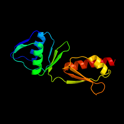
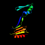
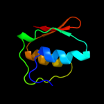
PDB header:membrane protein Chain: A: PDB Molecule:escc; PDBTitle: periplasmic domain of the outer membrane secretin escc from2 enteropathogenic e.coli (epec)
Confidence and coverage
Confidence:
99.9%
Coverage:
34%
140 residues ( 34% of your sequence) have been modelled with 99.9% confidence by the single highest scoring template.
You may wish to submit your sequence to Phyrealarm. This will automatically scan your sequence every week for new potential templates as they appear in the Phyre2 library.
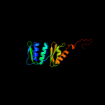
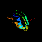
Region: 23 - 176 Aligned: 149 Modelled: 154 Confidence: 99.8% Identity: 12% PDB header:protein transport Chain: D: PDB Molecule:type 2 secretion system, secretin gspd; PDBTitle: the crystal structure of enterotoxigenic escherichia coli gspc-gspd2 complex from the type ii secretion system
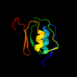
Region: 20 - 176 Aligned: 131 Modelled: 136 Confidence: 99.8% Identity: 31% PDB header:transport protein Chain: A: PDB Molecule:protein transport protein hofq; PDBTitle: structure of the extra-membranous domain of the secretin2 hofq from actinobacillus actinomycetemcomitans
Region: 24 - 198 Aligned: 151 Modelled: 157 Confidence: 99.7% Identity: 14% PDB header:protein transport Chain: A: PDB Molecule:general secretion pathway protein gspd; PDBTitle: crystal structure of the n-terminal domain of the secretin gspd from2 etec determined with the assistance of a nanobody
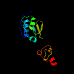
Region: 26 - 100 Aligned: 75 Modelled: 75 Confidence: 97.3% Identity: 16% PDB header:membrane protein, metal transport Chain: A: PDB Molecule:iron(iii) dicitrate transport protein feca; PDBTitle: solution nmr structure of the periplasmic signaling domain2 of the outer membrane iron transporter feca from3 escherichia coli.