| Secondary structure and disorder prediction | |
| | |
1 | . | . | . | . | . | . | . | . | 10 | . | . | . | . | . | . | . | . | . | 20 | . | . | . | . | . | . | . | . | . | 30 | . | . | . | . | . |
| Sequence | |
M | T | L | A | Q | F | A | M | I | F | W | H | D | L | A | A | P | I | L | A | G | I | I | T | A | A | I | V | S | W | W | R | N | R | K |
| Secondary structure | |
|
|  | | | | | | | | | | | | | | | | | | | | | | | | | | | | | | |
|
|
| SS confidence | |
|
|
|
|
|
|
|
|
|
|
|
|
|
|
|
|
|
|
|
|
|
|
|
|
|
|
|
|
|
|
|
|
|
|
|
| Disorder | |
? | ? |
|
|
|
|
|
|
|
|
|
|
|
|
|
|
|
|
|
|
|
|
|
|
|
|
|
|
|
|
|
| ? | ? | ? |
| Disorder confidence | |
|
|
|
|
|
|
|
|
|
|
|
|
|
|
|
|
|
|
|
|
|
|
|
|
|
|
|
|
|
|
|
|
|
|
|
| |
| Confidence Key |
| High(9) | |
|
|
|
|
|
|
|
|
|
Low (0) |
| ? | Disordered |
| Alpha helix |
  | Beta strand |
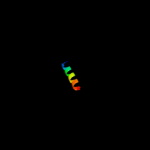
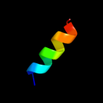
Hover over an aligned region to see model and summary info
Please note, only up to the top 20 hits are modelled to reduce computer load
|
| 1 |
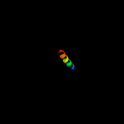
|
PDB 2hg5 chain D
Region: 18 - 35
Aligned: 18
Modelled: 18
Confidence: 10.7%
Identity: 11%
PDB header:membrane protein
Chain: D: PDB Molecule:kcsa channel;
PDBTitle: cs+ complex of a k channel with an amide to ester substitution in the2 selectivity filter
Phyre2
| 2 |
|
PDB 1no7 chain A
Region: 18 - 34
Aligned: 17
Modelled: 17
Confidence: 9.7%
Identity: 35%
Fold: Major capsid protein VP5
Superfamily: Major capsid protein VP5
Family: Major capsid protein VP5
Phyre2
| 3 |
|
PDB 1no7 chain A
Region: 18 - 34
Aligned: 17
Modelled: 17
Confidence: 9.7%
Identity: 35%
PDB header:viral protein
Chain: A: PDB Molecule:major capsid protein;
PDBTitle: structure of the large protease resistant upper domain of2 vp5, the major capsid protein of herpes simplex virus-1
Phyre2
| 4 |
|
PDB 3im4 chain C
Region: 13 - 29
Aligned: 17
Modelled: 17
Confidence: 6.7%
Identity: 47%
PDB header:structural protein, signaling protein
Chain: C: PDB Molecule:dual specificity a kinase-anchoring protein 2;
PDBTitle: crystal structure of camp-dependent protein kinase a2 regulatory subunit i alpha in complex with dual-specific a-3 kinase anchoring protein 2
Phyre2
| 5 |
|
PDB 3t41 chain B
Region: 14 - 27
Aligned: 14
Modelled: 14
Confidence: 5.6%
Identity: 36%
PDB header:hydrolase
Chain: B: PDB Molecule:epidermin leader peptide processing serine protease epip;
PDBTitle: 1.95 angstrom resolution crystal structure of epidermin leader peptide2 processing serine protease (epip) s393a mutant from staphylococcus3 aureus
Phyre2
| 6 |
|
PDB 1r0r chain E
Region: 14 - 27
Aligned: 14
Modelled: 14
Confidence: 5.5%
Identity: 29%
Fold: Subtilisin-like
Superfamily: Subtilisin-like
Family: Subtilases
Phyre2
|
| Detailed template information | |
Due to computational demand, binding site predictions are not run for batch jobs
If you want to predict binding sites, please manually submit your model of choice to 3DLigandSite
Phyre is for academic use only
| Please cite: Protein structure prediction on
the web: a case study using the Phyre server |
| Kelley LA and Sternberg MJE. Nature Protocols
4, 363 - 371 (2009) [pdf] [Import into BibTeX] |
| |
| If you use the binding site
predictions from 3DLigandSite, please also cite: |
| 3DLigandSite: predicting ligand-binding sites using similar structures. |
| Wass MN, Kelley LA and Sternberg
MJ Nucleic Acids Research 38, W469-73 (2010) [PubMed] |
| |
|
|
|
|