Phyre Home Retrieve Phyre Job Id
Emaill.a.kelley@imperial.ac.uk
DescriptionP76342
DateThu Jan 5 12:22:01 GMT 2012
Unique Job IDa21e475de29d8825

Summary 

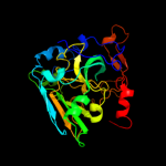
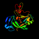
Top model
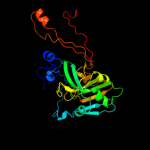
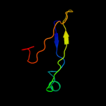
Image coloured by rainbow N → C terminus
Model (left) based on template d1xdya_
Top template information
Fold:Oxidoreductase molybdopterin-binding domain
Superfamily:Oxidoreductase molybdopterin-binding domain
Family:Oxidoreductase molybdopterin-binding domain
Confidence and coverage
Confidence:100.0% Coverage: 79%
263 residues ( 79% of your sequence) have been modelled with 100.0% confidence by the single highest scoring template.
3D viewing
Interactive 3D view in Jmol

Sequence analysis 

Secondary structure and disorder prediction 

   1........10.........20.........30.........40.........50.........60
Sequence  MKKNQFLKESDVTAESVFFMKRRQVLKALGISATALSLPHAAHADLLSWFKGNDRPPAPA
Secondary structure 


SS confidence 


Disorder  ???????????????????


?

????????????????????
Disorder confidence 


   .........70.........80.........90.........100.........110.........120
Sequence  GKALEFSKPAAWQNNLPLTPADKVSGYNNFYEFGLDKADPAANAGSLKTDPWTLKISGEV
Secondary structure 


SS confidence 


Disorder 


?

?
???????????
??


Disorder confidence 


   .........130.........140.........150.........160.........170.........180
Sequence  AKPLTLDHDDLTRRFPLEERIYRMRCVEAWSMVVPWIGFPLHKLLALAEPTSNAKYVAFE
Secondary structure 


SS confidence 


Disorder 


Disorder confidence 


   .........190.........200.........210.........220.........230.........240
Sequence  TIYAPEQMPGQQDRFIGGGLKYPYVEGLRLDEAMHPLTLMTVGVYGKALPPQNGAPVRLI
Secondary structure 


SS confidence 


Disorder 


?


?
?


?


Disorder confidence 


   .........250.........260.........270.........280.........290.........300
Sequence  VPWKYGFKGIKSIVSIKLTRERPPTTWNLAAPDEYGFYANVNPYVDHPRWSQATERFIGS
Secondary structure 


SS confidence 


Disorder 


?????????
Disorder confidence 


   .........310.........320.........330....
Sequence  GGILDVQRQPTLLFNGYAAQVASLYRGLDLRENF
Secondary structure 


SS confidence 


Disorder  ???????????


???????
Disorder confidence 


Confidence Key
High(9)                    Low (0)
?Disordered
Alpha helix
Beta strand

Domain analysis 

Hover over an aligned region to see model and summary info

Please note, only up to the top 20 hits are modelled to reduce computer load

RankAligned region

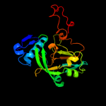
PDB 1xdy chain A

3D model

Region: 62 - 330
Aligned: 263
Modelled: 269
Confidence: 100.0%
Identity: 100%
Fold: Oxidoreductase molybdopterin-binding domain
Superfamily: Oxidoreductase molybdopterin-binding domain
Family: Oxidoreductase molybdopterin-binding domain

Phyre2

PDB 1xdy chain C

3D model

Region: 62 - 330
Aligned: 264
Modelled: 269
Confidence: 100.0%
Identity: 100%
PDB header:oxidoreductase
Chain: C: PDB Molecule:bacterial sulfite oxidase;
PDBTitle: structural and biochemical identification of a novel2 bacterial oxidoreductase, w-containing cofactor

Phyre2

PDB 2xts chain C

3D model

Region: 63 - 334
Aligned: 244
Modelled: 251
Confidence: 100.0%
Identity: 20%
PDB header:oxidoreductase/electron transport
Chain: C: PDB Molecule:sulfite dehydrogenase;
PDBTitle: crystal structure of the sulfane dehydrogenase soxcd from paracoccus2 pantotrophus

Phyre2

PDB 1sox chain B

3D model

Region: 52 - 334
Aligned: 263
Modelled: 264
Confidence: 100.0%
Identity: 21%
PDB header:oxidoreductase
Chain: B: PDB Molecule:sulfite oxidase;
PDBTitle: sulfite oxidase from chicken liver

Phyre2

PDB 2bih chain A

3D model

Region: 56 - 334
Aligned: 256
Modelled: 261
Confidence: 100.0%
Identity: 19%
PDB header:oxidoreductase
Chain: A: PDB Molecule:nitrate reductase [nadph];
PDBTitle: crystal structure of the molybdenum-containing nitrate2 reducing fragment of pichia angusta assimilatory nitrate3 reductase

Phyre2

PDB 2bpb chain A

3D model

Region: 68 - 334
Aligned: 248
Modelled: 256
Confidence: 100.0%
Identity: 24%
PDB header:oxidoreductase
Chain: A: PDB Molecule:sulfite\:cytochrome c oxidoreductase subunit a;
PDBTitle: sulfite dehydrogenase from starkeya novella

Phyre2

PDB 2bii chain A

3D model

Region: 83 - 334
Aligned: 229
Modelled: 238
Confidence: 100.0%
Identity: 19%
PDB header:oxidoreductase
Chain: A: PDB Molecule:nitrate reductase [nadph];
PDBTitle: crystal structure of nitrate-reducing fragment of2 assimilatory nitrate reductase from pichia angusta

Phyre2

PDB 2a9d chain B

3D model

Region: 63 - 334
Aligned: 253
Modelled: 259
Confidence: 100.0%
Identity: 19%
PDB header:oxidoreductase
Chain: B: PDB Molecule:sulfite oxidase;
PDBTitle: crystal structure of recombinant chicken sulfite oxidase with arg at2 residue 161

Phyre2

PDB 1ogp chain D

3D model

Region: 53 - 334
Aligned: 264
Modelled: 271
Confidence: 100.0%
Identity: 20%
PDB header:oxidoreductase
Chain: D: PDB Molecule:sulfite oxidase;
PDBTitle: the crystal structure of plant sulfite oxidase provides2 insight into sulfite oxidation in plants and animals

Phyre2

PDB 1ogp chain A domain 2

3D model

Region: 44 - 283
Aligned: 222
Modelled: 234
Confidence: 100.0%
Identity: 21%
Fold: Oxidoreductase molybdopterin-binding domain
Superfamily: Oxidoreductase molybdopterin-binding domain
Family: Oxidoreductase molybdopterin-binding domain

Phyre2

PDB 2a9d chain A domain 2

3D model

Region: 53 - 294
Aligned: 216
Modelled: 229
Confidence: 100.0%
Identity: 22%
Fold: Oxidoreductase molybdopterin-binding domain
Superfamily: Oxidoreductase molybdopterin-binding domain
Family: Oxidoreductase molybdopterin-binding domain

Phyre2

PDB 2e76 chain D

3D model

Region: 17 - 70
Aligned: 54
Modelled: 54
Confidence: 77.3%
Identity: 17%
PDB header:photosynthesis
Chain: D: PDB Molecule:cytochrome b6-f complex iron-sulfur subunit;
PDBTitle: crystal structure of the cytochrome b6f complex with tridecyl-2 stigmatellin (tds) from m.laminosus

Phyre2

PDB 1p84 chain E

3D model

Region: 2 - 42
Aligned: 41
Modelled: 41
Confidence: 53.8%
Identity: 22%
PDB header:oxidoreductase
Chain: E: PDB Molecule:ubiquinol-cytochrome c reductase iron-sulfur
PDBTitle: hdbt inhibited yeast cytochrome bc1 complex

Phyre2

PDB 2fyu chain E

3D model

Region: 20 - 41
Aligned: 22
Modelled: 22
Confidence: 34.5%
Identity: 14%
PDB header:oxidoreductase
Chain: E: PDB Molecule:ubiquinol-cytochrome c reductase iron-sulfur subunit,
PDBTitle: crystal structure of bovine heart mitochondrial bc1 with jg1442 inhibitor

Phyre2

PDB 1ogp chain A domain 1

3D model

Region: 279 - 334
Aligned: 48
Modelled: 48
Confidence: 28.0%
Identity: 15%
Fold: Immunoglobulin-like beta-sandwich
Superfamily: E set domains
Family: Molybdenum-containing oxidoreductases-like dimerisation domain

Phyre2

PDB 2fyn chain O

3D model

Region: 20 - 41
Aligned: 22
Modelled: 22
Confidence: 22.0%
Identity: 9%
PDB header:oxidoreductase
Chain: O: PDB Molecule:ubiquinol-cytochrome c reductase iron-sulfur
PDBTitle: crystal structure analysis of the double mutant rhodobacter2 sphaeroides bc1 complex

Phyre2

PDB 1r6o chain C domain 1

3D model

Region: 231 - 241
Aligned: 11
Modelled: 11
Confidence: 21.2%
Identity: 18%
Fold: ClpS-like
Superfamily: ClpS-like
Family: Adaptor protein ClpS (YljA)

Phyre2

PDB 2a9d chain A domain 1

3D model

Region: 279 - 334
Aligned: 48
Modelled: 52
Confidence: 13.5%
Identity: 21%
Fold: Immunoglobulin-like beta-sandwich
Superfamily: E set domains
Family: Molybdenum-containing oxidoreductases-like dimerisation domain

Phyre2

PDB 3h3g chain B

3D model

Region: 15 - 31
Aligned: 17
Modelled: 17
Confidence: 12.9%
Identity: 29%
PDB header:membrane protein
Chain: B: PDB Molecule:parathyroid hormone-related protein;
PDBTitle: crystal structure of the extracellular domain of the human parathyroid2 hormone receptor (pth1r) in complex with parathyroid hormone-related3 protein (pthrp)

Phyre2

PDB 2axw chain A domain 1

3D model

Region: 100 - 125
Aligned: 26
Modelled: 26
Confidence: 12.0%
Identity: 12%
Fold: Common fold of diphtheria toxin/transcription factors/cytochrome f
Superfamily: Bacterial adhesins
Family: Pilus subunits

Phyre2
1

d1xdya_
2

c1xdyC_
3

c2xtsC_
4

c1soxB_
5

c2bihA_
6

c2bpbA_
7

c2biiA_
8

c2a9dB_
9

c1ogpD_
10

d1ogpa2
11

d2a9da2
12

c2e76D_
13

c1p84E_
14

c2fyuE_
15

d1ogpa1
16

c2fynO_
17

d1r6oc1
18

d2a9da1
19

c3h3gB_
20

d2axwa1
21


22


23


24


25


26


27


28


29


30


31


32


33


34


35


36


37


38


Detailed template information 

#
Template Alignment Coverage3D Model Confidence
% i.d. Template Information
1d1xdya_


100.0 100 Fold:Oxidoreductase molybdopterin-binding domain
Superfamily:Oxidoreductase molybdopterin-binding domain
Family:Oxidoreductase molybdopterin-binding domain
2c1xdyC_


100.0 100 PDB header:oxidoreductase
Chain: C: PDB Molecule:bacterial sulfite oxidase;
PDBTitle: structural and biochemical identification of a novel2 bacterial oxidoreductase, w-containing cofactor
3c2xtsC_


100.0 20 PDB header:oxidoreductase/electron transport
Chain: C: PDB Molecule:sulfite dehydrogenase;
PDBTitle: crystal structure of the sulfane dehydrogenase soxcd from paracoccus2 pantotrophus
4c1soxB_


100.0 21 PDB header:oxidoreductase
Chain: B: PDB Molecule:sulfite oxidase;
PDBTitle: sulfite oxidase from chicken liver
5c2bihA_


100.0 19 PDB header:oxidoreductase
Chain: A: PDB Molecule:nitrate reductase [nadph];
PDBTitle: crystal structure of the molybdenum-containing nitrate2 reducing fragment of pichia angusta assimilatory nitrate3 reductase
6c2bpbA_


100.0 24 PDB header:oxidoreductase
Chain: A: PDB Molecule:sulfite\:cytochrome c oxidoreductase subunit a;
PDBTitle: sulfite dehydrogenase from starkeya novella
7c2biiA_


100.0 19 PDB header:oxidoreductase
Chain: A: PDB Molecule:nitrate reductase [nadph];
PDBTitle: crystal structure of nitrate-reducing fragment of2 assimilatory nitrate reductase from pichia angusta
8c2a9dB_


100.0 19 PDB header:oxidoreductase
Chain: B: PDB Molecule:sulfite oxidase;
PDBTitle: crystal structure of recombinant chicken sulfite oxidase with arg at2 residue 161
9c1ogpD_


100.0 20 PDB header:oxidoreductase
Chain: D: PDB Molecule:sulfite oxidase;
PDBTitle: the crystal structure of plant sulfite oxidase provides2 insight into sulfite oxidation in plants and animals
10d1ogpa2


100.0 21 Fold:Oxidoreductase molybdopterin-binding domain
Superfamily:Oxidoreductase molybdopterin-binding domain
Family:Oxidoreductase molybdopterin-binding domain
11d2a9da2


100.0 22 Fold:Oxidoreductase molybdopterin-binding domain
Superfamily:Oxidoreductase molybdopterin-binding domain
Family:Oxidoreductase molybdopterin-binding domain
12c2e76D_


77.3 17 PDB header:photosynthesis
Chain: D: PDB Molecule:cytochrome b6-f complex iron-sulfur subunit;
PDBTitle: crystal structure of the cytochrome b6f complex with tridecyl-2 stigmatellin (tds) from m.laminosus
13c1p84E_


53.8 22 PDB header:oxidoreductase
Chain: E: PDB Molecule:ubiquinol-cytochrome c reductase iron-sulfur
PDBTitle: hdbt inhibited yeast cytochrome bc1 complex
14c2fyuE_


34.5 14 PDB header:oxidoreductase
Chain: E: PDB Molecule:ubiquinol-cytochrome c reductase iron-sulfur subunit,
PDBTitle: crystal structure of bovine heart mitochondrial bc1 with jg1442 inhibitor
15d1ogpa1


28.0 15 Fold:Immunoglobulin-like beta-sandwich
Superfamily:E set domains
Family:Molybdenum-containing oxidoreductases-like dimerisation domain
16c2fynO_


22.0 9 PDB header:oxidoreductase
Chain: O: PDB Molecule:ubiquinol-cytochrome c reductase iron-sulfur
PDBTitle: crystal structure analysis of the double mutant rhodobacter2 sphaeroides bc1 complex
17d1r6oc1


21.2 18 Fold:ClpS-like
Superfamily:ClpS-like
Family:Adaptor protein ClpS (YljA)
18d2a9da1


13.5 21 Fold:Immunoglobulin-like beta-sandwich
Superfamily:E set domains
Family:Molybdenum-containing oxidoreductases-like dimerisation domain
19c3h3gB_


12.9 29 PDB header:membrane protein
Chain: B: PDB Molecule:parathyroid hormone-related protein;
PDBTitle: crystal structure of the extracellular domain of the human parathyroid2 hormone receptor (pth1r) in complex with parathyroid hormone-related3 protein (pthrp)
20d2axwa1


12.0 12 Fold:Common fold of diphtheria toxin/transcription factors/cytochrome f
Superfamily:Bacterial adhesins
Family:Pilus subunits
21d2evea1


not modelled 11.8 19 Fold:PUA domain-like
Superfamily:PUA domain-like
Family:Atu2648/PH1033-like
22c3dnjB_


not modelled 11.5 9 PDB header:peptide binding protein
Chain: B: PDB Molecule:atp-dependent clp protease adapter protein clps;
PDBTitle: the structure of the caulobacter crescentus clps protease2 adaptor protein in complex with a n-end rule peptide
23c3ffdP_


not modelled 10.7 31 PDB header:immune system/hormone
Chain: P: PDB Molecule:parathyroid hormone-related protein;
PDBTitle: structure of parathyroid hormone-related protein complexed2 to a neutralizing monoclonal antibody
24d1l7la_


not modelled 9.9 38 Fold:Galactose-binding domain-like
Superfamily:Galactose-binding domain-like
Family:PA-IL, galactose-binding lectin 1
25d1pd0a2


not modelled 9.4 16 Fold:Common fold of diphtheria toxin/transcription factors/cytochrome f
Superfamily:beta-sandwich domain of Sec23/24
Family:beta-sandwich domain of Sec23/24
26c2inpE_


not modelled 7.9 14 PDB header:oxidoreductase
Chain: E: PDB Molecule:phenol hydroxylase component pho;
PDBTitle: structure of the phenol hydroxylase-regulatory protein2 complex
27d2g2xa1


not modelled 7.8 27 Fold:PUA domain-like
Superfamily:PUA domain-like
Family:Atu2648/PH1033-like
28c1r8oA_


not modelled 7.1 22 PDB header:hydrolase inhibitor
Chain: A: PDB Molecule:kunitz trypsin inhibitor;
PDBTitle: crystal structure of an unusual kunitz-type trypsin inhibitor from2 copaifera langsdorffii seeds
29c3diiB_


not modelled 6.8 16 PDB header:oxidoreductase
Chain: B: PDB Molecule:short-chain dehydrogenase/reductase sdr;
PDBTitle: crystal structure of a carbohydrate specific scor enzyme2 from clostridium thermocellum, ligand-free form
30d1zud21


not modelled 6.3 9 Fold:beta-Grasp (ubiquitin-like)
Superfamily:MoaD/ThiS
Family:ThiS
31d2huha1


not modelled 6.2 18 Fold:C2 domain-like
Superfamily:Smr-associated domain-like
Family:Smr-associated domain
32c2fvnA_


not modelled 6.0 12 PDB header:cell adhesion
Chain: A: PDB Molecule:protein afad;
PDBTitle: the fibrillar tip complex of the afa/dr adhesins from2 pathogen e. coli displays synergistic binding to 5 1 and v3 3 integrins
33d1qo7a_


not modelled 5.7 30 Fold:alpha/beta-Hydrolases
Superfamily:alpha/beta-Hydrolases
Family:Epoxide hydrolase
34d2ar1a1


not modelled 5.5 18 Fold:PUA domain-like
Superfamily:PUA domain-like
Family:Atu2648/PH1033-like
35d1pgw21


not modelled 5.5 22 Fold:Nucleoplasmin-like/VP (viral coat and capsid proteins)
Superfamily:Positive stranded ssRNA viruses
Family:Comoviridae-like VP
36d2hkja1


not modelled 5.3 22 Fold:S13-like H2TH domain
Superfamily:S13-like H2TH domain
Family:Topoisomerase VI-B subunit middle domain
37d1pgl21


not modelled 5.3 22 Fold:Nucleoplasmin-like/VP (viral coat and capsid proteins)
Superfamily:Positive stranded ssRNA viruses
Family:Comoviridae-like VP
38d1ny721


not modelled 5.2 26 Fold:Nucleoplasmin-like/VP (viral coat and capsid proteins)
Superfamily:Positive stranded ssRNA viruses
Family:Comoviridae-like VP

Binding site prediction 

Due to computational demand, binding site predictions are not run for batch jobs

If you want to predict binding sites, please manually submit your model of choice to 3DLigandSite


Phyre is for academic use only

Please cite: Protein structure prediction on the web: a case study using the Phyre server
Kelley LA and Sternberg MJE. Nature Protocols 4, 363 - 371 (2009) [pdf] [Import into BibTeX]
 
If you use the binding site predictions from 3DLigandSite, please also cite:
3DLigandSite: predicting ligand-binding sites using similar structures.
Wass MN, Kelley LA and Sternberg MJ Nucleic Acids Research 38, W469-73 (2010) [PubMed]
 
© Structural Bioinformatics Group
Imperial College London
Lawrence Kelley, Benjamin Jefferys
Disclaimer
Terms and Conditions
Component software
Template detection: HHpred 1.51
Secondary structure prediction: Psi-pred 2.5
Disorder prediction: Disopred 2.4
Transmembrane prediction: Memsat_SVM
Multi-template modelling and ab initio: Poing 1.0