Phyre Home Retrieve Phyre Job Id
Emaill.a.kelley@imperial.ac.uk
DescriptionP58094
DateThu Jan 5 12:06:38 GMT 2012
Unique Job ID9be46d66b3ac865b

Summary 

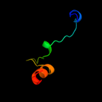
Top model
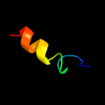
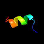
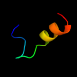
Image coloured by rainbow N → C terminus
Model (left) based on template c3r7kB_
Top template information
PDB header:oxidoreductase
Chain: B: PDB Molecule:probable acyl coa dehydrogenase;
PDBTitle: crystal structure of a probable acyl coa dehydrogenase from2 mycobacterium abscessus atcc 19977 / dsm 44196
Confidence and coverage
Confidence: 32.0% Coverage: 33%
18 residues ( 33% of your sequence) have been modelled with 32.0% confidence by the single highest scoring template.
You may wish to submit your sequence to Phyrealarm. This will automatically scan your sequence every week for new potential templates as they appear in the Phyre2 library.
Please note: You must be registered and logged in to use Phyrealarm.
3D viewing
Interactive 3D view in Jmol

Sequence analysis 

Secondary structure and disorder prediction 

   1........10.........20.........30.........40.........50.....
Sequence  MVGQEQLESSPLCQHSDNETETKRECSVVIPDDWQLTSQQQAFIELFAEDDQPKQ
Secondary structure 


SS confidence 


Disorder  ???


?


?

???????????


?


?


???????
Disorder confidence 


Confidence Key
High(9)                    Low (0)
?Disordered
Alpha helix
Beta strand

Domain analysis 

Hover over an aligned region to see model and summary info

Please note, only up to the top 20 hits are modelled to reduce computer load

RankAligned region

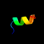
PDB 3r7k chain B

3D model

Region: 31 - 49
Aligned: 18
Modelled: 19
Confidence: 32.0%
Identity: 22%
PDB header:oxidoreductase
Chain: B: PDB Molecule:probable acyl coa dehydrogenase;
PDBTitle: crystal structure of a probable acyl coa dehydrogenase from2 mycobacterium abscessus atcc 19977 / dsm 44196

Phyre2

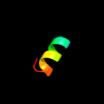
PDB 1vlb chain A domain 1

3D model

Region: 35 - 48
Aligned: 14
Modelled: 14
Confidence: 30.1%
Identity: 21%
Fold: CO dehydrogenase ISP C-domain like
Superfamily: CO dehydrogenase ISP C-domain like
Family: CO dehydrogenase ISP C-domain like

Phyre2

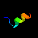
PDB 1v97 chain A domain 1

3D model

Region: 35 - 48
Aligned: 14
Modelled: 14
Confidence: 28.9%
Identity: 14%
Fold: CO dehydrogenase ISP C-domain like
Superfamily: CO dehydrogenase ISP C-domain like
Family: CO dehydrogenase ISP C-domain like

Phyre2

PDB 1ffv chain A domain 1

3D model

Region: 32 - 48
Aligned: 17
Modelled: 17
Confidence: 28.6%
Identity: 18%
Fold: CO dehydrogenase ISP C-domain like
Superfamily: CO dehydrogenase ISP C-domain like
Family: CO dehydrogenase ISP C-domain like

Phyre2

PDB 2cmp chain A

3D model

Region: 35 - 47
Aligned: 13
Modelled: 13
Confidence: 26.4%
Identity: 38%
PDB header:terminase
Chain: A: PDB Molecule:terminase small subunit;
PDBTitle: crystal structure of the dna binding domain of g1p small2 terminase subunit from bacteriophage sf6

Phyre2

PDB 1ffu chain A

3D model

Region: 9 - 48
Aligned: 36
Modelled: 40
Confidence: 23.4%
Identity: 17%
PDB header:hydrolase
Chain: A: PDB Molecule:cuts, iron-sulfur protein of carbon monoxide
PDBTitle: carbon monoxide dehydrogenase from hydrogenophaga2 pseudoflava which lacks the mo-pyranopterin moiety of the3 molybdenum cofactor

Phyre2

PDB 1jro chain A domain 1

3D model

Region: 35 - 48
Aligned: 14
Modelled: 14
Confidence: 20.5%
Identity: 36%
Fold: CO dehydrogenase ISP C-domain like
Superfamily: CO dehydrogenase ISP C-domain like
Family: CO dehydrogenase ISP C-domain like

Phyre2

PDB 1dgj chain A domain 1

3D model

Region: 35 - 48
Aligned: 14
Modelled: 14
Confidence: 20.4%
Identity: 36%
Fold: CO dehydrogenase ISP C-domain like
Superfamily: CO dehydrogenase ISP C-domain like
Family: CO dehydrogenase ISP C-domain like

Phyre2

PDB 1zxi chain A domain 1

3D model

Region: 35 - 48
Aligned: 14
Modelled: 14
Confidence: 19.1%
Identity: 21%
Fold: CO dehydrogenase ISP C-domain like
Superfamily: CO dehydrogenase ISP C-domain like
Family: CO dehydrogenase ISP C-domain like

Phyre2

PDB 1rm6 chain C domain 1

3D model

Region: 35 - 48
Aligned: 14
Modelled: 14
Confidence: 17.5%
Identity: 36%
Fold: CO dehydrogenase ISP C-domain like
Superfamily: CO dehydrogenase ISP C-domain like
Family: CO dehydrogenase ISP C-domain like

Phyre2

PDB 3b9j chain I

3D model

Region: 35 - 48
Aligned: 14
Modelled: 14
Confidence: 16.8%
Identity: 14%
PDB header:oxidoreductase
Chain: I: PDB Molecule:xanthine oxidase;
PDBTitle: structure of xanthine oxidase with 2-hydroxy-6-methylpurine

Phyre2

PDB 1t3q chain A domain 1

3D model

Region: 35 - 48
Aligned: 14
Modelled: 14
Confidence: 15.4%
Identity: 29%
Fold: CO dehydrogenase ISP C-domain like
Superfamily: CO dehydrogenase ISP C-domain like
Family: CO dehydrogenase ISP C-domain like

Phyre2

PDB 1t3q chain D

3D model

Region: 35 - 48
Aligned: 14
Modelled: 14
Confidence: 14.7%
Identity: 29%
PDB header:oxidoreductase
Chain: D: PDB Molecule:quinoline 2-oxidoreductase small subunit;
PDBTitle: crystal structure of quinoline 2-oxidoreductase from pseudomonas2 putida 86

Phyre2

PDB 3hrd chain H

3D model

Region: 35 - 48
Aligned: 14
Modelled: 14
Confidence: 13.8%
Identity: 36%
PDB header:oxidoreductase
Chain: H: PDB Molecule:nicotinate dehydrogenase small fes subunit;
PDBTitle: crystal structure of nicotinate dehydrogenase

Phyre2

PDB 1rm6 chain F

3D model

Region: 27 - 48
Aligned: 22
Modelled: 22
Confidence: 13.6%
Identity: 23%
PDB header:oxidoreductase
Chain: F: PDB Molecule:4-hydroxybenzoyl-coa reductase gamma subunit;
PDBTitle: structure of 4-hydroxybenzoyl-coa reductase from thauera2 aromatica

Phyre2

PDB 1lj2 chain B

3D model

Region: 38 - 47
Aligned: 10
Modelled: 10
Confidence: 13.5%
Identity: 50%
PDB header:viral protein/ translation
Chain: B: PDB Molecule:nonstructural rna-binding protein 34;
PDBTitle: recognition of eif4g by rotavirus nsp3 reveals a basis for2 mrna circularization

Phyre2

PDB 2jp7 chain A

3D model

Region: 35 - 49
Aligned: 15
Modelled: 15
Confidence: 13.2%
Identity: 13%
PDB header:translation
Chain: A: PDB Molecule:mrna export factor mex67;
PDBTitle: nmr structure of the mex67 uba domain

Phyre2

PDB 1n62 chain A domain 1

3D model

Region: 35 - 48
Aligned: 14
Modelled: 14
Confidence: 12.0%
Identity: 21%
Fold: CO dehydrogenase ISP C-domain like
Superfamily: CO dehydrogenase ISP C-domain like
Family: CO dehydrogenase ISP C-domain like

Phyre2

PDB 1n60 chain D

3D model

Region: 35 - 48
Aligned: 14
Modelled: 14
Confidence: 10.5%
Identity: 21%
PDB header:oxidoreductase
Chain: D: PDB Molecule:carbon monoxide dehydrogenase small chain;
PDBTitle: crystal structure of the cu,mo-co dehydrogenase (codh); cyanide-2 inactivated form

Phyre2

PDB 3eub chain J

3D model

Region: 35 - 48
Aligned: 14
Modelled: 14
Confidence: 9.8%
Identity: 14%
PDB header:oxidoreductase
Chain: J: PDB Molecule:xanthine dehydrogenase/oxidase;
PDBTitle: crystal structure of desulfo-xanthine oxidase with xanthine

Phyre2
1

c3r7kB_
2

d1vlba1
3

d1v97a1
4

d1ffva1
5

c2cmpA_
6

c1ffuA_
7

d1jroa1
8

d1dgja1
9

d1zxia1
10

d1rm6c1
11

c3b9jI_
12

d1t3qa1
13

c1t3qD_
14

c3hrdH_
15

c1rm6F_
16

c1lj2B_
17

c2jp7A_
18

d1n62a1
19

c1n60D_
20

c3eubJ_
21


22


23


24


25


26


27


28


29


30


Detailed template information 

#
Template Alignment Coverage3D Model Confidence
% i.d. Template Information
1c3r7kB_


32.0 22 PDB header:oxidoreductase
Chain: B: PDB Molecule:probable acyl coa dehydrogenase;
PDBTitle: crystal structure of a probable acyl coa dehydrogenase from2 mycobacterium abscessus atcc 19977 / dsm 44196
2d1vlba1


30.1 21 Fold:CO dehydrogenase ISP C-domain like
Superfamily:CO dehydrogenase ISP C-domain like
Family:CO dehydrogenase ISP C-domain like
3d1v97a1


28.9 14 Fold:CO dehydrogenase ISP C-domain like
Superfamily:CO dehydrogenase ISP C-domain like
Family:CO dehydrogenase ISP C-domain like
4d1ffva1


28.6 18 Fold:CO dehydrogenase ISP C-domain like
Superfamily:CO dehydrogenase ISP C-domain like
Family:CO dehydrogenase ISP C-domain like
5c2cmpA_


26.4 38 PDB header:terminase
Chain: A: PDB Molecule:terminase small subunit;
PDBTitle: crystal structure of the dna binding domain of g1p small2 terminase subunit from bacteriophage sf6
6c1ffuA_


23.4 17 PDB header:hydrolase
Chain: A: PDB Molecule:cuts, iron-sulfur protein of carbon monoxide
PDBTitle: carbon monoxide dehydrogenase from hydrogenophaga2 pseudoflava which lacks the mo-pyranopterin moiety of the3 molybdenum cofactor
7d1jroa1


20.5 36 Fold:CO dehydrogenase ISP C-domain like
Superfamily:CO dehydrogenase ISP C-domain like
Family:CO dehydrogenase ISP C-domain like
8d1dgja1


20.4 36 Fold:CO dehydrogenase ISP C-domain like
Superfamily:CO dehydrogenase ISP C-domain like
Family:CO dehydrogenase ISP C-domain like
9d1zxia1


19.1 21 Fold:CO dehydrogenase ISP C-domain like
Superfamily:CO dehydrogenase ISP C-domain like
Family:CO dehydrogenase ISP C-domain like
10d1rm6c1


17.5 36 Fold:CO dehydrogenase ISP C-domain like
Superfamily:CO dehydrogenase ISP C-domain like
Family:CO dehydrogenase ISP C-domain like
11c3b9jI_


16.8 14 PDB header:oxidoreductase
Chain: I: PDB Molecule:xanthine oxidase;
PDBTitle: structure of xanthine oxidase with 2-hydroxy-6-methylpurine
12d1t3qa1


15.4 29 Fold:CO dehydrogenase ISP C-domain like
Superfamily:CO dehydrogenase ISP C-domain like
Family:CO dehydrogenase ISP C-domain like
13c1t3qD_


14.7 29 PDB header:oxidoreductase
Chain: D: PDB Molecule:quinoline 2-oxidoreductase small subunit;
PDBTitle: crystal structure of quinoline 2-oxidoreductase from pseudomonas2 putida 86
14c3hrdH_


13.8 36 PDB header:oxidoreductase
Chain: H: PDB Molecule:nicotinate dehydrogenase small fes subunit;
PDBTitle: crystal structure of nicotinate dehydrogenase
15c1rm6F_


13.6 23 PDB header:oxidoreductase
Chain: F: PDB Molecule:4-hydroxybenzoyl-coa reductase gamma subunit;
PDBTitle: structure of 4-hydroxybenzoyl-coa reductase from thauera2 aromatica
16c1lj2B_


13.5 50 PDB header:viral protein/ translation
Chain: B: PDB Molecule:nonstructural rna-binding protein 34;
PDBTitle: recognition of eif4g by rotavirus nsp3 reveals a basis for2 mrna circularization
17c2jp7A_


13.2 13 PDB header:translation
Chain: A: PDB Molecule:mrna export factor mex67;
PDBTitle: nmr structure of the mex67 uba domain
18d1n62a1


12.0 21 Fold:CO dehydrogenase ISP C-domain like
Superfamily:CO dehydrogenase ISP C-domain like
Family:CO dehydrogenase ISP C-domain like
19c1n60D_


10.5 21 PDB header:oxidoreductase
Chain: D: PDB Molecule:carbon monoxide dehydrogenase small chain;
PDBTitle: crystal structure of the cu,mo-co dehydrogenase (codh); cyanide-2 inactivated form
20c3eubJ_


9.8 14 PDB header:oxidoreductase
Chain: J: PDB Molecule:xanthine dehydrogenase/oxidase;
PDBTitle: crystal structure of desulfo-xanthine oxidase with xanthine
21c2qguA_


not modelled 8.8 33 PDB header:structural genomics, unknown function
Chain: A: PDB Molecule:probable signal peptide protein;
PDBTitle: three-dimensional structure of the phospholipid-binding protein from2 ralstonia solanacearum q8xv73_ralsq in complex with a phospholipid at3 the resolution 1.53 a. northeast structural genomics consortium4 target rsr89
22c3t97B_


not modelled 8.5 36 PDB header:protein transport
Chain: B: PDB Molecule:nuclear pore complex protein nup54;
PDBTitle: molecular architecture of the transport channel of the nuclear pore2 complex: nup62/nup54
23d1hd6a_


not modelled 8.1 25 Fold:Protozoan pheromone-like
Superfamily:Protozoan pheromone proteins
Family:Protozoan pheromone proteins
24d1mzba_


not modelled 7.0 5 Fold:DNA/RNA-binding 3-helical bundle
Superfamily:"Winged helix" DNA-binding domain
Family:FUR-like
25c3i38F_


not modelled 6.9 22 PDB header:chaperone
Chain: F: PDB Molecule:putative chaperone dnaj;
PDBTitle: structure of a putative chaperone protein dnaj from klebsiella2 pneumoniae subsp. pneumoniae mgh 78578
26d1al3a_


not modelled 6.8 22 Fold:Periplasmic binding protein-like II
Superfamily:Periplasmic binding protein-like II
Family:Phosphate binding protein-like
27c1al3A_


not modelled 6.8 22 PDB header:transcription regulation
Chain: A: PDB Molecule:cys regulon transcriptional activator cysb;
PDBTitle: cofactor binding fragment of cysb from klebsiella aerogenes
28c2xznT_


not modelled 6.8 19 PDB header:ribosome
Chain: T: PDB Molecule:rps19e;
PDBTitle: crystal structure of the eukaryotic 40s ribosomal2 subunit in complex with initiation factor 1. this file3 contains the 40s subunit and initiation factor for4 molecule 2
29c3fiaA_


not modelled 6.0 19 PDB header:protein binding
Chain: A: PDB Molecule:intersectin-1;
PDBTitle: crystal structure of the eh 1 domain from human intersectin-2 1 protein. northeast structural genomics consortium target3 hr3646e.
30c3kxeD_


not modelled 5.9 50 PDB header:protein binding
Chain: D: PDB Molecule:antitoxin protein pard-1;
PDBTitle: a conserved mode of protein recognition and binding in a2 pard-pare toxin-antitoxin complex

Binding site prediction 

Due to computational demand, binding site predictions are not run for batch jobs

If you want to predict binding sites, please manually submit your model of choice to 3DLigandSite


Phyre is for academic use only

Please cite: Protein structure prediction on the web: a case study using the Phyre server
Kelley LA and Sternberg MJE. Nature Protocols 4, 363 - 371 (2009) [pdf] [Import into BibTeX]
 
If you use the binding site predictions from 3DLigandSite, please also cite:
3DLigandSite: predicting ligand-binding sites using similar structures.
Wass MN, Kelley LA and Sternberg MJ Nucleic Acids Research 38, W469-73 (2010) [PubMed]
 
© Structural Bioinformatics Group
Imperial College London
Lawrence Kelley, Benjamin Jefferys
Disclaimer
Terms and Conditions
Component software
Template detection: HHpred 1.51
Secondary structure prediction: Psi-pred 2.5
Disorder prediction: Disopred 2.4
Transmembrane prediction: Memsat_SVM
Multi-template modelling and ab initio: Poing 1.0