Phyre Home Retrieve Phyre Job Id
Emaill.a.kelley@imperial.ac.uk
DescriptionP0AF54
DateThu Jan 5 11:25:15 GMT 2012
Unique Job ID979c06f115f3f880

Summary 

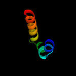
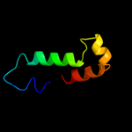
Top model
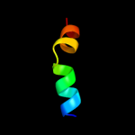
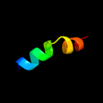
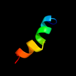
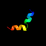
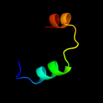
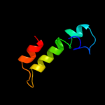
Image coloured by rainbow N → C terminus
Model (left) based on template c2dqbB_
Top template information
PDB header:hydrolase, dna binding protein
Chain: B: PDB Molecule:deoxyguanosinetriphosphate triphosphohydrolase, putative;
PDBTitle: crystal structure of dntp triphosphohydrolase from thermus2 thermophilus hb8, which is homologous to dgtp triphosphohydrolase
Confidence and coverage
Confidence: 51.4% Coverage: 34%
35 residues ( 34% of your sequence) have been modelled with 51.4% confidence by the single highest scoring template.
You may wish to submit your sequence to Phyrealarm. This will automatically scan your sequence every week for new potential templates as they appear in the Phyre2 library.
Please note: You must be registered and logged in to use Phyrealarm.
3D viewing
Interactive 3D view in Jmol

Sequence analysis 

Secondary structure and disorder prediction 

   1........10.........20.........30.........40.........50.........60
Sequence  MNGTIYQRIEDNAHFRELVEKRQRFATILSIIMLAVYIGFILLIAFAPGWLGTPLNPNTS
Secondary structure 


SS confidence 


Disorder  ??


?


??
??

Disorder confidence 


   .........70.........80.........90.........100....
Sequence  VTRGIPIGVGVIVISFVLTGIYIWRANGEFDRLNNEVLHEVQAS
Secondary structure 

SS confidence 


Disorder 


????
Disorder confidence 


Confidence Key
High(9)                    Low (0)
?Disordered
Alpha helix
Beta strand

Domain analysis 

Hover over an aligned region to see model and summary info

Please note, only up to the top 20 hits are modelled to reduce computer load

RankAligned region

PDB 2dqb chain B

3D model

Region: 5 - 39
Aligned: 35
Modelled: 35
Confidence: 51.4%
Identity: 29%
PDB header:hydrolase, dna binding protein
Chain: B: PDB Molecule:deoxyguanosinetriphosphate triphosphohydrolase, putative;
PDBTitle: crystal structure of dntp triphosphohydrolase from thermus2 thermophilus hb8, which is homologous to dgtp triphosphohydrolase

Phyre2

PDB 2pgs chain A

3D model

Region: 5 - 43
Aligned: 39
Modelled: 35
Confidence: 19.7%
Identity: 23%
PDB header:hydrolase
Chain: A: PDB Molecule:putative deoxyguanosinetriphosphate triphosphohydrolase;
PDBTitle: crystal structure of a putative deoxyguanosinetriphosphate2 triphosphohydrolase from pseudomonas syringae pv. phaseolicola 1448a

Phyre2

PDB 3lo3 chain E

3D model

Region: 2 - 24
Aligned: 23
Modelled: 23
Confidence: 17.7%
Identity: 9%
PDB header:structure genomics, unknown function
Chain: E: PDB Molecule:uncharacterized conserved protein;
PDBTitle: the crystal structure of a conserved functionally unknown2 protein from colwellia psychrerythraea 34h.

Phyre2

PDB 3fh6 chain F

3D model

Region: 2 - 91
Aligned: 90
Modelled: 90
Confidence: 16.3%
Identity: 12%
PDB header:transport protein
Chain: F: PDB Molecule:maltose transport system permease protein malf;
PDBTitle: crystal structure of the resting state maltose transporter from e.2 coli

Phyre2

PDB 2fiu chain A domain 1

3D model

Region: 2 - 24
Aligned: 23
Modelled: 23
Confidence: 13.5%
Identity: 9%
Fold: Ferredoxin-like
Superfamily: Dimeric alpha+beta barrel
Family: Atu0297-like

Phyre2

PDB 2o6i chain A domain 1

3D model

Region: 2 - 20
Aligned: 19
Modelled: 19
Confidence: 12.6%
Identity: 11%
Fold: HD-domain/PDEase-like
Superfamily: HD-domain/PDEase-like
Family: HD domain

Phyre2

PDB 2o6i chain A

3D model

Region: 2 - 20
Aligned: 19
Modelled: 19
Confidence: 12.6%
Identity: 11%
PDB header:hydrolase
Chain: A: PDB Molecule:hd domain protein;
PDBTitle: structure of an enterococcus faecalis hd domain phosphohydrolase

Phyre2

PDB 2q14 chain A

3D model

Region: 2 - 20
Aligned: 19
Modelled: 19
Confidence: 12.2%
Identity: 11%
PDB header:hydrolase
Chain: A: PDB Molecule:phosphohydrolase;
PDBTitle: crystal structure of phosphohydrolase (bt4208) from bacteroides2 thetaiotaomicron vpi-5482 at 2.20 a resolution

Phyre2

PDB 2hek chain A domain 1

3D model

Region: 3 - 22
Aligned: 20
Modelled: 20
Confidence: 11.7%
Identity: 25%
Fold: HD-domain/PDEase-like
Superfamily: HD-domain/PDEase-like
Family: HD domain

Phyre2

PDB 2kjg chain A

3D model

Region: 9 - 34
Aligned: 26
Modelled: 26
Confidence: 11.3%
Identity: 19%
PDB header:metal binding protein
Chain: A: PDB Molecule:archaeal protein sso6904;
PDBTitle: solution structure of an archaeal protein sso6904 from2 hyperthermophilic sulfolobus solfataricus

Phyre2

PDB 3bg2 chain A

3D model

Region: 5 - 39
Aligned: 35
Modelled: 35
Confidence: 10.1%
Identity: 31%
PDB header:hydrolase
Chain: A: PDB Molecule:dgtp triphosphohydrolase;
PDBTitle: crystal structure of deoxyguanosinetriphosphate triphosphohydrolase2 from flavobacterium sp. med217

Phyre2

PDB 1tu7 chain A domain 1

3D model

Region: 3 - 29
Aligned: 27
Modelled: 27
Confidence: 9.6%
Identity: 26%
Fold: GST C-terminal domain-like
Superfamily: GST C-terminal domain-like
Family: Glutathione S-transferase (GST), C-terminal domain

Phyre2

PDB 3sjb chain C

3D model

Region: 82 - 97
Aligned: 16
Modelled: 16
Confidence: 9.0%
Identity: 31%
PDB header:hydrolase/transport protein
Chain: C: PDB Molecule:golgi to er traffic protein 1;
PDBTitle: crystal structure of s. cerevisiae get3 in the open state in complex2 with get1 cytosolic domain

Phyre2

PDB 1q59 chain A

3D model

Region: 52 - 99
Aligned: 45
Modelled: 48
Confidence: 8.6%
Identity: 2%
Fold: Toxins' membrane translocation domains
Superfamily: Bcl-2 inhibitors of programmed cell death
Family: Bcl-2 inhibitors of programmed cell death

Phyre2

PDB 3u1n chain C

3D model

Region: 3 - 22
Aligned: 20
Modelled: 20
Confidence: 8.3%
Identity: 25%
PDB header:hydrolase
Chain: C: PDB Molecule:sam domain and hd domain-containing protein 1;
PDBTitle: structure of the catalytic core of human samhd1

Phyre2

PDB 3qnq chain D

3D model

Region: 48 - 102
Aligned: 53
Modelled: 55
Confidence: 7.8%
Identity: 9%
PDB header:membrane protein, transport protein
Chain: D: PDB Molecule:pts system, cellobiose-specific iic component;
PDBTitle: crystal structure of the transporter chbc, the iic component from the2 n,n'-diacetylchitobiose-specific phosphotransferase system

Phyre2

PDB 1w6k chain A domain 2

3D model

Region: 78 - 101
Aligned: 24
Modelled: 24
Confidence: 7.5%
Identity: 21%
Fold: alpha/alpha toroid
Superfamily: Terpenoid cyclases/Protein prenyltransferases
Family: Terpene synthases

Phyre2

PDB 2o2f chain A

3D model

Region: 51 - 101
Aligned: 45
Modelled: 51
Confidence: 7.5%
Identity: 13%
PDB header:apoptosis
Chain: A: PDB Molecule:apoptosis regulator bcl-2;
PDBTitle: solution structure of the anti-apoptotic protein bcl-2 in2 complex with an acyl-sulfonamide-based ligand

Phyre2

PDB 3sja chain G

3D model

Region: 82 - 99
Aligned: 18
Modelled: 18
Confidence: 7.4%
Identity: 28%
PDB header:hydrolase/transport protein
Chain: G: PDB Molecule:golgi to er traffic protein 1;
PDBTitle: crystal structure of s. cerevisiae get3 in the open state in complex2 with get1 cytosolic domain

Phyre2

PDB 1pv7 chain A

3D model

Region: 22 - 76
Aligned: 54
Modelled: 55
Confidence: 6.5%
Identity: 13%
Fold: MFS general substrate transporter
Superfamily: MFS general substrate transporter
Family: LacY-like proton/sugar symporter

Phyre2
1

c2dqbB_
2

c2pgsA_
3

c3lo3E_
4

c3fh6F_
5

d2fiua1
6

d2o6ia1
7

c2o6iA_
8

c2q14A_
9

d2heka1
10

c2kjgA_
11

c3bg2A_
12

d1tu7a1
13

c3sjbC_
14

d1q59a_
15

c3u1nC_
16

c3qnqD_
17

d1w6ka2
18

c2o2fA_
19

c3sjaG_
20

d1pv7a_
21


22


23


24


25


26


27


28


29


Detailed template information 

#
Template Alignment Coverage3D Model Confidence
% i.d. Template Information
1c2dqbB_


51.4 29 PDB header:hydrolase, dna binding protein
Chain: B: PDB Molecule:deoxyguanosinetriphosphate triphosphohydrolase, putative;
PDBTitle: crystal structure of dntp triphosphohydrolase from thermus2 thermophilus hb8, which is homologous to dgtp triphosphohydrolase
2c2pgsA_


19.7 23 PDB header:hydrolase
Chain: A: PDB Molecule:putative deoxyguanosinetriphosphate triphosphohydrolase;
PDBTitle: crystal structure of a putative deoxyguanosinetriphosphate2 triphosphohydrolase from pseudomonas syringae pv. phaseolicola 1448a
3c3lo3E_


17.7 9 PDB header:structure genomics, unknown function
Chain: E: PDB Molecule:uncharacterized conserved protein;
PDBTitle: the crystal structure of a conserved functionally unknown2 protein from colwellia psychrerythraea 34h.
4c3fh6F_


16.3 12 PDB header:transport protein
Chain: F: PDB Molecule:maltose transport system permease protein malf;
PDBTitle: crystal structure of the resting state maltose transporter from e.2 coli
5d2fiua1


13.5 9 Fold:Ferredoxin-like
Superfamily:Dimeric alpha+beta barrel
Family:Atu0297-like
6d2o6ia1


12.6 11 Fold:HD-domain/PDEase-like
Superfamily:HD-domain/PDEase-like
Family:HD domain
7c2o6iA_


12.6 11 PDB header:hydrolase
Chain: A: PDB Molecule:hd domain protein;
PDBTitle: structure of an enterococcus faecalis hd domain phosphohydrolase
8c2q14A_


12.2 11 PDB header:hydrolase
Chain: A: PDB Molecule:phosphohydrolase;
PDBTitle: crystal structure of phosphohydrolase (bt4208) from bacteroides2 thetaiotaomicron vpi-5482 at 2.20 a resolution
9d2heka1


11.7 25 Fold:HD-domain/PDEase-like
Superfamily:HD-domain/PDEase-like
Family:HD domain
10c2kjgA_


11.3 19 PDB header:metal binding protein
Chain: A: PDB Molecule:archaeal protein sso6904;
PDBTitle: solution structure of an archaeal protein sso6904 from2 hyperthermophilic sulfolobus solfataricus
11c3bg2A_


10.1 31 PDB header:hydrolase
Chain: A: PDB Molecule:dgtp triphosphohydrolase;
PDBTitle: crystal structure of deoxyguanosinetriphosphate triphosphohydrolase2 from flavobacterium sp. med217
12d1tu7a1


9.6 26 Fold:GST C-terminal domain-like
Superfamily:GST C-terminal domain-like
Family:Glutathione S-transferase (GST), C-terminal domain
13c3sjbC_


9.0 31 PDB header:hydrolase/transport protein
Chain: C: PDB Molecule:golgi to er traffic protein 1;
PDBTitle: crystal structure of s. cerevisiae get3 in the open state in complex2 with get1 cytosolic domain
14d1q59a_


8.6 2 Fold:Toxins' membrane translocation domains
Superfamily:Bcl-2 inhibitors of programmed cell death
Family:Bcl-2 inhibitors of programmed cell death
15c3u1nC_


8.3 25 PDB header:hydrolase
Chain: C: PDB Molecule:sam domain and hd domain-containing protein 1;
PDBTitle: structure of the catalytic core of human samhd1
16c3qnqD_


7.8 9 PDB header:membrane protein, transport protein
Chain: D: PDB Molecule:pts system, cellobiose-specific iic component;
PDBTitle: crystal structure of the transporter chbc, the iic component from the2 n,n'-diacetylchitobiose-specific phosphotransferase system
17d1w6ka2


7.5 21 Fold:alpha/alpha toroid
Superfamily:Terpenoid cyclases/Protein prenyltransferases
Family:Terpene synthases
18c2o2fA_


7.5 13 PDB header:apoptosis
Chain: A: PDB Molecule:apoptosis regulator bcl-2;
PDBTitle: solution structure of the anti-apoptotic protein bcl-2 in2 complex with an acyl-sulfonamide-based ligand
19c3sjaG_


7.4 28 PDB header:hydrolase/transport protein
Chain: G: PDB Molecule:golgi to er traffic protein 1;
PDBTitle: crystal structure of s. cerevisiae get3 in the open state in complex2 with get1 cytosolic domain
20d1pv7a_


6.5 13 Fold:MFS general substrate transporter
Superfamily:MFS general substrate transporter
Family:LacY-like proton/sugar symporter
21c1sseB_


not modelled 6.4 23 PDB header:transcription activator
Chain: B: PDB Molecule:ap-1 like transcription factor yap1;
PDBTitle: solution structure of the oxidized form of the yap1 redox2 domain
22d1vpua_


not modelled 6.1 25 Fold:HIV-1 VPU cytoplasmic domain
Superfamily:HIV-1 VPU cytoplasmic domain
Family:HIV-1 VPU cytoplasmic domain
23d2gw6a1


not modelled 5.5 17 Fold:Restriction endonuclease-like
Superfamily:tRNA-intron endonuclease catalytic domain-like
Family:tRNA-intron endonuclease catalytic domain-like
24d1tuwa_


not modelled 5.5 15 Fold:Ferredoxin-like
Superfamily:Dimeric alpha+beta barrel
Family:Polyketide synthesis cyclase
25c2vofA_


not modelled 5.4 4 PDB header:apoptosis
Chain: A: PDB Molecule:bcl-2-related protein a1;
PDBTitle: structure of mouse a1 bound to the puma bh3-domain
26c1ei0A_


not modelled 5.3 24 PDB header:cell cycle
Chain: A: PDB Molecule:p8mtcp1;
PDBTitle: nmr structure of the alpha-helical hairpin of p8mtcp1
27c3a0hX_


not modelled 5.3 23 PDB header:electron transport
Chain: X: PDB Molecule:photosystem ii reaction center protein x;
PDBTitle: crystal structure of i-substituted photosystem ii complex
28c3a0hx_


not modelled 5.3 23 PDB header:electron transport
Chain: X: PDB Molecule:photosystem ii reaction center protein x;
PDBTitle: crystal structure of i-substituted photosystem ii complex
29c1s5lx_


not modelled 5.1 25 PDB header:photosynthesis
Chain: X: PDB Molecule:photosystem ii psbx protein;
PDBTitle: architecture of the photosynthetic oxygen evolving center

Binding site prediction 

Due to computational demand, binding site predictions are not run for batch jobs

If you want to predict binding sites, please manually submit your model of choice to 3DLigandSite


Phyre is for academic use only

Please cite: Protein structure prediction on the web: a case study using the Phyre server
Kelley LA and Sternberg MJE. Nature Protocols 4, 363 - 371 (2009) [pdf] [Import into BibTeX]
 
If you use the binding site predictions from 3DLigandSite, please also cite:
3DLigandSite: predicting ligand-binding sites using similar structures.
Wass MN, Kelley LA and Sternberg MJ Nucleic Acids Research 38, W469-73 (2010) [PubMed]
 
© Structural Bioinformatics Group
Imperial College London
Lawrence Kelley, Benjamin Jefferys
Disclaimer
Terms and Conditions
Component software
Template detection: HHpred 1.51
Secondary structure prediction: Psi-pred 2.5
Disorder prediction: Disopred 2.4
Transmembrane prediction: Memsat_SVM
Multi-template modelling and ab initio: Poing 1.0