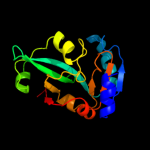
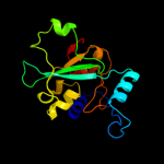
1 c2dxaA_
100.0
97
PDB header: translationChain: A: PDB Molecule: protein ybak;PDBTitle: crystal structure of trans editing enzyme prox from e.coli
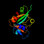
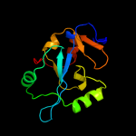
2 d1dbxa_
100.0
62
Fold: YbaK/ProRS associated domainSuperfamily: YbaK/ProRS associated domainFamily: YbaK/ProRS associated domain3 d1vjfa_
100.0
14
Fold: YbaK/ProRS associated domainSuperfamily: YbaK/ProRS associated domainFamily: YbaK/ProRS associated domain4 d1wdva_
100.0
21
Fold: YbaK/ProRS associated domainSuperfamily: YbaK/ProRS associated domainFamily: YbaK/ProRS associated domain5 d1vkia_
100.0
21
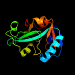
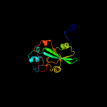
Fold: YbaK/ProRS associated domainSuperfamily: YbaK/ProRS associated domainFamily: YbaK/ProRS associated domain6 c2cx5B_
100.0
23
PDB header: translationChain: B: PDB Molecule: a putative trans-editing enzyme;PDBTitle: crystal structure of a putative trans-editing enzyme for2 prolyl trna synthetase
7 c3op6B_
100.0
18
PDB header: unknown functionChain: B: PDB Molecule: uncharacterized protein;PDBTitle: crystal structure of an oligo-nucleotide binding protein (lpg1207)2 from legionella pneumophila subsp. pneumophila str. philadelphia 1 at3 2.00 a resolution
8 c3memA_
100.0
18
PDB header: signaling proteinChain: A: PDB Molecule: putative signal transduction protein;PDBTitle: crystal structure of a putative signal transduction protein2 (maqu_0641) from marinobacter aquaeolei vt8 at 2.25 a resolution
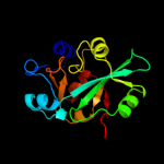
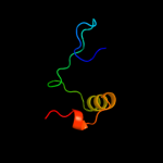
9 c2j3mA_
98.5
23
PDB header: ligaseChain: A: PDB Molecule: prolyl-trna synthetase;PDBTitle: prolyl-trna synthetase from enterococcus faecalis complexed2 with atp, manganese and prolinol
10 d1j9ba_
86.2
9
Fold: Thioredoxin foldSuperfamily: Thioredoxin-likeFamily: ArsC-like11 c3gkxB_
83.9
19
PDB header: structural genomics, unknown functionChain: B: PDB Molecule: putative arsc family related protein;PDBTitle: crystal structure of putative arsc family related protein from2 bacteroides fragilis
12 d1z3ea1
82.2
17
Fold: Thioredoxin foldSuperfamily: Thioredoxin-likeFamily: ArsC-like13 c3fz4A_
81.5
12
PDB header: oxidoreductaseChain: A: PDB Molecule: putative arsenate reductase;PDBTitle: the crystal structure of a possible arsenate reductase from2 streptococcus mutans ua159
14 c3l78A_
81.3
14
PDB header: transcriptionChain: A: PDB Molecule: regulatory protein spx;PDBTitle: the crystal structure of smu.1142c from streptococcus mutans ua159
15 c3rdwB_
69.1
23
PDB header: oxidoreductaseChain: B: PDB Molecule: putative arsenate reductase;PDBTitle: putative arsenate reductase from yersinia pestis
16 c2kokA_
66.6
22
PDB header: oxidoreductaseChain: A: PDB Molecule: arsenate reductase;PDBTitle: solution structure of an arsenate reductase (arsc) related protein2 from brucella melitensis. seattle structural genomics center for3 infectious disease target braba.00007.a.
17 c3f0iA_
64.0
26
PDB header: oxidoreductaseChain: A: PDB Molecule: arsenate reductase;PDBTitle: arsenate reductase from vibrio cholerae.
18 d1rw1a_
60.5
17
Fold: Thioredoxin foldSuperfamily: Thioredoxin-likeFamily: ArsC-like19 c3lgcA_
46.9
24
PDB header: unknown functionChain: A: PDB Molecule: glutaredoxin 1;PDBTitle: crystal structure of glutaredoxin 1 from francisella2 tularensis
20 d1fova_
41.8
19
Fold: Thioredoxin foldSuperfamily: Thioredoxin-likeFamily: Thioltransferase21 c3nznA_
not modelled
34.5
20
PDB header: oxidoreductaseChain: A: PDB Molecule: glutaredoxin;PDBTitle: the crystal structure of the glutaredoxin from methanosarcina mazei2 go1
22 d1t1va_
not modelled
34.3
17
Fold: Thioredoxin foldSuperfamily: Thioredoxin-likeFamily: SH3BGR (SH3-binding, glutamic acid-rich protein-like)23 c2khpA_
not modelled
29.7
16
PDB header: electron transportChain: A: PDB Molecule: glutaredoxin;PDBTitle: solution structure of glutaredoxin from brucella melitensis
24 c3ic4A_
not modelled
24.8
10
PDB header: oxidoreductaseChain: A: PDB Molecule: glutaredoxin (grx-1);PDBTitle: the crystal structure of the glutaredoxin(grx-1) from archaeoglobus2 fulgidus
25 d1nm3a1
not modelled
23.1
30
Fold: Thioredoxin foldSuperfamily: Thioredoxin-likeFamily: Thioltransferase26 d1h75a_
not modelled
23.1
15
Fold: Thioredoxin foldSuperfamily: Thioredoxin-likeFamily: Thioltransferase27 c3h8qB_
not modelled
22.5
3
PDB header: oxidoreductaseChain: B: PDB Molecule: thioredoxin reductase 3;PDBTitle: crystal structure of glutaredoxin domain of human thioredoxin2 reductase 3
28 c2ct6A_
not modelled
21.5
33
PDB header: structural genomics, unknown functionChain: A: PDB Molecule: sh3 domain-binding glutamic acid-rich-likePDBTitle: solution structure of the sh3 domain-binding glutamic acid-2 rich-like protein 2
29 c2e7pC_
not modelled
21.2
11
PDB header: electron transportChain: C: PDB Molecule: glutaredoxin;PDBTitle: crystal structure of the holo form of glutaredoxin c1 from populus2 tremula x tremuloides
30 c1nm3B_
not modelled
20.9
22
PDB header: electron transportChain: B: PDB Molecule: protein hi0572;PDBTitle: crystal structure of heamophilus influenza hybrid-prx5
31 c1u6tA_
not modelled
18.7
29
PDB header: protein binding, signaling proteinChain: A: PDB Molecule: sh3 domain-binding glutamic acid-rich-likePDBTitle: crystal structure of the human sh3 binding glutamic-rich2 protein like
32 d1wika_
not modelled
16.0
5
Fold: Thioredoxin foldSuperfamily: Thioredoxin-likeFamily: Thioltransferase33 d1r7ha_
not modelled
15.8
15
Fold: Thioredoxin foldSuperfamily: Thioredoxin-likeFamily: Thioltransferase34 d1abaa_
not modelled
14.1
15
Fold: Thioredoxin foldSuperfamily: Thioredoxin-likeFamily: Thioltransferase35 c2ht9A_
not modelled
11.6
19
PDB header: oxidoreductaseChain: A: PDB Molecule: glutaredoxin-2;PDBTitle: the structure of dimeric human glutaredoxin 2
36 c1ykaA_
not modelled
11.4
13
PDB header: electron transportChain: A: PDB Molecule: monothiol glutaredoxin ydhd;PDBTitle: solution structure of grx4, a monothiol glutaredoxin from2 e. coli.
37 d2obba1
not modelled
9.2
13
Fold: HAD-likeSuperfamily: HAD-likeFamily: BT0820-like38 c3qmxA_
not modelled
8.9
29
PDB header: electron transportChain: A: PDB Molecule: glutaredoxin a;PDBTitle: x-ray crystal structure of synechocystis sp. pcc 6803 glutaredoxin a
39 d1gmua2
not modelled
7.5
8
Fold: Ferredoxin-likeSuperfamily: Urease metallochaperone UreE, C-terminal domainFamily: Urease metallochaperone UreE, C-terminal domain40 d1jqga2
not modelled
7.0
5
Fold: Ferredoxin-likeSuperfamily: Protease propeptides/inhibitorsFamily: Pancreatic carboxypeptidase, activation domain41 c3kc2A_
not modelled
6.9
30
PDB header: hydrolaseChain: A: PDB Molecule: uncharacterized protein ykr070w;PDBTitle: crystal structure of mitochondrial had-like phosphatase from2 saccharomyces cerevisiae
42 c2klxA_
not modelled
6.6
21
PDB header: oxidoreductaseChain: A: PDB Molecule: glutaredoxin;PDBTitle: solution structure of glutaredoxin from bartonella henselae str.2 houston
43 d2v94a1
not modelled
6.4
30
Fold: Ribosomal proteins S24e, L23 and L15eSuperfamily: Ribosomal proteins S24e, L23 and L15eFamily: Ribosomal protein S24e44 d1pz4a_
not modelled
5.8
7
Fold: SCP-likeSuperfamily: SCP-likeFamily: Sterol carrier protein, SCP45 d1ayea2
not modelled
5.5
22
Fold: Ferredoxin-likeSuperfamily: Protease propeptides/inhibitorsFamily: Pancreatic carboxypeptidase, activation domain46 d2boaa2
not modelled
5.5
6
Fold: Ferredoxin-likeSuperfamily: Protease propeptides/inhibitorsFamily: Pancreatic carboxypeptidase, activation domain47 d1pyta_
not modelled
5.4
22
Fold: Ferredoxin-likeSuperfamily: Protease propeptides/inhibitorsFamily: Pancreatic carboxypeptidase, activation domain48 d1nsaa2
not modelled
5.4
17
Fold: Ferredoxin-likeSuperfamily: Protease propeptides/inhibitorsFamily: Pancreatic carboxypeptidase, activation domain49 d1kwma2
not modelled
5.3
11
Fold: Ferredoxin-likeSuperfamily: Protease propeptides/inhibitorsFamily: Pancreatic carboxypeptidase, activation domain50 c1yj7A_
not modelled
5.3
21
PDB header: protein transportChain: A: PDB Molecule: escj;PDBTitle: crystal structure of enteropathogenic e.coli (epec) type iii secretion2 system protein escj
51 d1pcaa1
not modelled
5.1
22
Fold: Ferredoxin-likeSuperfamily: Protease propeptides/inhibitorsFamily: Pancreatic carboxypeptidase, activation domain