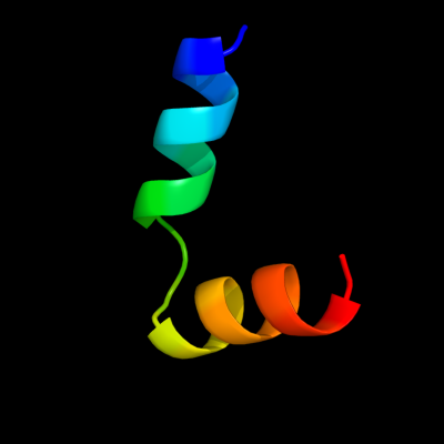
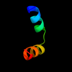
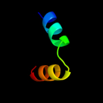
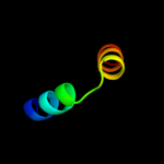
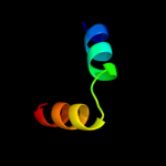
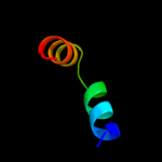
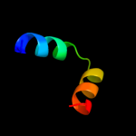
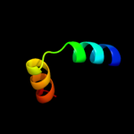
PDB header:ribosome Chain: D: PDB Molecule:50s ribosomal protein l7/l12; PDBTitle: crystal structure of ribosomal protein l12 from thermotoga maritima
Confidence and coverage
Confidence:
63.6%
Coverage:
37%
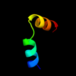
24 residues ( 37% of your sequence) have been modelled with 63.6% confidence by the single highest scoring template.
You may wish to submit your sequence to Phyrealarm. This will automatically scan your sequence every week for new potential templates as they appear in the Phyre2 library.
Please note: You must be registered and logged in to use Phyrealarm.
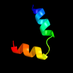
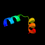
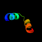
Region: 32 - 55 Aligned: 24 Modelled: 24 Confidence: 60.8% Identity: 33% PDB header:structural protein Chain: W: PDB Molecule:50s ribosomal protein l7/l12; PDBTitle: ribosomal protein l10-l12(ntd) complex, space group p212121,2 form a
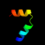
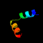
Region: 32 - 55 Aligned: 24 Modelled: 24 Confidence: 60.8% Identity: 33% PDB header:structural protein Chain: U: PDB Molecule:50s ribosomal protein l7/l12; PDBTitle: ribosomal protein l10-l12(ntd) complex, space group p212121,2 form a
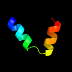
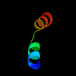
Region: 32 - 55 Aligned: 24 Modelled: 24 Confidence: 60.5% Identity: 33% PDB header:structural protein Chain: U: PDB Molecule:50s ribosomal protein l7/l12; PDBTitle: ribosomal protein l10-l12(ntd) complex, space group p212121,2 form b
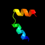
Region: 32 - 55 Aligned: 24 Modelled: 24 Confidence: 60.2% Identity: 33% PDB header:structural protein Chain: X: PDB Molecule:50s ribosomal protein l7/l12; PDBTitle: ribosomal protein l10-l12(ntd) complex, space group p212121,2 form b
Region: 32 - 55 Aligned: 24 Modelled: 24 Confidence: 60.1% Identity: 33% PDB header:structural protein Chain: Y: PDB Molecule:50s ribosomal protein l7/l12; PDBTitle: ribosomal protein l10-l12(ntd) complex, space group p212121,2 form b
Region: 32 - 55 Aligned: 24 Modelled: 24 Confidence: 60.1% Identity: 33% PDB header:structural protein Chain: W: PDB Molecule:50s ribosomal protein l7/l12; PDBTitle: ribosomal protein l10-l12(ntd) complex, space group p212121,2 form b
Region: 32 - 55 Aligned: 24 Modelled: 24 Confidence: 60.0% Identity: 33% PDB header:structural protein Chain: V: PDB Molecule:50s ribosomal protein l7/l12; PDBTitle: ribosomal protein l10-l12(ntd) complex, space group p212121,2 form b
Region: 32 - 55 Aligned: 24 Modelled: 24 Confidence: 59.9% Identity: 33% PDB header:structural protein Chain: V: PDB Molecule:50s ribosomal protein l7/l12; PDBTitle: ribosomal protein l10-l12(ntd) complex, space group p212121,2 form a
Region: 32 - 55 Aligned: 24 Modelled: 24 Confidence: 55.7% Identity: 33% PDB header:structural protein Chain: Z: PDB Molecule:50s ribosomal protein l7/l12; PDBTitle: ribosomal protein l10-l12(ntd) complex, space group p212121,2 form b
Region: 32 - 55 Aligned: 24 Modelled: 24 Confidence: 13.3% Identity: 33% PDB header:structural protein Chain: Y: PDB Molecule:50s ribosomal protein l7/l12; PDBTitle: ribosomal protein l10-l12(ntd) complex, space group p212121,2 form a
Phyre2
21
22
23
24
25
26
27
Detailed template information
Binding site prediction
Due to computational demand, binding site predictions are not run for batch jobs
If you want to predict binding sites, please manually submit your model of choice to 3DLigandSite
Phyre is for academic use only
Please cite: Protein structure prediction on
the web: a case study using the Phyre server
Kelley LA and Sternberg MJE. Nature Protocols
4, 363 - 371 (2009) [pdf] [Import into BibTeX]
If you use the binding site
predictions from 3DLigandSite, please also cite:
3DLigandSite: predicting ligand-binding sites using similar structures.
Wass MN, Kelley LA and Sternberg
MJ Nucleic Acids Research 38, W469-73 (2010) [PubMed]