Phyre Home Retrieve Phyre Job Id
Emaill.a.kelley@imperial.ac.uk
DescriptionP0AG59
DateThu Jan 5 11:28:11 GMT 2012
Unique Job ID8f434d413bd1fa17

Summary 

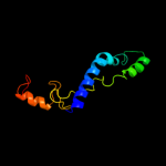
Top model
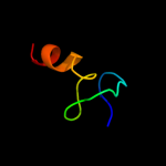
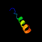
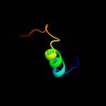
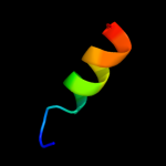
Image coloured by rainbow N → C terminus
Model (left) based on template d2qaln1
Top template information
Fold:Glucocorticoid receptor-like (DNA-binding domain)
Superfamily:Glucocorticoid receptor-like (DNA-binding domain)
Family:Ribosomal protein S14
Confidence and coverage
Confidence:100.0% Coverage: 95%
96 residues ( 95% of your sequence) have been modelled with 100.0% confidence by the single highest scoring template.
3D viewing
Interactive 3D view in Jmol

Sequence analysis 

Secondary structure and disorder prediction 

   1........10.........20.........30.........40.........50.........60
Sequence  MAKQSMKAREVKRVALADKYFAKRAELKAIISDVNASDEDRWNAVLKLQTLPRDSSPSRQ
Secondary structure 


SS confidence 


Disorder  ???


?
?
??


?


Disorder confidence 


   .........70.........80.........90.........100.
Sequence  RNRCRQTGRPHGFLRKFGLSRIKVREAAMRGEIPGLKKASW
Secondary structure 


SS confidence 


Disorder 


???????
Disorder confidence 


Confidence Key
High(9)                    Low (0)
?Disordered
Alpha helix
Beta strand

Domain analysis 

Hover over an aligned region to see model and summary info

Please note, only up to the top 20 hits are modelled to reduce computer load

RankAligned region

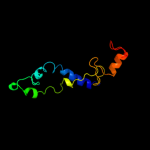
PDB 2qal chain N domain 1

3D model

Region: 2 - 101
Aligned: 96
Modelled: 100
Confidence: 100.0%
Identity: 100%
Fold: Glucocorticoid receptor-like (DNA-binding domain)
Superfamily: Glucocorticoid receptor-like (DNA-binding domain)
Family: Ribosomal protein S14

Phyre2

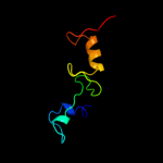
PDB 3bbn chain N

3D model

Region: 2 - 101
Aligned: 99
Modelled: 100
Confidence: 100.0%
Identity: 40%
PDB header:ribosome
Chain: N: PDB Molecule:ribosomal protein s14;
PDBTitle: homology model for the spinach chloroplast 30s subunit2 fitted to 9.4a cryo-em map of the 70s chlororibosome.

Phyre2

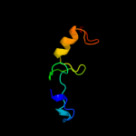
PDB 2gy9 chain N

3D model

Region: 41 - 101
Aligned: 61
Modelled: 61
Confidence: 99.9%
Identity: 100%
PDB header:ribosome
Chain: N: PDB Molecule:30s ribosomal subunit protein s14;
PDBTitle: structure of the 30s subunit of a pre-translocational e.2 coli ribosome obtained by fitting atomic models for rna and3 protein components into cryo-em map emd-1056

Phyre2

PDB 2uub chain N domain 1

3D model

Region: 48 - 101
Aligned: 54
Modelled: 54
Confidence: 99.9%
Identity: 41%
Fold: Glucocorticoid receptor-like (DNA-binding domain)
Superfamily: Glucocorticoid receptor-like (DNA-binding domain)
Family: Ribosomal protein S14

Phyre2

PDB 3jyv chain N

3D model

Region: 62 - 89
Aligned: 28
Modelled: 28
Confidence: 93.8%
Identity: 32%
PDB header:ribosome
Chain: N: PDB Molecule:40s ribosomal protein s29(a);
PDBTitle: structure of the 40s rrna and proteins and p/e trna for eukaryotic2 ribosome based on cryo-em map of thermomyces lanuginosus ribosome at3 8.9a resolution

Phyre2

PDB 2xzn chain N

3D model

Region: 62 - 89
Aligned: 28
Modelled: 28
Confidence: 93.7%
Identity: 32%
PDB header:ribosome
Chain: N: PDB Molecule:rps29e;
PDBTitle: crystal structure of the eukaryotic 40s ribosomal2 subunit in complex with initiation factor 1. this file3 contains the 40s subunit and initiation factor for4 molecule 2

Phyre2

PDB 2zkq chain N

3D model

Region: 62 - 88
Aligned: 27
Modelled: 27
Confidence: 91.8%
Identity: 33%
PDB header:ribosomal protein/rna
Chain: N: PDB Molecule:
PDBTitle: structure of a mammalian ribosomal 40s subunit within an2 80s complex obtained by docking homology models of the rna3 and proteins into an 8.7 a cryo-em map

Phyre2

PDB 1s1h chain N

3D model

Region: 64 - 89
Aligned: 26
Modelled: 26
Confidence: 75.3%
Identity: 35%
PDB header:ribosome
Chain: N: PDB Molecule:40s ribosomal protein s29-b;
PDBTitle: structure of the ribosomal 80s-eef2-sordarin complex from2 yeast obtained by docking atomic models for rna and protein3 components into a 11.7 a cryo-em map. this file, 1s1h,4 contains 40s subunit. the 60s ribosomal subunit is in file5 1s1i.

Phyre2

PDB 2bmf chain A domain 1

3D model

Region: 72 - 94
Aligned: 23
Modelled: 23
Confidence: 21.0%
Identity: 30%
Fold: P-loop containing nucleoside triphosphate hydrolases
Superfamily: P-loop containing nucleoside triphosphate hydrolases
Family: RNA helicase

Phyre2

PDB 2zhh chain A

3D model

Region: 75 - 100
Aligned: 26
Modelled: 26
Confidence: 20.1%
Identity: 12%
PDB header:transcription
Chain: A: PDB Molecule:redox-sensitive transcriptional activator soxr;
PDBTitle: crystal structure of soxr

Phyre2

PDB 1r8d chain A

3D model

Region: 75 - 100
Aligned: 26
Modelled: 26
Confidence: 14.5%
Identity: 8%
Fold: Putative DNA-binding domain
Superfamily: Putative DNA-binding domain
Family: DNA-binding N-terminal domain of transcription activators

Phyre2

PDB 2iea chain A domain 3

3D model

Region: 66 - 94
Aligned: 29
Modelled: 29
Confidence: 13.4%
Identity: 24%
Fold: TK C-terminal domain-like
Superfamily: TK C-terminal domain-like
Family: Transketolase C-terminal domain-like

Phyre2

PDB 3gpv chain A

3D model

Region: 75 - 100
Aligned: 26
Modelled: 24
Confidence: 13.0%
Identity: 19%
PDB header:transcription regulator
Chain: A: PDB Molecule:transcriptional regulator, merr family;
PDBTitle: crystal structure of a transcriptional regulator, merr2 family from bacillus thuringiensis

Phyre2

PDB 1qw1 chain A

3D model

Region: 72 - 97
Aligned: 26
Modelled: 26
Confidence: 12.9%
Identity: 12%
PDB header:gene regulation
Chain: A: PDB Molecule:diphtheria toxin repressor;
PDBTitle: solution structure of the c-terminal domain of dtxr2 residues 110-226

Phyre2

PDB 1yuz chain B

3D model

Region: 64 - 77
Aligned: 14
Modelled: 14
Confidence: 12.3%
Identity: 21%
PDB header:oxidoreductase
Chain: B: PDB Molecule:nigerythrin;
PDBTitle: partially reduced state of nigerythrin

Phyre2

PDB 3izb chain V

3D model

Region: 78 - 100
Aligned: 23
Modelled: 23
Confidence: 11.0%
Identity: 35%
PDB header:ribosome
Chain: V: PDB Molecule:40s ribosomal protein rps25 (s25e);
PDBTitle: localization of the small subunit ribosomal proteins into a 6.1 a2 cryo-em map of saccharomyces cerevisiae translating 80s ribosome

Phyre2

PDB 1y6u chain A

3D model

Region: 77 - 91
Aligned: 15
Modelled: 15
Confidence: 10.3%
Identity: 27%
PDB header:dna binding protein
Chain: A: PDB Molecule:excisionase from transposon tn916;
PDBTitle: the structure of the excisionase (xis) protein from2 conjugative transposon tn916 provides insights into the3 regulation of heterobivalent tyrosine recombinases

Phyre2

PDB 2xzm chain 8

3D model

Region: 79 - 100
Aligned: 22
Modelled: 22
Confidence: 9.9%
Identity: 27%
PDB header:ribosome
Chain: 8: PDB Molecule:rps25e,;
PDBTitle: crystal structure of the eukaryotic 40s ribosomal2 subunit in complex with initiation factor 1. this file3 contains the 40s subunit and initiation factor for4 molecule 1

Phyre2

PDB 2cp5 chain A domain 1

3D model

Region: 59 - 78
Aligned: 20
Modelled: 20
Confidence: 9.8%
Identity: 30%
Fold: SH3-like barrel
Superfamily: Cap-Gly domain
Family: Cap-Gly domain

Phyre2

PDB 3iz6 chain V

3D model

Region: 78 - 100
Aligned: 23
Modelled: 23
Confidence: 9.1%
Identity: 26%
PDB header:ribosome
Chain: V: PDB Molecule:40s ribosomal protein s25 (s25e);
PDBTitle: localization of the small subunit ribosomal proteins into a 5.5 a2 cryo-em map of triticum aestivum translating 80s ribosome

Phyre2
1

d2qaln1
2

c3bbnN_
3

c2gy9N_
4

d2uubn1
5

c3jyvN_
6

c2xznN_
7

c2zkqn_
8

c1s1hN_
9

d2bmfa1
10

c2zhhA_
11

d1r8da_
12

d2ieaa3
13

c3gpvA_
14

c1qw1A_
15

c1yuzB_
16

c3izbV_
17

c1y6uA_
18

c2xzm8_
19

d2cp5a1
20

c3iz6V_
21


22


23


24


25


26


27


28


29


30


31


32


33


34


35


36


37


38


Detailed template information 

#
Template Alignment Coverage3D Model Confidence
% i.d. Template Information
1d2qaln1


100.0 100 Fold:Glucocorticoid receptor-like (DNA-binding domain)
Superfamily:Glucocorticoid receptor-like (DNA-binding domain)
Family:Ribosomal protein S14
2c3bbnN_


100.0 40 PDB header:ribosome
Chain: N: PDB Molecule:ribosomal protein s14;
PDBTitle: homology model for the spinach chloroplast 30s subunit2 fitted to 9.4a cryo-em map of the 70s chlororibosome.
3c2gy9N_


99.9 100 PDB header:ribosome
Chain: N: PDB Molecule:30s ribosomal subunit protein s14;
PDBTitle: structure of the 30s subunit of a pre-translocational e.2 coli ribosome obtained by fitting atomic models for rna and3 protein components into cryo-em map emd-1056
4d2uubn1


99.9 41 Fold:Glucocorticoid receptor-like (DNA-binding domain)
Superfamily:Glucocorticoid receptor-like (DNA-binding domain)
Family:Ribosomal protein S14
5c3jyvN_


93.8 32 PDB header:ribosome
Chain: N: PDB Molecule:40s ribosomal protein s29(a);
PDBTitle: structure of the 40s rrna and proteins and p/e trna for eukaryotic2 ribosome based on cryo-em map of thermomyces lanuginosus ribosome at3 8.9a resolution
6c2xznN_


93.7 32 PDB header:ribosome
Chain: N: PDB Molecule:rps29e;
PDBTitle: crystal structure of the eukaryotic 40s ribosomal2 subunit in complex with initiation factor 1. this file3 contains the 40s subunit and initiation factor for4 molecule 2
7c2zkqn_


91.8 33 PDB header:ribosomal protein/rna
Chain: N: PDB Molecule:
PDBTitle: structure of a mammalian ribosomal 40s subunit within an2 80s complex obtained by docking homology models of the rna3 and proteins into an 8.7 a cryo-em map
8c1s1hN_


75.3 35 PDB header:ribosome
Chain: N: PDB Molecule:40s ribosomal protein s29-b;
PDBTitle: structure of the ribosomal 80s-eef2-sordarin complex from2 yeast obtained by docking atomic models for rna and protein3 components into a 11.7 a cryo-em map. this file, 1s1h,4 contains 40s subunit. the 60s ribosomal subunit is in file5 1s1i.
9d2bmfa1


21.0 30 Fold:P-loop containing nucleoside triphosphate hydrolases
Superfamily:P-loop containing nucleoside triphosphate hydrolases
Family:RNA helicase
10c2zhhA_


20.1 12 PDB header:transcription
Chain: A: PDB Molecule:redox-sensitive transcriptional activator soxr;
PDBTitle: crystal structure of soxr
11d1r8da_


14.5 8 Fold:Putative DNA-binding domain
Superfamily:Putative DNA-binding domain
Family:DNA-binding N-terminal domain of transcription activators
12d2ieaa3


13.4 24 Fold:TK C-terminal domain-like
Superfamily:TK C-terminal domain-like
Family:Transketolase C-terminal domain-like
13c3gpvA_


13.0 19 PDB header:transcription regulator
Chain: A: PDB Molecule:transcriptional regulator, merr family;
PDBTitle: crystal structure of a transcriptional regulator, merr2 family from bacillus thuringiensis
14c1qw1A_


12.9 12 PDB header:gene regulation
Chain: A: PDB Molecule:diphtheria toxin repressor;
PDBTitle: solution structure of the c-terminal domain of dtxr2 residues 110-226
15c1yuzB_


12.3 21 PDB header:oxidoreductase
Chain: B: PDB Molecule:nigerythrin;
PDBTitle: partially reduced state of nigerythrin
16c3izbV_


11.0 35 PDB header:ribosome
Chain: V: PDB Molecule:40s ribosomal protein rps25 (s25e);
PDBTitle: localization of the small subunit ribosomal proteins into a 6.1 a2 cryo-em map of saccharomyces cerevisiae translating 80s ribosome
17c1y6uA_


10.3 27 PDB header:dna binding protein
Chain: A: PDB Molecule:excisionase from transposon tn916;
PDBTitle: the structure of the excisionase (xis) protein from2 conjugative transposon tn916 provides insights into the3 regulation of heterobivalent tyrosine recombinases
18c2xzm8_


9.9 27 PDB header:ribosome
Chain: 8: PDB Molecule:rps25e,;
PDBTitle: crystal structure of the eukaryotic 40s ribosomal2 subunit in complex with initiation factor 1. this file3 contains the 40s subunit and initiation factor for4 molecule 1
19d2cp5a1


9.8 30 Fold:SH3-like barrel
Superfamily:Cap-Gly domain
Family:Cap-Gly domain
20c3iz6V_


9.1 26 PDB header:ribosome
Chain: V: PDB Molecule:40s ribosomal protein s25 (s25e);
PDBTitle: localization of the small subunit ribosomal proteins into a 5.5 a2 cryo-em map of triticum aestivum translating 80s ribosome
21d1n10a2


not modelled 8.7 17 Fold:Double psi beta-barrel
Superfamily:Barwin-like endoglucanases
Family:Pollen allergen PHL P 1 N-terminal domain
22d1nj1a2


not modelled 8.7 17 Fold:IF3-like
Superfamily:C-terminal domain of ProRS
Family:C-terminal domain of ProRS
23c3bboY_


not modelled 7.8 38 PDB header:ribosome
Chain: Y: PDB Molecule:ribosomal protein l28;
PDBTitle: homology model for the spinach chloroplast 50s subunit2 fitted to 9.4a cryo-em map of the 70s chlororibosome
24d1kcfa1


not modelled 7.4 25 Fold:LEM/SAP HeH motif
Superfamily:SAP domain
Family:SAP domain
25c1z4hA_


not modelled 7.3 14 PDB header:protein binding, dna binding protein
Chain: A: PDB Molecule:tor inhibition protein;
PDBTitle: the response regulator tori belongs to a new family of2 atypical excisionase
26c2fugC_


not modelled 7.1 38 PDB header:oxidoreductase
Chain: C: PDB Molecule:nadh-quinone oxidoreductase chain 3;
PDBTitle: crystal structure of the hydrophilic domain of respiratory complex i2 from thermus thermophilus
27d2qamz1


not modelled 6.8 38 Fold:L28p-like
Superfamily:L28p-like
Family:Ribosomal protein L28
28d1e0ea_


not modelled 6.8 18 Fold:DNA/RNA-binding 3-helical bundle
Superfamily:N-terminal Zn binding domain of HIV integrase
Family:N-terminal Zn binding domain of HIV integrase
29d1s7za_


not modelled 6.4 33 Fold:Another 3-helical bundle
Superfamily:B-form DNA mimic Ocr
Family:B-form DNA mimic Ocr
30c3hh0C_


not modelled 6.3 4 PDB header:transcription regulator
Chain: C: PDB Molecule:transcriptional regulator, merr family;
PDBTitle: crystal strucure of a transcriptional regulator, merr family2 from bacillus cereus
31d1whha_


not modelled 6.2 17 Fold:SH3-like barrel
Superfamily:Cap-Gly domain
Family:Cap-Gly domain
32d1wg2a_


not modelled 5.9 28 Fold:AN1-like Zinc finger
Superfamily:AN1-like Zinc finger
Family:AN1-like Zinc finger
33d1whja_


not modelled 5.5 30 Fold:SH3-like barrel
Superfamily:Cap-Gly domain
Family:Cap-Gly domain
34d1k6ya1


not modelled 5.4 14 Fold:DNA/RNA-binding 3-helical bundle
Superfamily:N-terminal Zn binding domain of HIV integrase
Family:N-terminal Zn binding domain of HIV integrase
35d2coza1


not modelled 5.4 25 Fold:SH3-like barrel
Superfamily:Cap-Gly domain
Family:Cap-Gly domain
36c2k0aA_


not modelled 5.3 21 PDB header:metal binding protein
Chain: A: PDB Molecule:pre-mrna-splicing factor rds3;
PDBTitle: 1h, 15n and 13c chemical shift assignments for rds3 protein
37d1wjfa_


not modelled 5.2 14 Fold:DNA/RNA-binding 3-helical bundle
Superfamily:N-terminal Zn binding domain of HIV integrase
Family:N-terminal Zn binding domain of HIV integrase
38d2coya1


not modelled 5.2 21 Fold:SH3-like barrel
Superfamily:Cap-Gly domain
Family:Cap-Gly domain

Binding site prediction 

Due to computational demand, binding site predictions are not run for batch jobs

If you want to predict binding sites, please manually submit your model of choice to 3DLigandSite


Phyre is for academic use only

Please cite: Protein structure prediction on the web: a case study using the Phyre server
Kelley LA and Sternberg MJE. Nature Protocols 4, 363 - 371 (2009) [pdf] [Import into BibTeX]
 
If you use the binding site predictions from 3DLigandSite, please also cite:
3DLigandSite: predicting ligand-binding sites using similar structures.
Wass MN, Kelley LA and Sternberg MJ Nucleic Acids Research 38, W469-73 (2010) [PubMed]
 
© Structural Bioinformatics Group
Imperial College London
Lawrence Kelley, Benjamin Jefferys
Disclaimer
Terms and Conditions
Component software
Template detection: HHpred 1.51
Secondary structure prediction: Psi-pred 2.5
Disorder prediction: Disopred 2.4
Transmembrane prediction: Memsat_SVM
Multi-template modelling and ab initio: Poing 1.0