Phyre Home Retrieve Phyre Job Id
Emaill.a.kelley@imperial.ac.uk
DescriptionP16686
DateThu Jan 5 11:35:32 GMT 2012
Unique Job ID8ef15d24a2311650

Summary 

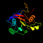
Top model
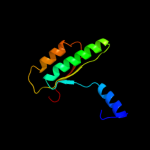
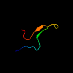
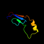
Image coloured by rainbow N → C terminus
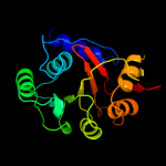
Model (left) based on template c2fsuA_
Top template information
PDB header:structural genomics, unknown function
Chain: A: PDB Molecule:protein phnh;
PDBTitle: crystal structure of the phnh protein from escherichia coli
Confidence and coverage
Confidence:100.0% Coverage: 88%
170 residues ( 88% of your sequence) have been modelled with 100.0% confidence by the single highest scoring template.
3D viewing
Interactive 3D view in Jmol

Sequence analysis 

Secondary structure and disorder prediction 

   1........10.........20.........30.........40.........50.........60
Sequence  MTLETAFMLPVQDAQHSFRRLLKAMSEPGVIVALHQLKRGWQPLNIATTSVLLTLADNDT
Secondary structure 


SS confidence 


Disorder  ??????


?????????


Disorder confidence 


   .........70.........80.........90.........100.........110.........120
Sequence  PVWLSTPLNNDIVNQSLRFHTNAPLVSQPEQATFAVTDEAISSEQLNALSTGTAVAPEAG
Secondary structure 


SS confidence 


Disorder 


??


?


Disorder confidence 


   .........130.........140.........150.........160.........170.........180
Sequence  ATLILQVASLSGGRMLRLTGAGIAEERMIAPQLPECILHELTERPHPFPLGIDLILTCGE
Secondary structure 


SS confidence 


Disorder 


?

?


?


Disorder confidence 


   .........190....
Sequence  RLLAIPRTTHVEVC
Secondary structure 


SS confidence 


Disorder 


?


??
Disorder confidence 


Confidence Key
High(9)                    Low (0)
?Disordered
Alpha helix
Beta strand

Domain analysis 

Hover over an aligned region to see model and summary info

Please note, only up to the top 20 hits are modelled to reduce computer load

RankAligned region

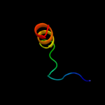
PDB 2fsu chain A

3D model

Region: 12 - 194
Aligned: 170
Modelled: 176
Confidence: 100.0%
Identity: 95%
PDB header:structural genomics, unknown function
Chain: A: PDB Molecule:protein phnh;
PDBTitle: crystal structure of the phnh protein from escherichia coli

Phyre2

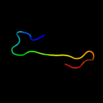
PDB 2fsu chain A domain 1

3D model

Region: 12 - 194
Aligned: 170
Modelled: 172
Confidence: 100.0%
Identity: 95%
Fold: PLP-dependent transferase-like
Superfamily: PhnH-like
Family: PhnH-like

Phyre2

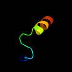
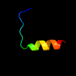
PDB 1i4a chain A

3D model

Region: 4 - 21
Aligned: 17
Modelled: 18
Confidence: 47.6%
Identity: 24%
Fold: Annexin
Superfamily: Annexin
Family: Annexin

Phyre2

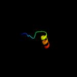
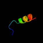
PDB 1w3w chain A

3D model

Region: 4 - 21
Aligned: 17
Modelled: 18
Confidence: 25.6%
Identity: 24%
PDB header:coagulation
Chain: A: PDB Molecule:annexin a8;
PDBTitle: the 2.1 angstroem resolution structure of annexin a8

Phyre2

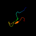
PDB 3s9x chain A

3D model

Region: 172 - 194
Aligned: 23
Modelled: 23
Confidence: 24.7%
Identity: 17%
PDB header:structural genomics, unknown function
Chain: A: PDB Molecule:asch domain;
PDBTitle: high resolution crystal structure of asch domain from lactobacillus2 crispatus jv v101

Phyre2

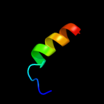
PDB 1t62 chain A

3D model

Region: 172 - 194
Aligned: 23
Modelled: 23
Confidence: 23.5%
Identity: 48%
Fold: PUA domain-like
Superfamily: PUA domain-like
Family: Hypothetical protein EF3133

Phyre2

PDB 1qjw chain A

3D model

Region: 42 - 130
Aligned: 89
Modelled: 89
Confidence: 22.3%
Identity: 19%
Fold: 7-stranded beta/alpha barrel
Superfamily: Glycosyl hydrolases family 6, cellulases
Family: Glycosyl hydrolases family 6, cellulases

Phyre2

PDB 2c7f chain A domain 1

3D model

Region: 116 - 149
Aligned: 34
Modelled: 34
Confidence: 21.4%
Identity: 9%
Fold: Glycosyl hydrolase domain
Superfamily: Glycosyl hydrolase domain
Family: Composite domain of glycosyl hydrolase families 5, 30, 39 and 51

Phyre2

PDB 3fdf chain A

3D model

Region: 125 - 154
Aligned: 30
Modelled: 30
Confidence: 19.4%
Identity: 17%
PDB header:structural genomics, unknown function
Chain: A: PDB Molecule:fr253;
PDBTitle: crystal structure of the serine phosphatase of rna2 polymerase ii ctd (ssu72 superfamily) from drosophila3 melanogaster. orthorhombic crystal form. northeast4 structural genomics consortium target fr253.

Phyre2

PDB 1dd1 chain A

3D model

Region: 90 - 142
Aligned: 53
Modelled: 53
Confidence: 18.7%
Identity: 11%
Fold: SMAD/FHA domain
Superfamily: SMAD/FHA domain
Family: SMAD domain

Phyre2

PDB 3o2q chain B

3D model

Region: 125 - 154
Aligned: 30
Modelled: 30
Confidence: 18.7%
Identity: 17%
PDB header:hydrolase
Chain: B: PDB Molecule:rna polymerase ii subunit a c-terminal domain phosphatase
PDBTitle: crystal structure of the human symplekin-ssu72-ctd phosphopeptide2 complex

Phyre2

PDB 1ygs chain A

3D model

Region: 90 - 142
Aligned: 53
Modelled: 53
Confidence: 14.9%
Identity: 11%
Fold: SMAD/FHA domain
Superfamily: SMAD/FHA domain
Family: SMAD domain

Phyre2

PDB 1hm6 chain A

3D model

Region: 1 - 21
Aligned: 20
Modelled: 21
Confidence: 14.4%
Identity: 15%
Fold: Annexin
Superfamily: Annexin
Family: Annexin

Phyre2

PDB 3o2s chain B

3D model

Region: 125 - 154
Aligned: 30
Modelled: 30
Confidence: 14.2%
Identity: 17%
PDB header:hydrolase
Chain: B: PDB Molecule:rna polymerase ii subunit a c-terminal domain phosphatase
PDBTitle: crystal structure of the human symplekin-ssu72 complex

Phyre2

PDB 1hvd chain A

3D model

Region: 1 - 21
Aligned: 20
Modelled: 21
Confidence: 12.7%
Identity: 20%
Fold: Annexin
Superfamily: Annexin
Family: Annexin

Phyre2

PDB 1ain chain A

3D model

Region: 4 - 21
Aligned: 17
Modelled: 18
Confidence: 12.0%
Identity: 18%
Fold: Annexin
Superfamily: Annexin
Family: Annexin

Phyre2

PDB 1avc chain A domain 1

3D model

Region: 1 - 21
Aligned: 20
Modelled: 21
Confidence: 11.4%
Identity: 25%
Fold: Annexin
Superfamily: Annexin
Family: Annexin

Phyre2

PDB 2nn6 chain H domain 3

3D model

Region: 166 - 182
Aligned: 17
Modelled: 17
Confidence: 11.3%
Identity: 29%
Fold: Eukaryotic type KH-domain (KH-domain type I)
Superfamily: Eukaryotic type KH-domain (KH-domain type I)
Family: Eukaryotic type KH-domain (KH-domain type I)

Phyre2

PDB 1m9i chain A domain 1

3D model

Region: 1 - 21
Aligned: 20
Modelled: 21
Confidence: 10.9%
Identity: 25%
Fold: Annexin
Superfamily: Annexin
Family: Annexin

Phyre2

PDB 1avc chain A

3D model

Region: 5 - 21
Aligned: 16
Modelled: 17
Confidence: 10.7%
Identity: 31%
PDB header:calcium/phospholipid-binding protein
Chain: A: PDB Molecule:annexin vi;
PDBTitle: bovine annexin vi (calcium-bound)

Phyre2
1

c2fsuA_
2

d2fsua1
3

d1i4aa_
4

c1w3wA_
5

c3s9xA_
6

d1t62a_
7

d1qjwa_
8

d2c7fa1
9

c3fdfA_
10

d1dd1a_
11

c3o2qB_
12

d1ygsa_
13

d1hm6a_
14

c3o2sB_
15

d1hvda_
16

d1aina_
17

d1avca1
18

d2nn6h3
19

d1m9ia1
20

c1avcA_
21


22


23


24


25


26


27


28


29


30


31


32


33


34


Detailed template information 

#
Template Alignment Coverage3D Model Confidence
% i.d. Template Information
1c2fsuA_


100.0 95 PDB header:structural genomics, unknown function
Chain: A: PDB Molecule:protein phnh;
PDBTitle: crystal structure of the phnh protein from escherichia coli
2d2fsua1


100.0 95 Fold:PLP-dependent transferase-like
Superfamily:PhnH-like
Family:PhnH-like
3d1i4aa_


47.6 24 Fold:Annexin
Superfamily:Annexin
Family:Annexin
4c1w3wA_


25.6 24 PDB header:coagulation
Chain: A: PDB Molecule:annexin a8;
PDBTitle: the 2.1 angstroem resolution structure of annexin a8
5c3s9xA_


24.7 17 PDB header:structural genomics, unknown function
Chain: A: PDB Molecule:asch domain;
PDBTitle: high resolution crystal structure of asch domain from lactobacillus2 crispatus jv v101
6d1t62a_


23.5 48 Fold:PUA domain-like
Superfamily:PUA domain-like
Family:Hypothetical protein EF3133
7d1qjwa_


22.3 19 Fold:7-stranded beta/alpha barrel
Superfamily:Glycosyl hydrolases family 6, cellulases
Family:Glycosyl hydrolases family 6, cellulases
8d2c7fa1


21.4 9 Fold:Glycosyl hydrolase domain
Superfamily:Glycosyl hydrolase domain
Family:Composite domain of glycosyl hydrolase families 5, 30, 39 and 51
9c3fdfA_


19.4 17 PDB header:structural genomics, unknown function
Chain: A: PDB Molecule:fr253;
PDBTitle: crystal structure of the serine phosphatase of rna2 polymerase ii ctd (ssu72 superfamily) from drosophila3 melanogaster. orthorhombic crystal form. northeast4 structural genomics consortium target fr253.
10d1dd1a_


18.7 11 Fold:SMAD/FHA domain
Superfamily:SMAD/FHA domain
Family:SMAD domain
11c3o2qB_


18.7 17 PDB header:hydrolase
Chain: B: PDB Molecule:rna polymerase ii subunit a c-terminal domain phosphatase
PDBTitle: crystal structure of the human symplekin-ssu72-ctd phosphopeptide2 complex
12d1ygsa_


14.9 11 Fold:SMAD/FHA domain
Superfamily:SMAD/FHA domain
Family:SMAD domain
13d1hm6a_


14.4 15 Fold:Annexin
Superfamily:Annexin
Family:Annexin
14c3o2sB_


14.2 17 PDB header:hydrolase
Chain: B: PDB Molecule:rna polymerase ii subunit a c-terminal domain phosphatase
PDBTitle: crystal structure of the human symplekin-ssu72 complex
15d1hvda_


12.7 20 Fold:Annexin
Superfamily:Annexin
Family:Annexin
16d1aina_


12.0 18 Fold:Annexin
Superfamily:Annexin
Family:Annexin
17d1avca1


11.4 25 Fold:Annexin
Superfamily:Annexin
Family:Annexin
18d2nn6h3


11.3 29 Fold:Eukaryotic type KH-domain (KH-domain type I)
Superfamily:Eukaryotic type KH-domain (KH-domain type I)
Family:Eukaryotic type KH-domain (KH-domain type I)
19d1m9ia1


10.9 25 Fold:Annexin
Superfamily:Annexin
Family:Annexin
20c1avcA_


10.7 31 PDB header:calcium/phospholipid-binding protein
Chain: A: PDB Molecule:annexin vi;
PDBTitle: bovine annexin vi (calcium-bound)
21d1m9ia2


not modelled 10.5 20 Fold:Annexin
Superfamily:Annexin
Family:Annexin
22c2hjhB_


not modelled 10.5 26 PDB header:hydrolase
Chain: B: PDB Molecule:nad-dependent histone deacetylase sir2;
PDBTitle: crystal structure of the sir2 deacetylase
23d1axna_


not modelled 10.0 20 Fold:Annexin
Superfamily:Annexin
Family:Annexin
24d2pofa1


not modelled 9.9 16 Fold:HIT-like
Superfamily:HIT-like
Family:CDH-like
25c3c8oB_


not modelled 9.3 5 PDB header:hydrolase regulator
Chain: B: PDB Molecule:regulator of ribonuclease activity a;
PDBTitle: the crystal structure of rraa from pao1
26d1alaa_


not modelled 8.3 20 Fold:Annexin
Superfamily:Annexin
Family:Annexin
27d1avca2


not modelled 8.0 20 Fold:Annexin
Superfamily:Annexin
Family:Annexin
28d1dm5a_


not modelled 7.2 15 Fold:Annexin
Superfamily:Annexin
Family:Annexin
29d1khxa_


not modelled 6.9 17 Fold:SMAD/FHA domain
Superfamily:SMAD/FHA domain
Family:SMAD domain
30c1khxA_


not modelled 6.9 17 PDB header:transcription
Chain: A: PDB Molecule:smad2;
PDBTitle: crystal structure of a phosphorylated smad2
31c1q46A_


not modelled 6.1 10 PDB header:translation
Chain: A: PDB Molecule:translation initiation factor 2 alpha subunit;
PDBTitle: crystal structure of the eif2 alpha subunit from2 saccharomyces cerevisia
32c2kl7A_


not modelled 6.0 44 PDB header:signaling protein, structural protein
Chain: A: PDB Molecule:fibulin-4;
PDBTitle: solution nmr structure of the egf-like 1 domain of human2 fibulin-4. northeast structural genomics target hr6275
33d1w7ba_


not modelled 6.0 20 Fold:Annexin
Superfamily:Annexin
Family:Annexin
34d2ie7a1


not modelled 5.8 20 Fold:Annexin
Superfamily:Annexin
Family:Annexin

Binding site prediction 

Due to computational demand, binding site predictions are not run for batch jobs

If you want to predict binding sites, please manually submit your model of choice to 3DLigandSite


Phyre is for academic use only

Please cite: Protein structure prediction on the web: a case study using the Phyre server
Kelley LA and Sternberg MJE. Nature Protocols 4, 363 - 371 (2009) [pdf] [Import into BibTeX]
 
If you use the binding site predictions from 3DLigandSite, please also cite:
3DLigandSite: predicting ligand-binding sites using similar structures.
Wass MN, Kelley LA and Sternberg MJ Nucleic Acids Research 38, W469-73 (2010) [PubMed]
 
© Structural Bioinformatics Group
Imperial College London
Lawrence Kelley, Benjamin Jefferys
Disclaimer
Terms and Conditions
Component software
Template detection: HHpred 1.51
Secondary structure prediction: Psi-pred 2.5
Disorder prediction: Disopred 2.4
Transmembrane prediction: Memsat_SVM
Multi-template modelling and ab initio: Poing 1.0