| Secondary structure and disorder prediction | |
| | |
1 | . | . | . | . | . | . | . | . | 10 | . | . | . | . | . | . | . | . | . | 20 | . | . | . | . | . | . | . | . | . | 30 | . | . | . | . | . | . | . | . | . | 40 | . | . | . | . | . | . | . | . | . | 50 | . | . | . | . | . | . | . | . | . | 60 |
| Sequence | |
M | M | K | K | F | I | A | P | L | L | A | L | L | V | S | G | C | Q | I | D | P | Y | T | H | A | P | T | L | T | S | T | D | W | Y | D | V | G | M | E | D | A | I | S | G | S | A | I | K | D | D | D | A | F | S | D | S | Q | A | D | R |
| Secondary structure | |
|  | | | | | | | | | | | | | | | |
|
|
|
|
|
|
| | | | |
|
|
|
| | | | | | | | | | |
|
|
|
|
|
|
| | | |
|
|
|
|
|
|
| |
| SS confidence | |
|
|
|
|
|
|
|
|
|
|
|
|
|
|
|
|
|
|
|
|
|
|
|
|
|
|
|
|
|
|
|
|
|
|
|
|
|
|
|
|
|
|
|
|
|
|
|
|
|
|
|
|
|
|
|
|
|
|
|
|
| Disorder | |
? | ? |
|
|
|
|
|
|
|
|
|
|
|
|
|
| ? | ? | ? | ? | ? | ? | ? | ? | ? | ? | ? | ? | ? | ? | ? | ? |
|
|
|
|
|
|
|
|
|
|
| ? | ? |
|
| ? | ? |
|
|
|
| ? | ? |
| ? | ? | ? |
|
| Disorder confidence | |
|
|
|
|
|
|
|
|
|
|
|
|
|
|
|
|
|
|
|
|
|
|
|
|
|
|
|
|
|
|
|
|
|
|
|
|
|
|
|
|
|
|
|
|
|
|
|
|
|
|
|
|
|
|
|
|
|
|
|
|
| |
| | |
. | . | . | . | . | . | . | . | . | 70 | . | . | . | . | . | . | . | . | . | 80 | . | . | . | . | . | . | . | . | . | 90 | . | . | . | . | . | . | . | . | . | 100 | . | . | . | . | . | . | . | . | . | 110 | . | . | . | . | . | . | . | . | . | 120 |
| Sequence | |
G | L | Y | L | K | G | Y | A | E | G | Q | K | K | T | C | Q | T | D | F | T | Y | A | R | G | L | S | G | K | S | F | P | A | S | C | N | N | V | E | N | A | S | Q | L | H | E | V | W | Q | K | G | A | D | E | N | A | S | T | I | R | L |
| Secondary structure | |
| | | | | | | | | | | | | |
|
| | | | | | | | | |
|
|
|
|
|
|
|
|
|
|
|
|
|
| | | | | | | | | | | | | | | | | | | |
|
|
| SS confidence | |
|
|
|
|
|
|
|
|
|
|
|
|
|
|
|
|
|
|
|
|
|
|
|
|
|
|
|
|
|
|
|
|
|
|
|
|
|
|
|
|
|
|
|
|
|
|
|
|
|
|
|
|
|
|
|
|
|
|
|
|
| Disorder | |
|
|
|
|
|
|
|
|
|
|
|
|
|
|
|
|
|
|
|
|
|
|
|
|
| ? |
|
|
| ? | ? | ? | ? | ? | ? |
|
| ? | ? |
|
|
|
|
|
|
|
|
|
|
|
|
| ? | ? | ? | ? | ? | ? | ? | ? |
| Disorder confidence | |
|
|
|
|
|
|
|
|
|
|
|
|
|
|
|
|
|
|
|
|
|
|
|
|
|
|
|
|
|
|
|
|
|
|
|
|
|
|
|
|
|
|
|
|
|
|
|
|
|
|
|
|
|
|
|
|
|
|
|
|
| |
| | |
. |
| Sequence | |
N |
| Secondary structure | |
|
| SS confidence | |
|
| Disorder | |
? |
| Disorder confidence | |
|
| |
| Confidence Key |
| High(9) | |
|
|
|
|
|
|
|
|
|
Low (0) |
| ? | Disordered |
| Alpha helix |
  | Beta strand |
Hover over an aligned region to see model and summary info
Please note, only up to the top 20 hits are modelled to reduce computer load
|
| 1 |
|
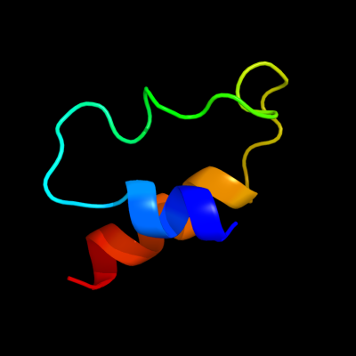
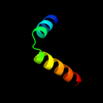
PDB 2jrm chain A
Region: 34 - 74
Aligned: 39
Modelled: 41
Confidence: 68.1%
Identity: 15%
PDB header:structural genomics, unknown function
Chain: A: PDB Molecule:ribosome modulation factor;
PDBTitle: solution nmr structure of ribosome modulation factor vp1593 from2 vibrio parahaemolyticus. northeast structural genomics target vpr55
Phyre2
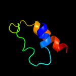
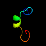
| 2 |
|
PDB 2ae3 chain A
Region: 44 - 74
Aligned: 31
Modelled: 31
Confidence: 11.9%
Identity: 16%
PDB header:hydrolase
Chain: A: PDB Molecule:glutaryl 7-aminocephalosporanic acid acylase;
PDBTitle: glutaryl 7-aminocephalosporanic acid acylase: mutational study of2 activation mechanism
Phyre2
| 3 |
|
PDB 2khg chain A
Region: 37 - 49
Aligned: 13
Modelled: 13
Confidence: 7.5%
Identity: 38%
PDB header:antimicrobial protein
Chain: A: PDB Molecule:plnj;
PDBTitle: plantaricin j in tfe
Phyre2
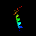
| 4 |
|
PDB 2ijr chain A domain 1
Region: 3 - 30
Aligned: 26
Modelled: 26
Confidence: 6.8%
Identity: 15%
Fold: Api92-like
Superfamily: Api92-like
Family: Api92-like
Phyre2
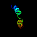
| 5 |
|
PDB 2wyb chain A
Region: 44 - 74
Aligned: 31
Modelled: 31
Confidence: 6.0%
Identity: 16%
PDB header:hydrolase
Chain: A: PDB Molecule:acyl-homoserine lactone acylase pvdq subunit
PDBTitle: the quorum quenching n-acyl homoserine lactone acylase pvdq2 with a covalently bound dodecanoic acid
Phyre2
| 6 |
|
PDB 2fxt chain A domain 1
Region: 60 - 82
Aligned: 22
Modelled: 23
Confidence: 5.6%
Identity: 23%
Fold: Cystatin-like
Superfamily: NTF2-like
Family: TIM44-like
Phyre2
|
| Detailed template information | |
Due to computational demand, binding site predictions are not run for batch jobs
If you want to predict binding sites, please manually submit your model of choice to 3DLigandSite
Phyre is for academic use only
| Please cite: Protein structure prediction on
the web: a case study using the Phyre server |
| Kelley LA and Sternberg MJE. Nature Protocols
4, 363 - 371 (2009) [pdf] [Import into BibTeX] |
| |
| If you use the binding site
predictions from 3DLigandSite, please also cite: |
| 3DLigandSite: predicting ligand-binding sites using similar structures. |
| Wass MN, Kelley LA and Sternberg
MJ Nucleic Acids Research 38, W469-73 (2010) [PubMed] |
| |
|
|
|
|