| Secondary structure and disorder prediction | |
| | |
1 | . | . | . | . | . | . | . | . | 10 | . | . | . | . | . | . | . | . | . | 20 | . | . | . | . |
| Sequence | |
M | P | G | K | V | Q | D | F | F | L | C | S | L | L | L | C | I | V | S | A | G | W | C | G |
| Secondary structure | |
|
|  | | | | | | | | | | | | | | | | |
|
|
|
|
|
| SS confidence | |
|
|
|
|
|
|
|
|
|
|
|
|
|
|
|
|
|
|
|
|
|
|
|
|
| Disorder | |
? | ? | ? | ? |
|
|
|
|
|
|
|
|
|
|
|
|
|
| ? | ? | ? | ? | ? | ? |
| Disorder confidence | |
|
|
|
|
|
|
|
|
|
|
|
|
|
|
|
|
|
|
|
|
|
|
|
|
| |
| Confidence Key |
| High(9) | |
|
|
|
|
|
|
|
|
|
Low (0) |
| ? | Disordered |
| Alpha helix |
  | Beta strand |
Hover over an aligned region to see model and summary info
Please note, only up to the top 20 hits are modelled to reduce computer load
|
| 1 |
|
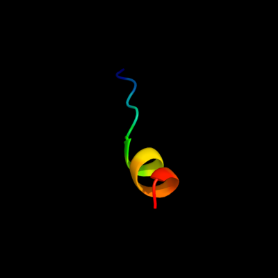
PDB 1zll chain E
Region: 4 - 18
Aligned: 15
Modelled: 15
Confidence: 7.5%
Identity: 67%
PDB header:membrane protein/signaling protein
Chain: E: PDB Molecule:cardiac phospholamban;
PDBTitle: nmr structure of unphosphorylated human phospholamban2 pentamer
Phyre2
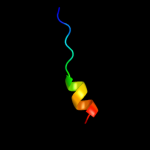
| 2 |
|
PDB 2p7v chain A
Region: 4 - 21
Aligned: 16
Modelled: 18
Confidence: 7.1%
Identity: 50%
PDB header:transcription
Chain: A: PDB Molecule:regulator of sigma d;
PDBTitle: crystal structure of the escherichia coli regulator of sigma 70, rsd,2 in complex with sigma 70 domain 4
Phyre2
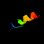
| 3 |
|
PDB 1cmr chain A
Region: 19 - 23
Aligned: 5
Modelled: 5
Confidence: 5.9%
Identity: 80%
Fold: Knottins (small inhibitors, toxins, lectins)
Superfamily: Scorpion toxin-like
Family: Short-chain scorpion toxins
Phyre2
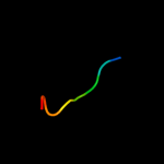
| 4 |
|
PDB 1nj1 chain A domain 2
Region: 17 - 25
Aligned: 9
Modelled: 9
Confidence: 5.4%
Identity: 56%
Fold: IF3-like
Superfamily: C-terminal domain of ProRS
Family: C-terminal domain of ProRS
Phyre2
| 5 |
|
PDB 1en2 chain A domain 2
Region: 21 - 24
Aligned: 4
Modelled: 4
Confidence: 5.2%
Identity: 100%
Fold: Knottins (small inhibitors, toxins, lectins)
Superfamily: Plant lectins/antimicrobial peptides
Family: Hevein-like agglutinin (lectin) domain
Phyre2
|
| Detailed template information | |
Due to computational demand, binding site predictions are not run for batch jobs
If you want to predict binding sites, please manually submit your model of choice to 3DLigandSite
Phyre is for academic use only
| Please cite: Protein structure prediction on
the web: a case study using the Phyre server |
| Kelley LA and Sternberg MJE. Nature Protocols
4, 363 - 371 (2009) [pdf] [Import into BibTeX] |
| |
| If you use the binding site
predictions from 3DLigandSite, please also cite: |
| 3DLigandSite: predicting ligand-binding sites using similar structures. |
| Wass MN, Kelley LA and Sternberg
MJ Nucleic Acids Research 38, W469-73 (2010) [PubMed] |
| |
|
|
|
|