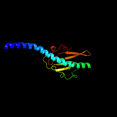
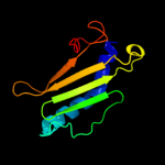
1 c3sokB_
99.9
22
PDB header: cell adhesionChain: B: PDB Molecule: fimbrial protein;PDBTitle: dichelobacter nodosus pilin fima
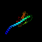
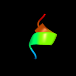
2 d1oqwa_
99.9
27
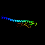
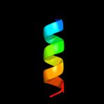
Fold: Pili subunitsSuperfamily: Pili subunitsFamily: Pilin3 d2pila_
99.9
30
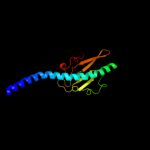
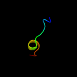
Fold: Pili subunitsSuperfamily: Pili subunitsFamily: Pilin4 d1qvea_
99.0
21
Fold: Pili subunitsSuperfamily: Pili subunitsFamily: Pilin5 c3jyzA_
99.0
18
PDB header: structural proteinChain: A: PDB Molecule: type iv pilin structural subunit;PDBTitle: crystal structure of pseudomonas aeruginosa (strain:2 pa110594) typeiv pilin in space group p41212
6 d1x6za1
98.6
17
Fold: Pili subunitsSuperfamily: Pili subunitsFamily: Pilin7 c3fu1B_
93.9
19
PDB header: protein transportChain: B: PDB Molecule: general secretion pathway protein g;PDBTitle: crystal structure of the major pseudopilin from the type 2 secretion2 system of vibrio cholerae
8 d1t92a_
93.7
17
Fold: Pili subunitsSuperfamily: Pili subunitsFamily: Pseudopilin9 c2kepA_
91.9
15
PDB header: transport proteinChain: A: PDB Molecule: general secretion pathway protein g;PDBTitle: solution structure of xcpt, the main component of the type 22 secretion system of pseudomonas aeruginosa
10 c1bttA_
41.7
31
PDB header: transmembrane proteinChain: A: PDB Molecule: band 3 anion transport protein;PDBTitle: the solution structures of the first and second2 transmembrane-spanning segments of band 3
11 c1btsA_
41.0
33
PDB header: transmembrane proteinChain: A: PDB Molecule: band 3 anion transport protein;PDBTitle: the solution structures of the first and second2 transmembrane-spanning segments of band 3
12 c4a18U_
39.6
64
PDB header: ribosomeChain: U: PDB Molecule: rpl13;PDBTitle: t.thermophila 60s ribosomal subunit in complex with initiation2 factor 6. this file contains 26s rrna and proteins of molecule 1
13 c3u5eL_
38.5
55
PDB header: ribosomeChain: L: PDB Molecule: 60s ribosomal protein l13-a;PDBTitle: the structure of the eukaryotic ribosome at 3.0 resolution
14 d3ehbb2
36.4
31
Fold: Transmembrane helix hairpinSuperfamily: Cytochrome c oxidase subunit II-like, transmembrane regionFamily: Cytochrome c oxidase subunit II-like, transmembrane region15 d1v54b2
29.5
20
Fold: Transmembrane helix hairpinSuperfamily: Cytochrome c oxidase subunit II-like, transmembrane regionFamily: Cytochrome c oxidase subunit II-like, transmembrane region16 d1fftb2
28.9
13
Fold: Transmembrane helix hairpinSuperfamily: Cytochrome c oxidase subunit II-like, transmembrane regionFamily: Cytochrome c oxidase subunit II-like, transmembrane region17 c3sojA_
28.3
17
PDB header: cell adhesionChain: A: PDB Molecule: pile;PDBTitle: francisella tularensis pilin pile
18 c2kxeA_
14.7
71
PDB header: transferaseChain: A: PDB Molecule: dna polymerase ii small subunit;PDBTitle: n-terminal domain of the dp1 subunit of an archaeal d-family dna2 polymerase
19 c1afoB_
14.7
13
PDB header: integral membrane proteinChain: B: PDB Molecule: glycophorin a;PDBTitle: dimeric transmembrane domain of human glycophorin a, nmr,2 20 structures
20 d1r89a1
14.2
62
Fold: PAP/OAS1 substrate-binding domainSuperfamily: PAP/OAS1 substrate-binding domainFamily: Archaeal tRNA CCA-adding enzyme substrate-binding domain21 c2wsfG_
not modelled
13.9
25
PDB header: photosynthesisChain: G: PDB Molecule: photosystem i reaction center subunit v,PDBTitle: improved model of plant photosystem i
22 c1xmeB_
not modelled
13.2
13
PDB header: oxidoreductaseChain: B: PDB Molecule: cytochrome c oxidase polypeptide ii;PDBTitle: structure of recombinant cytochrome ba3 oxidase from thermus2 thermophilus
23 c1qleB_
not modelled
13.2
25
PDB header: oxidoreductase/immune systemChain: B: PDB Molecule: cytochrome c oxidase polypeptide ii;PDBTitle: cryo-structure of the paracoccus denitrificans four-subunit2 cytochrome c oxidase in the completely oxidized state3 complexed with an antibody fv fragment
24 c1ar1B_
not modelled
13.2
25
PDB header: complex (oxidoreductase/antibody)Chain: B: PDB Molecule: cytochrome c oxidase;PDBTitle: structure at 2.7 angstrom resolution of the paracoccus2 denitrificans two-subunit cytochrome c oxidase complexed3 with an antibody fv fragment
25 d3dtub2
not modelled
12.8
25
Fold: Transmembrane helix hairpinSuperfamily: Cytochrome c oxidase subunit II-like, transmembrane regionFamily: Cytochrome c oxidase subunit II-like, transmembrane region26 c2kadB_
not modelled
11.8
47
PDB header: membrane proteinChain: B: PDB Molecule: transmembrane peptide of matrix protein 2;PDBTitle: magic-angle-spinning solid-state nmr structure of influenza2 a m2 transmembrane domain
27 c2kadC_
not modelled
11.8
47
PDB header: membrane proteinChain: C: PDB Molecule: transmembrane peptide of matrix protein 2;PDBTitle: magic-angle-spinning solid-state nmr structure of influenza2 a m2 transmembrane domain
28 c2kadD_
not modelled
11.8
47
PDB header: membrane proteinChain: D: PDB Molecule: transmembrane peptide of matrix protein 2;PDBTitle: magic-angle-spinning solid-state nmr structure of influenza2 a m2 transmembrane domain
29 c2kadA_
not modelled
11.8
47
PDB header: membrane proteinChain: A: PDB Molecule: transmembrane peptide of matrix protein 2;PDBTitle: magic-angle-spinning solid-state nmr structure of influenza2 a m2 transmembrane domain
30 d1m56d_
not modelled
11.2
21
Fold: Single transmembrane helixSuperfamily: Bacterial aa3 type cytochrome c oxidase subunit IVFamily: Bacterial aa3 type cytochrome c oxidase subunit IV31 d1r3jc_
not modelled
10.6
9
Fold: Voltage-gated potassium channelsSuperfamily: Voltage-gated potassium channelsFamily: Voltage-gated potassium channels32 c2o01G_
not modelled
10.2
25
PDB header: photosynthesisChain: G: PDB Molecule: photosystem i reaction center subunit v,PDBTitle: the structure of a plant photosystem i supercomplex at 3.42 angstrom resolution
33 c2ljcA_
not modelled
10.0
47
PDB header: transport protein/inhibitorChain: A: PDB Molecule: m2 protein, bm2 protein chimera;PDBTitle: structure of the influenza am2-bm2 chimeric channel bound to2 rimantadine
34 d1oqva_
not modelled
9.7
15
Fold: Pili subunitsSuperfamily: Pili subunitsFamily: TcpA-like pilin35 d1tcra2
not modelled
9.4
29
Fold: Immunoglobulin-like beta-sandwichSuperfamily: ImmunoglobulinFamily: C1 set domains (antibody constant domain-like)36 c2kb1A_
not modelled
8.1
12
PDB header: membrane proteinChain: A: PDB Molecule: wsk3;PDBTitle: nmr studies of a channel protein without membrane:2 structure and dynamics of water-solubilized kcsa
37 c2wscK_
not modelled
7.0
50
PDB header: photosynthesisChain: K: PDB Molecule: photosystem i reaction center subunit psak,PDBTitle: improved model of plant photosystem i
38 d1az3a_
not modelled
6.9
38
Fold: Restriction endonuclease-likeSuperfamily: Restriction endonuclease-likeFamily: Restriction endonuclease EcoRV39 c2vvyC_
not modelled
6.6
14
PDB header: viral proteinChain: C: PDB Molecule: protein b15;PDBTitle: structure of vaccinia virus protein b14
40 c1mp6A_
not modelled
6.6
47
PDB header: membrane proteinChain: A: PDB Molecule: matrix protein m2;PDBTitle: structure of the transmembrane region of the m2 protein h+2 channel by solid state nmr spectroscopy
41 c1nyjB_
not modelled
6.6
47
PDB header: viral proteinChain: B: PDB Molecule: matrix protein m2;PDBTitle: the closed state structure of m2 protein h+ channel by2 solid state nmr spectroscopy
42 c2kqtA_
not modelled
6.6
47
PDB header: transport proteinChain: A: PDB Molecule: m2 protein;PDBTitle: solid-state nmr structure of the m2 transmembrane peptide of the2 influenza a virus in dmpc lipid bilayers bound to deuterated3 amantadine
43 c2kqtC_
not modelled
6.6
47
PDB header: transport proteinChain: C: PDB Molecule: m2 protein;PDBTitle: solid-state nmr structure of the m2 transmembrane peptide of the2 influenza a virus in dmpc lipid bilayers bound to deuterated3 amantadine
44 c1nyjC_
not modelled
6.6
47
PDB header: viral proteinChain: C: PDB Molecule: matrix protein m2;PDBTitle: the closed state structure of m2 protein h+ channel by2 solid state nmr spectroscopy
45 c2kqtB_
not modelled
6.6
47
PDB header: transport proteinChain: B: PDB Molecule: m2 protein;PDBTitle: solid-state nmr structure of the m2 transmembrane peptide of the2 influenza a virus in dmpc lipid bilayers bound to deuterated3 amantadine
46 c1nyjD_
not modelled
6.6
47
PDB header: viral proteinChain: D: PDB Molecule: matrix protein m2;PDBTitle: the closed state structure of m2 protein h+ channel by2 solid state nmr spectroscopy
47 c1nyjA_
not modelled
6.6
47
PDB header: viral proteinChain: A: PDB Molecule: matrix protein m2;PDBTitle: the closed state structure of m2 protein h+ channel by2 solid state nmr spectroscopy
48 c2kqtD_
not modelled
6.6
47
PDB header: transport proteinChain: D: PDB Molecule: m2 protein;PDBTitle: solid-state nmr structure of the m2 transmembrane peptide of the2 influenza a virus in dmpc lipid bilayers bound to deuterated3 amantadine
49 c2j7aC_
not modelled
6.4
7
PDB header: oxidoreductaseChain: C: PDB Molecule: cytochrome c quinol dehydrogenase nrfh;PDBTitle: crystal structure of cytochrome c nitrite reductase nrfha2 complex from desulfovibrio vulgaris
50 c2dcoA_
not modelled
6.4
25
PDB header: membrane proteinChain: A: PDB Molecule: s1p4 first extracellular loop peptidomimetic;PDBTitle: s1p4 first extracellular loop peptidomimetic
51 d2a9ha1
not modelled
6.3
10
Fold: Voltage-gated potassium channelsSuperfamily: Voltage-gated potassium channelsFamily: Voltage-gated potassium channels52 c3lw5K_
not modelled
6.1
26
PDB header: photosynthesisChain: K: PDB Molecule: photosystem i reaction center subunit x psak;PDBTitle: improved model of plant photosystem i
53 c3f5dA_
not modelled
6.0
10
PDB header: structural genomics, unknown functionChain: A: PDB Molecule: protein ydea;PDBTitle: crystal structure of a protein of unknown function from2 bacillus subtilis
54 c1spfA_
not modelled
5.8
25
PDB header: lipoprotein(surface film)Chain: A: PDB Molecule: pulmonary surfactant-associated polypeptide c;PDBTitle: the nmr structure of the pulmonary surfactant-associated2 polypeptide sp-c in an apolar solvent contains a valyl-3 rich alpha-helix
55 c1tiiC_
not modelled
5.7
50
PDB header: enterotoxinChain: C: PDB Molecule: heat labile enterotoxin type iib;PDBTitle: escherichia coli heat labile enterotoxin type iib
56 d1xmeb2
not modelled
5.5
50
Fold: Transmembrane helix hairpinSuperfamily: Cytochrome c oxidase subunit II-like, transmembrane regionFamily: Cytochrome c oxidase subunit II-like, transmembrane region57 c1b22A_
not modelled
5.4
20
PDB header: dna binding proteinChain: A: PDB Molecule: dna repair protein rad51;PDBTitle: rad51 (n-terminal domain)
58 d1b22a_
not modelled
5.4
20
Fold: SAM domain-likeSuperfamily: Rad51 N-terminal domain-likeFamily: DNA repair protein Rad51, N-terminal domain59 d2ab0a1
not modelled
5.3
20
Fold: Flavodoxin-likeSuperfamily: Class I glutamine amidotransferase-likeFamily: DJ-1/PfpI