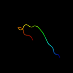
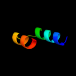
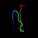
| 1 |
|
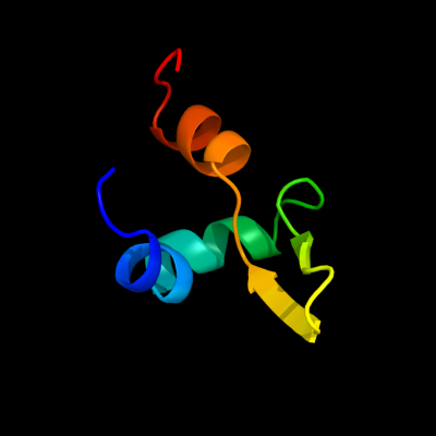
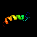
PDB 3bjq chain A
Region: 10 - 58
Aligned: 35
Modelled: 48
Confidence: 25.0%
Identity: 23%
PDB header:viral protein
Chain: A: PDB Molecule:phage-related protein;
PDBTitle: crystal structure of a phage-related protein (bb3626) from bordetella2 bronchiseptica rb50 at 2.05 a resolution
Phyre2
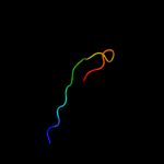
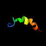
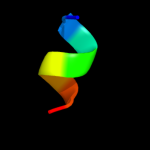
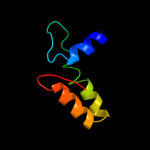
| 2 |
|
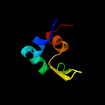
PDB 2e52 chain A
Region: 123 - 160
Aligned: 38
Modelled: 38
Confidence: 20.4%
Identity: 26%
PDB header:hydrolase/dna
Chain: A: PDB Molecule:type ii restriction enzyme hindiii;
PDBTitle: crystal structural analysis of hindiii restriction endonuclease in2 complex with cognate dna at 2.0 angstrom resolution
Phyre2
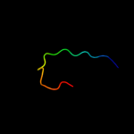
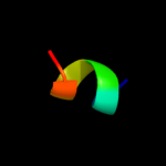
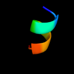
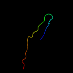
| 3 |
|
PDB 1m9z chain A
Region: 9 - 25
Aligned: 17
Modelled: 17
Confidence: 18.2%
Identity: 12%
Fold: Snake toxin-like
Superfamily: Snake toxin-like
Family: Extracellular domain of cell surface receptors
Phyre2
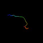
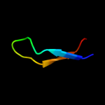
| 4 |
|
PDB 1ks6 chain A
Region: 9 - 25
Aligned: 17
Modelled: 17
Confidence: 18.0%
Identity: 29%
Fold: Snake toxin-like
Superfamily: Snake toxin-like
Family: Extracellular domain of cell surface receptors
Phyre2
| 5 |
|
PDB 2pjy chain B domain 1
Region: 9 - 25
Aligned: 17
Modelled: 17
Confidence: 17.7%
Identity: 12%
Fold: Snake toxin-like
Superfamily: Snake toxin-like
Family: Extracellular domain of cell surface receptors
Phyre2
| 6 |
|
PDB 1plo chain A
Region: 9 - 25
Aligned: 17
Modelled: 17
Confidence: 16.7%
Identity: 12%
Fold: Snake toxin-like
Superfamily: Snake toxin-like
Family: Extracellular domain of cell surface receptors
Phyre2
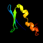
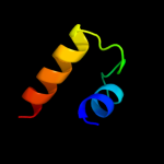
| 7 |
|
PDB 2kgm chain A
Region: 129 - 156
Aligned: 22
Modelled: 28
Confidence: 14.9%
Identity: 41%
PDB header:signaling protein
Chain: A: PDB Molecule:protein ste5;
PDBTitle: solution structure of ste5pm24 in sds micelle
Phyre2
| 8 |
|
PDB 2l4u chain A
Region: 129 - 156
Aligned: 22
Modelled: 28
Confidence: 11.8%
Identity: 41%
PDB header:signaling protein
Chain: A: PDB Molecule:24mer peptide from protein ste5;
PDBTitle: solution structure of ste5pm24 in the presence of sds micelle
Phyre2
| 9 |
|
PDB 1xk5 chain A
Region: 122 - 129
Aligned: 8
Modelled: 8
Confidence: 9.3%
Identity: 38%
PDB header:transport protein
Chain: A: PDB Molecule:snurportin-1;
PDBTitle: crystal structure of the m3g-cap-binding domain of2 snurportin1 in complex with a m3gpppg-cap dinucleotide
Phyre2
| 10 |
|
PDB 1ukf chain A
Region: 21 - 68
Aligned: 47
Modelled: 48
Confidence: 9.0%
Identity: 19%
Fold: Cysteine proteinases
Superfamily: Cysteine proteinases
Family: Avirulence protein Avrpph3
Phyre2
| 11 |
|
PDB 2fcw chain A domain 1
Region: 102 - 126
Aligned: 25
Modelled: 25
Confidence: 8.9%
Identity: 32%
Fold: RAP domain-like
Superfamily: RAP domain-like
Family: RAP domain
Phyre2
| 12 |
|
PDB 3gb8 chain B
Region: 122 - 129
Aligned: 8
Modelled: 8
Confidence: 8.0%
Identity: 38%
PDB header:transport protein
Chain: B: PDB Molecule:snurportin-1;
PDBTitle: crystal structure of crm1/snurportin-1 complex
Phyre2
| 13 |
|
PDB 3gjx chain E
Region: 122 - 129
Aligned: 8
Modelled: 8
Confidence: 7.6%
Identity: 38%
PDB header:protein transport
Chain: E: PDB Molecule:snurportin-1;
PDBTitle: crystal structure of the nuclear export complex crm1-2 snurportin1-rangtp
Phyre2
| 14 |
|
PDB 3h7h chain A
Region: 30 - 46
Aligned: 17
Modelled: 17
Confidence: 7.3%
Identity: 41%
PDB header:transcription
Chain: A: PDB Molecule:transcription elongation factor spt4;
PDBTitle: crystal structure of the human transcription elongation factor dsif,2 hspt4/hspt5 (176-273)
Phyre2
| 15 |
|
PDB 2zxr chain A
Region: 5 - 23
Aligned: 18
Modelled: 19
Confidence: 6.3%
Identity: 44%
PDB header:hydrolase
Chain: A: PDB Molecule:single-stranded dna specific exonuclease recj;
PDBTitle: crystal structure of recj in complex with mg2+ from thermus2 thermophilus hb8
Phyre2
| 16 |
|
PDB 2fok chain A domain 1
Region: 26 - 85
Aligned: 50
Modelled: 60
Confidence: 6.2%
Identity: 16%
Fold: DNA/RNA-binding 3-helical bundle
Superfamily: "Winged helix" DNA-binding domain
Family: Restriction endonuclease FokI, N-terminal (recognition) domain
Phyre2
| 17 |
|
PDB 1txk chain A
Region: 109 - 133
Aligned: 25
Modelled: 25
Confidence: 6.2%
Identity: 28%
PDB header:biosynthetic protein
Chain: A: PDB Molecule:glucans biosynthesis protein g;
PDBTitle: crystal structure of escherichia coli opgg
Phyre2
| 18 |
|
PDB 1f44 chain A domain 1
Region: 99 - 132
Aligned: 33
Modelled: 34
Confidence: 6.1%
Identity: 21%
Fold: SAM domain-like
Superfamily: lambda integrase-like, N-terminal domain
Family: lambda integrase-like, N-terminal domain
Phyre2
| 19 |
|
PDB 1j0s chain A
Region: 5 - 46
Aligned: 42
Modelled: 42
Confidence: 6.1%
Identity: 12%
Fold: beta-Trefoil
Superfamily: Cytokine
Family: Interleukin-1 (IL-1)
Phyre2
| 20 |
|
PDB 1txk chain A domain 2
Region: 109 - 133
Aligned: 25
Modelled: 25
Confidence: 6.1%
Identity: 28%
Fold: Supersandwich
Superfamily: Galactose mutarotase-like
Family: MdoG-like
Phyre2
| 21 |
|
| 22 |
|
| 23 |
|