Phyre Home Retrieve Phyre Job Id
Emaill.a.kelley@imperial.ac.uk
DescriptionP0AAU7
DateThu Jan 5 11:13:52 GMT 2012
Unique Job ID83a5f0df84b3c75a

Summary 

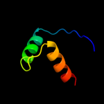
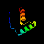
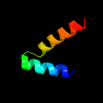
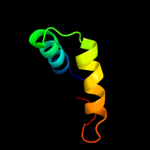
Top model
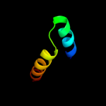
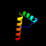
Image coloured by rainbow N → C terminus
Model (left) based on template c1q5vB_
Top template information
PDB header:transcription
Chain: B: PDB Molecule:nickel responsive regulator;
PDBTitle: apo-nikr
Confidence and coverage
Confidence: 95.3% Coverage: 41%
40 residues ( 41% of your sequence) have been modelled with 95.3% confidence by the single highest scoring template.
You may wish to submit your sequence to Phyrealarm. This will automatically scan your sequence every week for new potential templates as they appear in the Phyre2 library.
3D viewing
Interactive 3D view in Jmol

Sequence analysis 

Secondary structure and disorder prediction 

   1........10.........20.........30.........40.........50.........60
Sequence  MAKEQTDRTTLDLFAHERRPGRPKTNPLSRDEQLRINKRNQLKRDKVRGLKRVELKLNAE
Secondary structure 


SS confidence 


Disorder  ???????


????????????????

????


???


Disorder confidence 


   .........70.........80.........90.......
Sequence  AVEALNELAESRNMSRSELIEEMLMQQLAALRSQGIV
Secondary structure 


SS confidence 


Disorder 


????


?????
Disorder confidence 


Confidence Key
High(9)                    Low (0)
?Disordered
Alpha helix
Beta strand

Domain analysis 

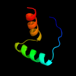
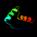
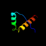
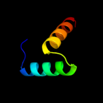
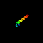
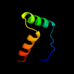
Hover over an aligned region to see model and summary info

Please note, only up to the top 20 hits are modelled to reduce computer load

RankAligned region

PDB 1q5v chain B

3D model

Region: 50 - 89
Aligned: 40
Modelled: 40
Confidence: 95.3%
Identity: 35%
PDB header:transcription
Chain: B: PDB Molecule:nickel responsive regulator;
PDBTitle: apo-nikr

Phyre2

PDB 2bj3 chain D

3D model

Region: 50 - 89
Aligned: 40
Modelled: 40
Confidence: 95.2%
Identity: 20%
PDB header:transcription
Chain: D: PDB Molecule:nickel responsive regulator;
PDBTitle: nikr-apo

Phyre2

PDB 2bj7 chain A domain 1

3D model

Region: 50 - 89
Aligned: 40
Modelled: 40
Confidence: 95.2%
Identity: 20%
Fold: Ribbon-helix-helix
Superfamily: Ribbon-helix-helix
Family: CopG-like

Phyre2

PDB 2ca9 chain B

3D model

Region: 50 - 92
Aligned: 43
Modelled: 43
Confidence: 94.6%
Identity: 23%
PDB header:transcriptional regulation
Chain: B: PDB Molecule:putative nickel-responsive regulator;
PDBTitle: apo-nikr from helicobacter pylori in closed trans-2 conformation

Phyre2

PDB 2hza chain A domain 1

3D model

Region: 51 - 89
Aligned: 39
Modelled: 39
Confidence: 93.1%
Identity: 36%
Fold: Ribbon-helix-helix
Superfamily: Ribbon-helix-helix
Family: CopG-like

Phyre2

PDB 2hza chain B domain 1

3D model

Region: 51 - 89
Aligned: 39
Modelled: 39
Confidence: 92.8%
Identity: 36%
Fold: Ribbon-helix-helix
Superfamily: Ribbon-helix-helix
Family: CopG-like

Phyre2

PDB 2k5j chain B

3D model

Region: 50 - 89
Aligned: 40
Modelled: 40
Confidence: 89.9%
Identity: 33%
PDB header:structural genomics, unknown function
Chain: B: PDB Molecule:uncharacterized protein yiif;
PDBTitle: solution structure of protein yiif from shigella flexneri2 serotype 5b (strain 8401) . northeast structural genomics3 consortium target sft1

Phyre2

PDB 2cpg chain A

3D model

Region: 51 - 88
Aligned: 38
Modelled: 38
Confidence: 72.3%
Identity: 26%
Fold: Ribbon-helix-helix
Superfamily: Ribbon-helix-helix
Family: CopG-like

Phyre2

PDB 1y9b chain A domain 1

3D model

Region: 52 - 80
Aligned: 29
Modelled: 29
Confidence: 29.3%
Identity: 14%
Fold: Ribbon-helix-helix
Superfamily: Ribbon-helix-helix
Family: VCA0319-like

Phyre2

PDB 2kwp chain A

3D model

Region: 58 - 92
Aligned: 35
Modelled: 35
Confidence: 25.7%
Identity: 14%
PDB header:transcription
Chain: A: PDB Molecule:transcription elongation protein nusa;
PDBTitle: solution structure of the aminoterminal domain of e. coli nusa

Phyre2

PDB 3kk4 chain B

3D model

Region: 55 - 90
Aligned: 36
Modelled: 36
Confidence: 22.3%
Identity: 19%
PDB header:structural genomics, unknown function
Chain: B: PDB Molecule:uncharacterized protein bp1543;
PDBTitle: uncharacterized protein bp1543 from bordetella pertussis tohama i

Phyre2

PDB 2kkm chain A

3D model

Region: 17 - 50
Aligned: 30
Modelled: 34
Confidence: 18.9%
Identity: 37%
PDB header:translation
Chain: A: PDB Molecule:translation machinery-associated protein 16;
PDBTitle: solution nmr structure of yeast protein yor252w [residues2 38-178]: northeast structural genomics consortium target3 yt654

Phyre2

PDB 1svm chain A

3D model

Region: 67 - 92
Aligned: 26
Modelled: 26
Confidence: 17.5%
Identity: 12%
Fold: P-loop containing nucleoside triphosphate hydrolases
Superfamily: P-loop containing nucleoside triphosphate hydrolases
Family: Extended AAA-ATPase domain

Phyre2

PDB 2k29 chain A

3D model

Region: 50 - 89
Aligned: 40
Modelled: 40
Confidence: 15.4%
Identity: 18%
PDB header:transcription
Chain: A: PDB Molecule:antitoxin relb;
PDBTitle: structure of the dbd domain of e. coli antitoxin relb

Phyre2

PDB 1l2f chain A

3D model

Region: 58 - 92
Aligned: 35
Modelled: 35
Confidence: 14.7%
Identity: 31%
PDB header:transcription
Chain: A: PDB Molecule:n utilization substance protein a;
PDBTitle: crystal structure of nusa from thermotoga maritima: a2 structure-based role of the n-terminal domain

Phyre2

PDB 2k9i chain B

3D model

Region: 57 - 89
Aligned: 33
Modelled: 33
Confidence: 12.9%
Identity: 21%
PDB header:dna binding protein
Chain: B: PDB Molecule:plasmid prn1, complete sequence;
PDBTitle: nmr structure of plasmid copy control protein orf56 from2 sulfolobus islandicus

Phyre2

PDB 1t6s chain A domain 2

3D model

Region: 56 - 81
Aligned: 26
Modelled: 26
Confidence: 12.5%
Identity: 19%
Fold: DNA/RNA-binding 3-helical bundle
Superfamily: "Winged helix" DNA-binding domain
Family: ScpB/YpuH-like

Phyre2

PDB 1qx8 chain A

3D model

Region: 61 - 71
Aligned: 11
Modelled: 11
Confidence: 11.8%
Identity: 55%
Fold: ROP-like
Superfamily: ROP protein
Family: ROP protein

Phyre2

PDB 2keb chain A

3D model

Region: 55 - 96
Aligned: 41
Modelled: 42
Confidence: 11.6%
Identity: 17%
PDB header:dna binding protein
Chain: A: PDB Molecule:dna polymerase subunit alpha b;
PDBTitle: nmr solution structure of the n-terminal domain of the dna polymerase2 alpha p68 subunit

Phyre2

PDB 2rbf chain B

3D model

Region: 52 - 93
Aligned: 42
Modelled: 42
Confidence: 10.8%
Identity: 19%
PDB header:oxidoreductase/dna
Chain: B: PDB Molecule:bifunctional protein puta;
PDBTitle: structure of the ribbon-helix-helix domain of escherichia coli puta2 (puta52) complexed with operator dna (o2)

Phyre2
1

c1q5vB_
2

c2bj3D_
3

d2bj7a1
4

c2ca9B_
5

d2hzaa1
6

d2hzab1
7

c2k5jB_
8

d2cpga_
9

d1y9ba1
10

c2kwpA_
11

c3kk4B_
12

c2kkmA_
13

d1svma_
14

c2k29A_
15

c1l2fA_
16

c2k9iB_
17

d1t6sa2
18

d1qx8a_
19

c2kebA_
20

c2rbfB_
21


22


23


24


25


26


27


28


29


30


31


32


Detailed template information 

#
Template Alignment Coverage3D Model Confidence
% i.d. Template Information
1c1q5vB_


95.3 35 PDB header:transcription
Chain: B: PDB Molecule:nickel responsive regulator;
PDBTitle: apo-nikr
2c2bj3D_


95.2 20 PDB header:transcription
Chain: D: PDB Molecule:nickel responsive regulator;
PDBTitle: nikr-apo
3d2bj7a1


95.2 20 Fold:Ribbon-helix-helix
Superfamily:Ribbon-helix-helix
Family:CopG-like
4c2ca9B_


94.6 23 PDB header:transcriptional regulation
Chain: B: PDB Molecule:putative nickel-responsive regulator;
PDBTitle: apo-nikr from helicobacter pylori in closed trans-2 conformation
5d2hzaa1


93.1 36 Fold:Ribbon-helix-helix
Superfamily:Ribbon-helix-helix
Family:CopG-like
6d2hzab1


92.8 36 Fold:Ribbon-helix-helix
Superfamily:Ribbon-helix-helix
Family:CopG-like
7c2k5jB_


89.9 33 PDB header:structural genomics, unknown function
Chain: B: PDB Molecule:uncharacterized protein yiif;
PDBTitle: solution structure of protein yiif from shigella flexneri2 serotype 5b (strain 8401) . northeast structural genomics3 consortium target sft1
8d2cpga_


72.3 26 Fold:Ribbon-helix-helix
Superfamily:Ribbon-helix-helix
Family:CopG-like
9d1y9ba1


29.3 14 Fold:Ribbon-helix-helix
Superfamily:Ribbon-helix-helix
Family:VCA0319-like
10c2kwpA_


25.7 14 PDB header:transcription
Chain: A: PDB Molecule:transcription elongation protein nusa;
PDBTitle: solution structure of the aminoterminal domain of e. coli nusa
11c3kk4B_


22.3 19 PDB header:structural genomics, unknown function
Chain: B: PDB Molecule:uncharacterized protein bp1543;
PDBTitle: uncharacterized protein bp1543 from bordetella pertussis tohama i
12c2kkmA_


18.9 37 PDB header:translation
Chain: A: PDB Molecule:translation machinery-associated protein 16;
PDBTitle: solution nmr structure of yeast protein yor252w [residues2 38-178]: northeast structural genomics consortium target3 yt654
13d1svma_


17.5 12 Fold:P-loop containing nucleoside triphosphate hydrolases
Superfamily:P-loop containing nucleoside triphosphate hydrolases
Family:Extended AAA-ATPase domain
14c2k29A_


15.4 18 PDB header:transcription
Chain: A: PDB Molecule:antitoxin relb;
PDBTitle: structure of the dbd domain of e. coli antitoxin relb
15c1l2fA_


14.7 31 PDB header:transcription
Chain: A: PDB Molecule:n utilization substance protein a;
PDBTitle: crystal structure of nusa from thermotoga maritima: a2 structure-based role of the n-terminal domain
16c2k9iB_


12.9 21 PDB header:dna binding protein
Chain: B: PDB Molecule:plasmid prn1, complete sequence;
PDBTitle: nmr structure of plasmid copy control protein orf56 from2 sulfolobus islandicus
17d1t6sa2


12.5 19 Fold:DNA/RNA-binding 3-helical bundle
Superfamily:"Winged helix" DNA-binding domain
Family:ScpB/YpuH-like
18d1qx8a_


11.8 55 Fold:ROP-like
Superfamily:ROP protein
Family:ROP protein
19c2kebA_


11.6 17 PDB header:dna binding protein
Chain: A: PDB Molecule:dna polymerase subunit alpha b;
PDBTitle: nmr solution structure of the n-terminal domain of the dna polymerase2 alpha p68 subunit
20c2rbfB_


10.8 19 PDB header:oxidoreductase/dna
Chain: B: PDB Molecule:bifunctional protein puta;
PDBTitle: structure of the ribbon-helix-helix domain of escherichia coli puta2 (puta52) complexed with operator dna (o2)
21c2jvdA_


not modelled 10.6 30 PDB header:structural genomics, unknown function
Chain: A: PDB Molecule:upf0291 protein ynzc;
PDBTitle: solution nmr structure of the folded n-terminal fragment of2 upf0291 protein ynzc from bacillus subtilis. northeast3 structural genomics target sr384-1-46
22c2x7lP_


not modelled 9.1 33 PDB header:immune system
Chain: P: PDB Molecule:hiv rev;
PDBTitle: implications of the hiv-1 rev dimer structure at 3.2a2 resolution for multimeric binding to the rev response3 element
23d1myla_


not modelled 8.7 20 Fold:Ribbon-helix-helix
Superfamily:Ribbon-helix-helix
Family:Arc/Mnt-like phage repressors
24d1mnta_


not modelled 8.3 16 Fold:Ribbon-helix-helix
Superfamily:Ribbon-helix-helix
Family:Arc/Mnt-like phage repressors
25c3h87D_


not modelled 8.1 27 PDB header:toxin/antitoxin
Chain: D: PDB Molecule:putative uncharacterized protein;
PDBTitle: rv0301 rv0300 toxin antitoxin complex from mycobacterium tuberculosis
26c2hepA_


not modelled 7.4 29 PDB header:structural genomics, unknown function
Chain: A: PDB Molecule:upf0291 protein ynzc;
PDBTitle: solution nmr structure of the upf0291 protein ynzc from2 bacillus subtilis. northeast structural genomics target3 sr384.
27d2hepa1


not modelled 7.4 29 Fold:Long alpha-hairpin
Superfamily:YnzC-like
Family:YznC-like
28c3lphD_


not modelled 6.8 33 PDB header:viral protein
Chain: D: PDB Molecule:protein rev;
PDBTitle: crystal structure of the hiv-1 rev dimer
29d1b28a_


not modelled 6.5 19 Fold:Ribbon-helix-helix
Superfamily:Ribbon-helix-helix
Family:Arc/Mnt-like phage repressors
30c2a8vA_


not modelled 6.1 24 PDB header:protein/rna
Chain: A: PDB Molecule:rna binding domain of rho transcription
PDBTitle: rho transcription termination factor/rna complex
31d2cz4a1


not modelled 5.4 19 Fold:Ferredoxin-like
Superfamily:GlnB-like
Family:Prokaryotic signal transducing protein
32c2r0vB_


not modelled 5.1 31 PDB header:transcription
Chain: B: PDB Molecule:chromatin structure-remodeling complex protein
PDBTitle: structure of the rsc4 tandem bromodomain acetylated at k25

Binding site prediction 

Due to computational demand, binding site predictions are not run for batch jobs

If you want to predict binding sites, please manually submit your model of choice to 3DLigandSite


Phyre is for academic use only

Please cite: Protein structure prediction on the web: a case study using the Phyre server
Kelley LA and Sternberg MJE. Nature Protocols 4, 363 - 371 (2009) [pdf] [Import into BibTeX]
 
If you use the binding site predictions from 3DLigandSite, please also cite:
3DLigandSite: predicting ligand-binding sites using similar structures.
Wass MN, Kelley LA and Sternberg MJ Nucleic Acids Research 38, W469-73 (2010) [PubMed]
 
© Structural Bioinformatics Group
Imperial College London
Lawrence Kelley, Benjamin Jefferys
Disclaimer
Terms and Conditions
Component software
Template detection: HHpred 1.51
Secondary structure prediction: Psi-pred 2.5
Disorder prediction: Disopred 2.4
Transmembrane prediction: Memsat_SVM
Multi-template modelling and ab initio: Poing 1.0