Phyre Home Retrieve Phyre Job Id
Emaill.a.kelley@imperial.ac.uk
DescriptionP75938
DateThu Jan 5 12:16:12 GMT 2012
Unique Job ID81d82c895378527b

Summary 

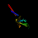
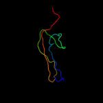
Top model
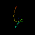
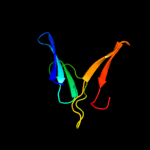
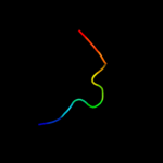
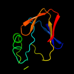
Image coloured by rainbow N → C terminus
Model (left) based on template c3a69A_
Top template information
PDB header:motor protein
Chain: A: PDB Molecule:flagellar hook protein flge;
PDBTitle: atomic model of the bacterial flagellar hook based on2 docking an x-ray derived structure and terminal two alpha-3 helices into an 7.1 angstrom resolution cryoem map
Confidence and coverage
Confidence:100.0% Coverage: 74%
185 residues ( 74% of your sequence) have been modelled with 100.0% confidence by the single highest scoring template.
You may wish to submit your sequence to Phyrealarm. This will automatically scan your sequence every week for new potential templates as they appear in the Phyre2 library.
3D viewing
Interactive 3D view in Jmol

Sequence analysis 

Secondary structure and disorder prediction 

   1........10.........20.........30.........40.........50.........60
Sequence  MDHAIYTAMGAASQTLNQQAVTASNLANASTPGFRAQLNALRAVPVEGLSLPTRTLVTAS
Secondary structure 


SS confidence 


Disorder  ???


?


?

??


????????????
??
Disorder confidence 


   .........70.........80.........90.........100.........110.........120
Sequence  TPGADMTPGKMDYTSRPLDVALQQDGWLAVQTADGSEGYTRNGSIQVDPTGQLTIQGHPV
Secondary structure 


SS confidence 


Disorder  ?????


?
?


?
Disorder confidence 


   .........130.........140.........150.........160.........170.........180
Sequence  IGEAGPIAVPEGAEITIAADGTISALNPGDPANTVAPVGRLKLVKATGSEVQRGDDGIFR
Secondary structure 


SS confidence 


Disorder 


?


????
??


??????????
??


?????????????
Disorder confidence 


   .........190.........200.........210.........220.........230.........240
Sequence  LSAETQATRGPVLQADPTLRVMSGVLEGSNVNAVAAMSDMIASARRFEMQMKVISSVDDN
Secondary structure 


SS confidence 


Disorder  ???????????????????


????


Disorder confidence 


   .........250.
Sequence  AGRANQLLSMS
Secondary structure 


SS confidence 


Disorder 


????
Disorder confidence 


Confidence Key
High(9)                    Low (0)
?Disordered
Alpha helix
Beta strand

Domain analysis 

Hover over an aligned region to see model and summary info

Please note, only up to the top 20 hits are modelled to reduce computer load

RankAligned region

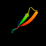
PDB 3a69 chain A

3D model

Region: 4 - 248
Aligned: 185
Modelled: 196
Confidence: 100.0%
Identity: 26%
PDB header:motor protein
Chain: A: PDB Molecule:flagellar hook protein flge;
PDBTitle: atomic model of the bacterial flagellar hook based on2 docking an x-ray derived structure and terminal two alpha-3 helices into an 7.1 angstrom resolution cryoem map

Phyre2

PDB 1wlg chain A

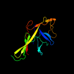
3D model

Region: 78 - 209
Aligned: 130
Modelled: 132
Confidence: 99.9%
Identity: 28%
Fold: Flagellar hook protein flgE
Superfamily: Flagellar hook protein flgE
Family: Flagellar hook protein flgE

Phyre2

PDB 1ucu chain A

3D model

Region: 210 - 249
Aligned: 40
Modelled: 40
Confidence: 91.7%
Identity: 13%
Fold: Phase 1 flagellin
Superfamily: Phase 1 flagellin
Family: Phase 1 flagellin

Phyre2

PDB 1ory chain B

3D model

Region: 210 - 249
Aligned: 40
Modelled: 39
Confidence: 65.5%
Identity: 18%
PDB header:chaperone
Chain: B: PDB Molecule:flagellin;
PDBTitle: flagellar export chaperone in complex with its cognate binding partner

Phyre2

PDB 2czr chain A domain 1

3D model

Region: 83 - 125
Aligned: 43
Modelled: 43
Confidence: 34.6%
Identity: 26%
Fold: Restriction endonuclease-like
Superfamily: TBP-interacting protein-like
Family: TBP-interacting protein-like

Phyre2

PDB 3k8v chain B

3D model

Region: 211 - 231
Aligned: 21
Modelled: 21
Confidence: 31.6%
Identity: 0%
PDB header:structural protein
Chain: B: PDB Molecule:flagellin homolog;
PDBTitle: crysatl structure of a bacterial cell-surface flagellin n20c20

Phyre2

PDB 1b2p chain A

3D model

Region: 74 - 120
Aligned: 47
Modelled: 47
Confidence: 25.3%
Identity: 23%
Fold: beta-Prism II
Superfamily: alpha-D-mannose-specific plant lectins
Family: alpha-D-mannose-specific plant lectins

Phyre2

PDB 2d9s chain A

3D model

Region: 207 - 249
Aligned: 43
Modelled: 43
Confidence: 22.3%
Identity: 14%
PDB header:ligase
Chain: A: PDB Molecule:cbl e3 ubiquitin protein ligase;
PDBTitle: solution structure of rsgi ruh-049, a uba domain from mouse2 cdna

Phyre2

PDB 1kj1 chain D

3D model

Region: 94 - 150
Aligned: 57
Modelled: 57
Confidence: 20.8%
Identity: 12%
Fold: beta-Prism II
Superfamily: alpha-D-mannose-specific plant lectins
Family: alpha-D-mannose-specific plant lectins

Phyre2

PDB 2axw chain A domain 1

3D model

Region: 72 - 130
Aligned: 59
Modelled: 59
Confidence: 18.3%
Identity: 22%
Fold: Common fold of diphtheria toxin/transcription factors/cytochrome f
Superfamily: Bacterial adhesins
Family: Pilus subunits

Phyre2

PDB 1eej chain A domain 2

3D model

Region: 85 - 120
Aligned: 36
Modelled: 36
Confidence: 14.5%
Identity: 22%
Fold: Cystatin-like
Superfamily: DsbC/DsbG N-terminal domain-like
Family: DsbC/DsbG N-terminal domain-like

Phyre2

PDB 3mez chain B

3D model

Region: 75 - 120
Aligned: 46
Modelled: 46
Confidence: 12.9%
Identity: 15%
PDB header:sugar binding protein
Chain: B: PDB Molecule:mannose-specific lectin 3 chain 2;
PDBTitle: x-ray structural analysis of a mannose specific lectin from dutch2 crocus (crocus vernus)

Phyre2

PDB 1lvo chain A

3D model

Region: 25 - 35
Aligned: 11
Modelled: 11
Confidence: 12.3%
Identity: 36%
Fold: Trypsin-like serine proteases
Superfamily: Trypsin-like serine proteases
Family: Viral cysteine protease of trypsin fold

Phyre2

PDB 3afo chain B

3D model

Region: 78 - 163
Aligned: 80
Modelled: 85
Confidence: 12.0%
Identity: 21%
PDB header:transferase
Chain: B: PDB Molecule:nadh kinase pos5;
PDBTitle: crystal structure of yeast nadh kinase complexed with nadh

Phyre2

PDB 2q6f chain B

3D model

Region: 25 - 35
Aligned: 11
Modelled: 11
Confidence: 11.9%
Identity: 45%
PDB header:hydrolase
Chain: B: PDB Molecule:infectious bronchitis virus (ibv) main protease;
PDBTitle: crystal structure of infectious bronchitis virus (ibv) main2 protease in complex with a michael acceptor inhibitor n3

Phyre2

PDB 1t3b chain A domain 2

3D model

Region: 85 - 110
Aligned: 26
Modelled: 26
Confidence: 11.8%
Identity: 15%
Fold: Cystatin-like
Superfamily: DsbC/DsbG N-terminal domain-like
Family: DsbC/DsbG N-terminal domain-like

Phyre2

PDB 2fvn chain A

3D model

Region: 72 - 134
Aligned: 63
Modelled: 63
Confidence: 11.0%
Identity: 21%
PDB header:cell adhesion
Chain: A: PDB Molecule:protein afad;
PDBTitle: the fibrillar tip complex of the afa/dr adhesins from2 pathogen e. coli displays synergistic binding to 5 1 and v3 3 integrins

Phyre2

PDB 2oqk chain A

3D model

Region: 80 - 101
Aligned: 22
Modelled: 22
Confidence: 10.1%
Identity: 18%
PDB header:translation
Chain: A: PDB Molecule:putative translation initiation factor eif-1a;
PDBTitle: crystal structure of putative cryptosporidium parvum translation2 initiation factor eif-1a

Phyre2

PDB 2duc chain A domain 1

3D model

Region: 25 - 35
Aligned: 11
Modelled: 11
Confidence: 10.1%
Identity: 27%
Fold: Trypsin-like serine proteases
Superfamily: Trypsin-like serine proteases
Family: Viral cysteine protease of trypsin fold

Phyre2

PDB 3d23 chain A

3D model

Region: 25 - 35
Aligned: 11
Modelled: 11
Confidence: 9.5%
Identity: 36%
PDB header:hydrolase
Chain: A: PDB Molecule:3c-like proteinase;
PDBTitle: main protease of hcov-hku1

Phyre2
1

c3a69A_
2

d1wlga_
3

d1ucua_
4

c1oryB_
5

d2czra1
6

c3k8vB_
7

d1b2pa_
8

c2d9sA_
9

d1kj1d_
10

d2axwa1
11

d1eeja2
12

c3mezB_
13

d1lvoa_
14

c3afoB_
15

c2q6fB_
16

d1t3ba2
17

c2fvnA_
18

c2oqkA_
19

d2duca1
20

c3d23A_
21


22


23


24


25


26


27


28


29


30


31


32


33


34


35


36


37


38


39


40


41


42


43


44


45


46


47


48


49


50


51


Detailed template information 

#
Template Alignment Coverage3D Model Confidence
% i.d. Template Information
1c3a69A_


100.0 26 PDB header:motor protein
Chain: A: PDB Molecule:flagellar hook protein flge;
PDBTitle: atomic model of the bacterial flagellar hook based on2 docking an x-ray derived structure and terminal two alpha-3 helices into an 7.1 angstrom resolution cryoem map
2d1wlga_


99.9 28 Fold:Flagellar hook protein flgE
Superfamily:Flagellar hook protein flgE
Family:Flagellar hook protein flgE
3d1ucua_


91.7 13 Fold:Phase 1 flagellin
Superfamily:Phase 1 flagellin
Family:Phase 1 flagellin
4c1oryB_


65.5 18 PDB header:chaperone
Chain: B: PDB Molecule:flagellin;
PDBTitle: flagellar export chaperone in complex with its cognate binding partner
5d2czra1


34.6 26 Fold:Restriction endonuclease-like
Superfamily:TBP-interacting protein-like
Family:TBP-interacting protein-like
6c3k8vB_


31.6 0 PDB header:structural protein
Chain: B: PDB Molecule:flagellin homolog;
PDBTitle: crysatl structure of a bacterial cell-surface flagellin n20c20
7d1b2pa_


25.3 23 Fold:beta-Prism II
Superfamily:alpha-D-mannose-specific plant lectins
Family:alpha-D-mannose-specific plant lectins
8c2d9sA_


22.3 14 PDB header:ligase
Chain: A: PDB Molecule:cbl e3 ubiquitin protein ligase;
PDBTitle: solution structure of rsgi ruh-049, a uba domain from mouse2 cdna
9d1kj1d_


20.8 12 Fold:beta-Prism II
Superfamily:alpha-D-mannose-specific plant lectins
Family:alpha-D-mannose-specific plant lectins
10d2axwa1


18.3 22 Fold:Common fold of diphtheria toxin/transcription factors/cytochrome f
Superfamily:Bacterial adhesins
Family:Pilus subunits
11d1eeja2


14.5 22 Fold:Cystatin-like
Superfamily:DsbC/DsbG N-terminal domain-like
Family:DsbC/DsbG N-terminal domain-like
12c3mezB_


12.9 15 PDB header:sugar binding protein
Chain: B: PDB Molecule:mannose-specific lectin 3 chain 2;
PDBTitle: x-ray structural analysis of a mannose specific lectin from dutch2 crocus (crocus vernus)
13d1lvoa_


12.3 36 Fold:Trypsin-like serine proteases
Superfamily:Trypsin-like serine proteases
Family:Viral cysteine protease of trypsin fold
14c3afoB_


12.0 21 PDB header:transferase
Chain: B: PDB Molecule:nadh kinase pos5;
PDBTitle: crystal structure of yeast nadh kinase complexed with nadh
15c2q6fB_


11.9 45 PDB header:hydrolase
Chain: B: PDB Molecule:infectious bronchitis virus (ibv) main protease;
PDBTitle: crystal structure of infectious bronchitis virus (ibv) main2 protease in complex with a michael acceptor inhibitor n3
16d1t3ba2


11.8 15 Fold:Cystatin-like
Superfamily:DsbC/DsbG N-terminal domain-like
Family:DsbC/DsbG N-terminal domain-like
17c2fvnA_


11.0 21 PDB header:cell adhesion
Chain: A: PDB Molecule:protein afad;
PDBTitle: the fibrillar tip complex of the afa/dr adhesins from2 pathogen e. coli displays synergistic binding to 5 1 and v3 3 integrins
18c2oqkA_


10.1 18 PDB header:translation
Chain: A: PDB Molecule:putative translation initiation factor eif-1a;
PDBTitle: crystal structure of putative cryptosporidium parvum translation2 initiation factor eif-1a
19d2duca1


10.1 27 Fold:Trypsin-like serine proteases
Superfamily:Trypsin-like serine proteases
Family:Viral cysteine protease of trypsin fold
20c3d23A_


9.5 36 PDB header:hydrolase
Chain: A: PDB Molecule:3c-like proteinase;
PDBTitle: main protease of hcov-hku1
21d1g73a_


not modelled 9.1 11 Fold:Spectrin repeat-like
Superfamily:Smac/diablo
Family:Smac/diablo
22c2cazF_


not modelled 9.0 17 PDB header:protein transport
Chain: F: PDB Molecule:protein srn2;
PDBTitle: escrt-i core
23c2iyjA_


not modelled 8.8 23 PDB header:isomerase
Chain: A: PDB Molecule:thiol disulfide interchange protein dsbc;
PDBTitle: crystal structure of the n-terminal dimer domain of e.coli2 dsbc
24c3ec1A_


not modelled 8.5 15 PDB header:hydrolase, signaling protein
Chain: A: PDB Molecule:yqeh gtpase;
PDBTitle: structure of yqeh gtpase from geobacillus stearothermophilus2 (an atnos1 / atnoa1 ortholog)
25d1p9sa_


not modelled 8.1 27 Fold:Trypsin-like serine proteases
Superfamily:Trypsin-like serine proteases
Family:Viral cysteine protease of trypsin fold
26c3h2yA_


not modelled 8.1 18 PDB header:hydrolase
Chain: A: PDB Molecule:gtpase family protein;
PDBTitle: crystal structure of yqeh gtpase from bacillus anthracis with dgdp2 bound
27c3lyvF_


not modelled 8.0 19 PDB header:chaperone
Chain: F: PDB Molecule:ribosome-associated factor y;
PDBTitle: crystal structure of a domain of ribosome-associated factor y from2 streptococcus pyogenes serotype m6. northeast structural genomics3 consortium target id dr64a
28d1niga_


not modelled 8.0 29 Fold:Ferritin-like
Superfamily:Cobalamin adenosyltransferase-like
Family:Hypothetical protein Ta1238
29c1jzdA_


not modelled 7.7 26 PDB header:oxidoreductase
Chain: A: PDB Molecule:thiol:disulfide interchange protein dsbc;
PDBTitle: dsbc-dsbdalpha complex
30c2dk7A_


not modelled 7.3 21 PDB header:transcription
Chain: A: PDB Molecule:transcription elongation regulator 1;
PDBTitle: solution structure of ww domain in transcription elongation2 regulator 1
31d1vhoa1


not modelled 7.1 21 Fold:Domain of alpha and beta subunits of F1 ATP synthase-like
Superfamily:Aminopeptidase/glucanase lid domain
Family:Aminopeptidase/glucanase lid domain
32c2dgyA_


not modelled 7.1 18 PDB header:translation
Chain: A: PDB Molecule:mgc11102 protein;
PDBTitle: solution structure of the eukaryotic initiation factor 1a2 in mgc11102 protein
33c3r0eD_


not modelled 6.9 9 PDB header:sugar binding protein
Chain: D: PDB Molecule:lectin;
PDBTitle: structure of remusatia vivipara lectin
34c2kjzA_


not modelled 6.9 18 PDB header:unknown function
Chain: A: PDB Molecule:atc0852;
PDBTitle: solution nmr structure of protein atc0852 from agrobacterium2 tumefaciens. northeast structural genomics consortium (nesg) target3 att2.
35d1fewa_


not modelled 6.9 11 Fold:Spectrin repeat-like
Superfamily:Smac/diablo
Family:Smac/diablo
36c3bqxA_


not modelled 6.8 21 PDB header:structural genomics, unknown function
Chain: A: PDB Molecule:glyoxalase-related enzyme;
PDBTitle: high resolution crystal structure of a glyoxalase-related enzyme from2 fulvimarina pelagi
37d1pn0a1


not modelled 6.6 27 Fold:FAD/NAD(P)-binding domain
Superfamily:FAD/NAD(P)-binding domain
Family:FAD-linked reductases, N-terminal domain
38c2jnhA_


not modelled 6.5 18 PDB header:ligase
Chain: A: PDB Molecule:e3 ubiquitin-protein ligase cbl-b;
PDBTitle: solution structure of the uba domain from cbl-b
39c1yt5A_


not modelled 6.3 20 PDB header:transferase
Chain: A: PDB Molecule:inorganic polyphosphate/atp-nad kinase;
PDBTitle: crystal structure of nad kinase from thermotoga maritima
40c3ohgA_


not modelled 6.1 9 PDB header:structural genomics, unknown function
Chain: A: PDB Molecule:uncharacterized protein from duf2233 family;
PDBTitle: crystal structure of a protein with unknown function from duf22332 family (bacova_00430) from bacteroides ovatus at 1.80 a resolution
41c2an1D_


not modelled 5.9 14 PDB header:transferase
Chain: D: PDB Molecule:putative kinase;
PDBTitle: structural genomics, the crystal structure of a putative kinase from2 salmonella typhimurim lt2
42d1o6wa2


not modelled 5.7 39 Fold:WW domain-like
Superfamily:WW domain
Family:WW domain
43c3jr0B_


not modelled 5.7 38 PDB header:structural protein
Chain: B: PDB Molecule:gene 9 protein;
PDBTitle: crystal structure of phage sf6 gp26 c-terminal knob domain
44d1nrja_


not modelled 5.5 67 Fold:Profilin-like
Superfamily:SNARE-like
Family:SRP alpha N-terminal domain-like
45d1jt8a_


not modelled 5.5 22 Fold:OB-fold
Superfamily:Nucleic acid-binding proteins
Family:Cold shock DNA-binding domain-like
46d1m9sa4


not modelled 5.5 16 Fold:SH3-like barrel
Superfamily:Prokaryotic SH3-related domain
Family:GW domain
47c3bk2A_


not modelled 5.4 26 PDB header:hydrolase
Chain: A: PDB Molecule:metal dependent hydrolase;
PDBTitle: crystal structure analysis of the rnase j/ump complex
48c3kglB_


not modelled 5.2 13 PDB header:plant protein
Chain: B: PDB Molecule:cruciferin;
PDBTitle: crystal structure of procruciferin, 11s globulin from2 brassica napus
49c2dbhA_


not modelled 5.1 43 PDB header:signaling protein
Chain: A: PDB Molecule:tumor necrosis factor receptor superfamily
PDBTitle: solution structure of the carboxyl-terminal card-like2 domain in human tnfr-related death receptor-6
50c2gq1A_


not modelled 5.1 16 PDB header:hydrolase
Chain: A: PDB Molecule:fructose-1,6-bisphosphatase;
PDBTitle: crystal structure of recombinant type i fructose-1,6-bisphosphatase2 from escherichia coli complexed with sulfate ions
51d1d7qa_


not modelled 5.1 18 Fold:OB-fold
Superfamily:Nucleic acid-binding proteins
Family:Cold shock DNA-binding domain-like

Binding site prediction 

Due to computational demand, binding site predictions are not run for batch jobs

If you want to predict binding sites, please manually submit your model of choice to 3DLigandSite


Phyre is for academic use only

Please cite: Protein structure prediction on the web: a case study using the Phyre server
Kelley LA and Sternberg MJE. Nature Protocols 4, 363 - 371 (2009) [pdf] [Import into BibTeX]
 
If you use the binding site predictions from 3DLigandSite, please also cite:
3DLigandSite: predicting ligand-binding sites using similar structures.
Wass MN, Kelley LA and Sternberg MJ Nucleic Acids Research 38, W469-73 (2010) [PubMed]
 
© Structural Bioinformatics Group
Imperial College London
Lawrence Kelley, Benjamin Jefferys
Disclaimer
Terms and Conditions
Component software
Template detection: HHpred 1.51
Secondary structure prediction: Psi-pred 2.5
Disorder prediction: Disopred 2.4
Transmembrane prediction: Memsat_SVM
Multi-template modelling and ab initio: Poing 1.0