| Secondary structure and disorder prediction | |
 |
| | |
1 | . | . | . | . | . | . | . | . | 10 | . | . | . | . | . | . | . | . | . | 20 | . | . | . | . | . | . | . | . | . | 30 | . | . | . | . | . | . | . | . | . | 40 | . | . | . | . | . | . | . | . | . | 50 | . | . | . | . | . | . | . | . | . | 60 |
| Sequence | |
M | L | T | S | F | K | L | H | S | L | K | P | Y | T | L | K | S | S | M | I | L | E | I | I | T | Y | I | L | C | F | F | S | M | I | I | A | F | V | D | N | T | F | S | I | K | I | Y | N | I | T | A | I | V | C | L | L | S | L | I | L |
| Secondary structure | |
|  | | | | | | | |
|
|
|
|
| | | | | | | | | | | | | | | | | | | | | | | |
|
|
| | | | | | | | | | | | | | | | | | | | |
| SS confidence | |
|
|
|
|
|
|
|
|
|
|
|
|
|
|
|
|
|
|
|
|
|
|
|
|
|
|
|
|
|
|
|
|
|
|
|
|
|
|
|
|
|
|
|
|
|
|
|
|
|
|
|
|
|
|
|
|
|
|
|
|
| Disorder | |
? | ? | ? |
|
|
|
|
| ? | ? | ? | ? | ? | ? | ? | ? |
|
|
|
|
|
|
|
|
|
|
|
|
|
|
|
|
|
|
|
|
| ? | ? | ? | ? | ? |
|
|
|
|
|
|
|
|
|
|
|
|
|
|
|
|
|
|
| Disorder confidence | |
|
|
|
|
|
|
|
|
|
|
|
|
|
|
|
|
|
|
|
|
|
|
|
|
|
|
|
|
|
|
|
|
|
|
|
|
|
|
|
|
|
|
|
|
|
|
|
|
|
|
|
|
|
|
|
|
|
|
|
|
| |
| | |
. | . | . | . | . | . | . | . | . | 70 | . | . | . | . | . | . | . | . | . | 80 | . | . | . | . | . | . | . | . | . | 90 | . | . | . | . | . | . | . | . | . | 100 | . | . | . | . | . | . | . | . | . | 110 | . | . | . | . | . | . | . | . | . | 120 |
| Sequence | |
R | G | R | Q | E | N | Y | N | I | K | N | L | I | L | P | L | S | I | F | L | I | G | L | L | D | L | I | W | Y | S | A | F | K | V | D | N | S | P | F | R | A | T | Y | H | S | Y | L | N | T | A | K | I | F | I | F | G | S | F | I | V |
| Secondary structure | |
| | | | | | | | | | | | | | | | | | | | | | | | | | | | | | | |
|
|
|
|
| | | | | | | | | | | | | | | | | | | | | | | |
| SS confidence | |
|
|
|
|
|
|
|
|
|
|
|
|
|
|
|
|
|
|
|
|
|
|
|
|
|
|
|
|
|
|
|
|
|
|
|
|
|
|
|
|
|
|
|
|
|
|
|
|
|
|
|
|
|
|
|
|
|
|
|
|
| Disorder | |
| ? | ? | ? | ? | ? | ? |
| ? | ? | ? |
|
|
|
|
|
|
|
|
|
|
|
|
|
|
|
|
|
|
|
|
| ? | ? | ? | ? | ? |
|
|
|
|
|
|
|
|
|
|
|
|
|
|
|
|
|
|
|
|
|
|
|
| Disorder confidence | |
|
|
|
|
|
|
|
|
|
|
|
|
|
|
|
|
|
|
|
|
|
|
|
|
|
|
|
|
|
|
|
|
|
|
|
|
|
|
|
|
|
|
|
|
|
|
|
|
|
|
|
|
|
|
|
|
|
|
|
|
| |
| | |
. | . | . | . | . | . | . | . | . | 130 | . | . | . | . | . | . | . | . | . | 140 | . | . | . | . | . | . | . | . | . | 150 | . | . | . | . | . | . | . | . | . | 160 | . | . | . | . | . | . | . | . | . | 170 | . | . | . | . | . | . | . | . | . | 180 |
| Sequence | |
F | L | T | L | T | S | Q | L | K | S | K | K | E | S | V | L | Y | T | L | Y | S | L | S | F | L | I | A | G | Y | A | M | Y | I | N | S | I | H | E | N | D | R | I | S | F | G | V | G | T | A | T | G | A | A | Y | S | T | M | L | I | G |
| Secondary structure | |
| | | | | | | | | | | | | | | | | | | | | | | | | | | | | | | | | | | |
|
|
|
|
|
|
|
|
|
|
|
|
| | | | | | | | | | | |
| SS confidence | |
|
|
|
|
|
|
|
|
|
|
|
|
|
|
|
|
|
|
|
|
|
|
|
|
|
|
|
|
|
|
|
|
|
|
|
|
|
|
|
|
|
|
|
|
|
|
|
|
|
|
|
|
|
|
|
|
|
|
|
|
| Disorder | |
|
|
|
|
| ? | ? |
| ? |
|
|
|
|
|
|
|
|
|
|
|
|
|
|
|
|
|
|
|
|
|
|
|
|
|
|
|
| ? |
| ? | ? | ? | ? | ? |
| ? |
|
|
|
|
|
|
|
|
|
|
|
|
|
|
| Disorder confidence | |
|
|
|
|
|
|
|
|
|
|
|
|
|
|
|
|
|
|
|
|
|
|
|
|
|
|
|
|
|
|
|
|
|
|
|
|
|
|
|
|
|
|
|
|
|
|
|
|
|
|
|
|
|
|
|
|
|
|
|
|
| |
| | |
. | . | . | . | . | . | . | . | . | 190 | . | . | . | . | . | . | . | . | . | 200 | . | . | . | . | . | . | . | . | . | 210 | . | . | . | . | . | . | . | . | . | 220 | . | . | . | . | . | . | . | . | . | 230 | . | . | . | . | . | . | . | . | . | 240 |
| Sequence | |
I | V | S | G | V | A | I | L | Y | T | K | K | N | H | P | F | L | F | L | L | N | S | C | A | V | L | Y | V | L | A | L | T | Q | T | R | A | T | L | L | L | F | P | I | I | C | V | A | A | L | I | A | Y | Y | N | K | S | P | K | K | F |
| Secondary structure | |
| | | | | | | | |
|
|
| | | | | | | | | | | | | | | | | | | |
|
|
|
| | | | | | | | | | | | | | | | | | |
|
|
|
|
| | |
| SS confidence | |
|
|
|
|
|
|
|
|
|
|
|
|
|
|
|
|
|
|
|
|
|
|
|
|
|
|
|
|
|
|
|
|
|
|
|
|
|
|
|
|
|
|
|
|
|
|
|
|
|
|
|
|
|
|
|
|
|
|
|
|
| Disorder | |
|
|
|
|
|
|
|
|
| ? |
| ? |
|
|
|
|
|
|
|
|
|
|
|
|
|
|
|
|
|
|
|
|
|
|
|
|
|
|
|
|
|
|
|
|
|
|
|
|
|
|
|
|
| ? | ? | ? | ? | ? |
|
|
| Disorder confidence | |
|
|
|
|
|
|
|
|
|
|
|
|
|
|
|
|
|
|
|
|
|
|
|
|
|
|
|
|
|
|
|
|
|
|
|
|
|
|
|
|
|
|
|
|
|
|
|
|
|
|
|
|
|
|
|
|
|
|
|
|
| |
| | |
. | . | . | . | . | . | . | . | . | 250 | . | . | . | . | . | . | . | . | . | 260 | . | . | . | . | . | . | . | . | . | 270 | . | . | . | . | . | . | . | . | . | 280 | . | . | . | . | . | . | . | . | . | 290 | . | . | . | . | . | . | . | . | . | 300 |
| Sequence | |
T | S | S | I | V | L | L | I | A | I | L | A | S | I | V | I | I | F | N | K | P | I | Q | N | R | Y | N | E | A | L | N | D | L | N | S | Y | T | N | A | N | S | V | T | S | L | G | A | R | L | A | M | Y | E | I | G | L | N | I | F | I |
| Secondary structure | |
| | | | | | | | | | | | | | | | |
|
| | | | | | | | | | | | | | | | | | |
|
|
|
|
|
|
| | | | | | | | | | | | | | | | |
| SS confidence | |
|
|
|
|
|
|
|
|
|
|
|
|
|
|
|
|
|
|
|
|
|
|
|
|
|
|
|
|
|
|
|
|
|
|
|
|
|
|
|
|
|
|
|
|
|
|
|
|
|
|
|
|
|
|
|
|
|
|
|
|
| Disorder | |
|
|
|
|
|
|
|
|
|
|
|
|
|
|
|
|
|
|
|
|
|
|
|
|
|
|
|
|
|
|
|
|
|
|
|
| ? | ? | ? | ? | ? | ? |
|
|
|
|
|
|
|
|
|
|
|
|
|
|
|
|
|
|
| Disorder confidence | |
|
|
|
|
|
|
|
|
|
|
|
|
|
|
|
|
|
|
|
|
|
|
|
|
|
|
|
|
|
|
|
|
|
|
|
|
|
|
|
|
|
|
|
|
|
|
|
|
|
|
|
|
|
|
|
|
|
|
|
|
| |
| | |
. | . | . | . | . | . | . | . | . | 310 | . | . | . | . | . | . | . | . | . | 320 | . | . | . | . | . | . | . | . | . | 330 | . | . | . | . | . | . | . | . | . | 340 | . | . | . | . | . | . | . | . | . | 350 | . | . | . | . | . | . | . | . | . | 360 |
| Sequence | |
K | S | P | F | S | F | R | S | A | E | S | R | A | E | S | M | N | L | L | V | A | E | H | N | R | L | R | G | A | L | E | F | S | N | V | H | L | H | N | E | I | I | E | A | G | S | L | K | G | L | M | G | I | F | S | T | L | F | L | Y |
| Secondary structure | |
|
|
|
|  | |  |
|
|
|
| | | | | | | | | | |
|
|
|
|
|
|
|
|
|
|
|
|
|
|
|
| | | | | | | | | | |
|
| | | | | | | | | | | |
| SS confidence | |
|
|
|
|
|
|
|
|
|
|
|
|
|
|
|
|
|
|
|
|
|
|
|
|
|
|
|
|
|
|
|
|
|
|
|
|
|
|
|
|
|
|
|
|
|
|
|
|
|
|
|
|
|
|
|
|
|
|
|
|
| Disorder | |
|
|
|
|
|
|
|
|
|
|
|
|
|
|
|
|
|
|
|
|
| ? |
|
|
| ? |
|
|
|
| ? | ? |
|
|
|
|
|
|
|
|
|
|
|
|
|
|
|
|
|
|
|
|
|
|
|
|
|
|
|
|
| Disorder confidence | |
|
|
|
|
|
|
|
|
|
|
|
|
|
|
|
|
|
|
|
|
|
|
|
|
|
|
|
|
|
|
|
|
|
|
|
|
|
|
|
|
|
|
|
|
|
|
|
|
|
|
|
|
|
|
|
|
|
|
|
|
| |
| | |
. | . | . | . | . | . | . | . | . | 370 | . | . | . | . | . | . | . | . | . | 380 | . | . | . | . | . | . | . | . | . | 390 | . | . | . | . | . | . | . | . | . | 400 | . | . | . | . | . | . | . | . | . | 410 | . | . | . | . | . | . | . | . | . |
| Sequence | |
F | S | L | F | Y | I | A | Y | K | K | R | A | L | G | L | L | I | L | T | L | G | I | V | G | I | G | L | S | D | V | I | I | W | A | R | S | I | P | I | I | I | I | S | A | I | V | L | L | L | V | I | N | N | R | N | N | T | I | N |
| Secondary structure | |
| | | | | | | |
|
|
|
|
| | | | | | | | | | | | | | | | | | | |
|
|
|
| | | | | | | | | | | | | | | |
|
|
|
|
|
|
|
|
| SS confidence | |
|
|
|
|
|
|
|
|
|
|
|
|
|
|
|
|
|
|
|
|
|
|
|
|
|
|
|
|
|
|
|
|
|
|
|
|
|
|
|
|
|
|
|
|
|
|
|
|
|
|
|
|
|
|
|
|
|
|
|
| Disorder | |
|
|
|
|
|
|
|
|
|
|
|
|
|
|
|
|
|
|
|
|
|
|
|
|
|
|
|
|
|
|
|
|
|
|
|
|
|
|
|
|
|
|
|
|
|
|
|
|
|
|
|
| ? | ? | ? | ? | ? | ? | ? |
| Disorder confidence | |
|
|
|
|
|
|
|
|
|
|
|
|
|
|
|
|
|
|
|
|
|
|
|
|
|
|
|
|
|
|
|
|
|
|
|
|
|
|
|
|
|
|
|
|
|
|
|
|
|
|
|
|
|
|
|
|
|
|
|
| |
| Confidence Key |
| High(9) | |
|
|
|
|
|
|
|
|
|
Low (0) |
| ? | Disordered |
| Alpha helix |
| Beta strand |
Hover over an aligned region to see model and summary info
Please note, only up to the top 20 hits are modelled to reduce computer load
|
| 1 |
|
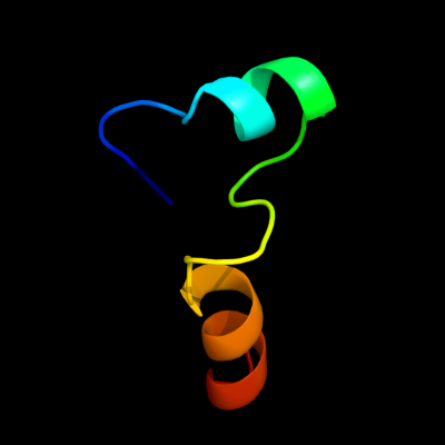
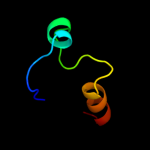
PDB 2d07 chain A
Region: 304 - 350
Aligned: 33
Modelled: 33
Confidence: 9.3%
Identity: 15%
PDB header:hydrolase
Chain: A: PDB Molecule:g/t mismatch-specific thymine dna glycosylase;
PDBTitle: crystal structure of sumo-3-modified thymine-dna glycosylase
Phyre2
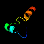
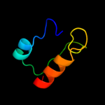
| 2 |
|
PDB 1mug chain A
Region: 304 - 350
Aligned: 33
Modelled: 33
Confidence: 8.9%
Identity: 12%
Fold: Uracil-DNA glycosylase-like
Superfamily: Uracil-DNA glycosylase-like
Family: Mug-like
Phyre2
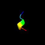
| 3 |
|
PDB 1a92 chain B
Region: 301 - 306
Aligned: 6
Modelled: 6
Confidence: 8.4%
Identity: 17%
PDB header:leucine zipper
Chain: B: PDB Molecule:delta antigen;
PDBTitle: oligomerization domain of hepatitis delta antigen
Phyre2
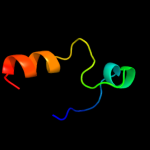
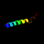
| 4 |
|
PDB 2rba chain B
Region: 304 - 350
Aligned: 33
Modelled: 33
Confidence: 7.6%
Identity: 12%
PDB header:hydrolase/dna
Chain: B: PDB Molecule:g/t mismatch-specific thymine dna glycosylase;
PDBTitle: structure of human thymine dna glycosylase bound to abasic and2 undamaged dna
Phyre2
| 5 |
|
PDB 1wra chain A domain 1
Region: 306 - 350
Aligned: 45
Modelled: 45
Confidence: 6.8%
Identity: 7%
Fold: Metallo-hydrolase/oxidoreductase
Superfamily: Metallo-hydrolase/oxidoreductase
Family: Pce catalytic domain-like
Phyre2
| 6 |
|
PDB 2knc chain A
Region: 344 - 373
Aligned: 30
Modelled: 30
Confidence: 6.4%
Identity: 17%
PDB header:cell adhesion
Chain: A: PDB Molecule:integrin alpha-iib;
PDBTitle: platelet integrin alfaiib-beta3 transmembrane-cytoplasmic2 heterocomplex
Phyre2
|
| Detailed template information | |
Due to computational demand, binding site predictions are not run for batch jobs
If you want to predict binding sites, please manually submit your model of choice to 3DLigandSite
Phyre is for academic use only
| Please cite: Protein structure prediction on
the web: a case study using the Phyre server |
| Kelley LA and Sternberg MJE. Nature Protocols
4, 363 - 371 (2009) [pdf] [Import into BibTeX] |
| |
| If you use the binding site
predictions from 3DLigandSite, please also cite: |
| 3DLigandSite: predicting ligand-binding sites using similar structures. |
| Wass MN, Kelley LA and Sternberg
MJ Nucleic Acids Research 38, W469-73 (2010) [PubMed] |
| |
|
|
|
|