Phyre Home Retrieve Phyre Job Id
Emaill.a.kelley@imperial.ac.uk
DescriptionP76544
DateThu Jan 5 12:24:19 GMT 2012
Unique Job ID7cb9a2a81d2c79ec

Summary 

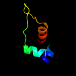
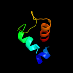
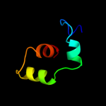
Top model
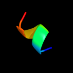
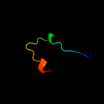
Image coloured by rainbow N → C terminus
Model (left) based on template d2csfa1
Top template information
Fold:lambda repressor-like DNA-binding domains
Superfamily:lambda repressor-like DNA-binding domains
Family:CUT domain
Confidence and coverage
Confidence: 65.6% Coverage: 44%
36 residues ( 44% of your sequence) have been modelled with 65.6% confidence by the single highest scoring template.
You may wish to submit your sequence to Phyrealarm. This will automatically scan your sequence every week for new potential templates as they appear in the Phyre2 library.
Please note: You must be registered and logged in to use Phyrealarm.
3D viewing
Interactive 3D view in Jmol

Sequence analysis 

Secondary structure and disorder prediction 

   1........10.........20.........30.........40.........50.........60
Sequence  MMVIRHECPSYCIAQKRVALREFSELVLGTLSLLLEQKTNGKCSASLYDCSEEEKLFVKR
Secondary structure 


SS confidence 


Disorder  ?


?


?
??????????????


Disorder confidence 


   .........70.........80.
Sequence  LKLIKADIHAQLKACDCDISE
Secondary structure 


SS confidence 


Disorder 


???????
Disorder confidence 


Confidence Key
High(9)                    Low (0)
?Disordered
Alpha helix
Beta strand

Domain analysis 

Hover over an aligned region to see model and summary info

Please note, only up to the top 20 hits are modelled to reduce computer load

RankAligned region

PDB 2csf chain A domain 1

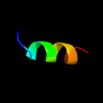
3D model

Region: 21 - 62
Aligned: 36
Modelled: 42
Confidence: 65.6%
Identity: 19%
Fold: lambda repressor-like DNA-binding domains
Superfamily: lambda repressor-like DNA-binding domains
Family: CUT domain

Phyre2

PDB 1wiz chain A

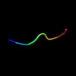
3D model

Region: 21 - 62
Aligned: 36
Modelled: 42
Confidence: 41.7%
Identity: 17%
Fold: lambda repressor-like DNA-binding domains
Superfamily: lambda repressor-like DNA-binding domains
Family: CUT domain

Phyre2

PDB 2o4a chain A domain 1

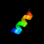
3D model

Region: 21 - 62
Aligned: 36
Modelled: 42
Confidence: 32.6%
Identity: 17%
Fold: lambda repressor-like DNA-binding domains
Superfamily: lambda repressor-like DNA-binding domains
Family: CUT domain

Phyre2

PDB 2w9y chain A

3D model

Region: 19 - 74
Aligned: 51
Modelled: 51
Confidence: 32.4%
Identity: 18%
PDB header:lipid transport
Chain: A: PDB Molecule:fatty acid/retinol binding protein protein 7,
PDBTitle: the structure of the lipid binding protein ce-far-7 from2 caenorhabditis elegans

Phyre2

PDB 1yse chain A

3D model

Region: 21 - 62
Aligned: 36
Modelled: 42
Confidence: 26.7%
Identity: 17%
PDB header:dna binding protein
Chain: A: PDB Molecule:dna-binding protein satb1;
PDBTitle: solution structure of the mar-binding domain of satb1

Phyre2

PDB 1x2l chain A domain 1

3D model

Region: 21 - 37
Aligned: 17
Modelled: 17
Confidence: 22.6%
Identity: 47%
Fold: lambda repressor-like DNA-binding domains
Superfamily: lambda repressor-like DNA-binding domains
Family: CUT domain

Phyre2

PDB 1wh8 chain A

3D model

Region: 21 - 37
Aligned: 17
Modelled: 17
Confidence: 17.5%
Identity: 53%
Fold: lambda repressor-like DNA-binding domains
Superfamily: lambda repressor-like DNA-binding domains
Family: CUT domain

Phyre2

PDB 1s7e chain A domain 2

3D model

Region: 21 - 37
Aligned: 17
Modelled: 17
Confidence: 15.1%
Identity: 53%
Fold: lambda repressor-like DNA-binding domains
Superfamily: lambda repressor-like DNA-binding domains
Family: CUT domain

Phyre2

PDB 2eo2 chain A

3D model

Region: 21 - 61
Aligned: 35
Modelled: 41
Confidence: 13.7%
Identity: 43%
PDB header:oxidoreductase
Chain: A: PDB Molecule:adult male hypothalamus cdna, riken full-length
PDBTitle: solution structure of the insertion region (510-573) of2 fthfs domain from mouse methylenetetrahydrofolate3 dehydrogenase (nadp+ dependent) 1-like protein

Phyre2

PDB 1wh6 chain A

3D model

Region: 21 - 62
Aligned: 36
Modelled: 42
Confidence: 12.4%
Identity: 28%
Fold: lambda repressor-like DNA-binding domains
Superfamily: lambda repressor-like DNA-binding domains
Family: CUT domain

Phyre2

PDB 1yqy chain A

3D model

Region: 46 - 63
Aligned: 18
Modelled: 18
Confidence: 11.2%
Identity: 56%
PDB header:hydrolase
Chain: A: PDB Molecule:lethal factor;
PDBTitle: structure of b. anthrax lethal factor in complex with a2 hydroxamate inhibitor

Phyre2

PDB 1sp7 chain A

3D model

Region: 8 - 17
Aligned: 10
Modelled: 10
Confidence: 11.0%
Identity: 60%
PDB header:structural protein
Chain: A: PDB Molecule:mini-collagen;
PDBTitle: structure of the cys-rich c-terminal domain of hydra2 minicollagen

Phyre2

PDB 3si5 chain X

3D model

Region: 56 - 62
Aligned: 7
Modelled: 7
Confidence: 10.6%
Identity: 71%
PDB header:cell cycle
Chain: X: PDB Molecule:protein casc5;
PDBTitle: kinetochore-bubr1 kinase complex

Phyre2

PDB 3si5 chain Y

3D model

Region: 56 - 62
Aligned: 7
Modelled: 7
Confidence: 10.4%
Identity: 71%
PDB header:cell cycle
Chain: Y: PDB Molecule:protein casc5;
PDBTitle: kinetochore-bubr1 kinase complex

Phyre2

PDB 1j7n chain A domain 3

3D model

Region: 51 - 63
Aligned: 13
Modelled: 13
Confidence: 10.1%
Identity: 69%
Fold: ADP-ribosylation
Superfamily: ADP-ribosylation
Family: ADP-ribosylating toxins

Phyre2

PDB 1axi chain B domain 1

3D model

Region: 5 - 11
Aligned: 7
Modelled: 7
Confidence: 9.3%
Identity: 57%
Fold: Immunoglobulin-like beta-sandwich
Superfamily: Fibronectin type III
Family: Fibronectin type III

Phyre2

PDB 2dt7 chain A

3D model

Region: 53 - 65
Aligned: 13
Modelled: 13
Confidence: 8.7%
Identity: 54%
PDB header:rna binding protein
Chain: A: PDB Molecule:splicing factor 3a subunit 3;
PDBTitle: solution structure of the second surp domain of human2 splicing factor sf3a120 in complex with a fragment of3 human splicing factor sf3a60

Phyre2

PDB 2wj8 chain N

3D model

Region: 38 - 59
Aligned: 22
Modelled: 22
Confidence: 7.4%
Identity: 36%
PDB header:rna binding protein/rna
Chain: N: PDB Molecule:nucleoprotein;
PDBTitle: respiratory syncitial virus ribonucleoprotein

Phyre2

PDB 1u2u chain B

3D model

Region: 11 - 21
Aligned: 11
Modelled: 11
Confidence: 7.3%
Identity: 55%
PDB header:transcription
Chain: B: PDB Molecule:general control protein gcn4;
PDBTitle: nmr solution structure of a designed heterodimeric leucine2 zipper

Phyre2

PDB 2w1o chain A

3D model

Region: 65 - 81
Aligned: 17
Modelled: 17
Confidence: 6.8%
Identity: 18%
PDB header:translation
Chain: A: PDB Molecule:60s acidic ribosomal protein p2;
PDBTitle: nmr structure of dimerization domain of human ribosomal2 protein p2

Phyre2
1

d2csfa1
2

d1wiza_
3

d2o4aa1
4

c2w9yA_
5

c1yseA_
6

d1x2la1
7

d1wh8a_
8

d1s7ea2
9

c2eo2A_
10

d1wh6a_
11

c1yqyA_
12

c1sp7A_
13

c3si5X_
14

c3si5Y_
15

d1j7na3
16

d1axib1
17

c2dt7A_
18

c2wj8N_
19

c1u2uB_
20

c2w1oA_
21


22


23


24


25


26


27


28


29


30


Detailed template information 

#
Template Alignment Coverage3D Model Confidence
% i.d. Template Information
1d2csfa1


65.6 19 Fold:lambda repressor-like DNA-binding domains
Superfamily:lambda repressor-like DNA-binding domains
Family:CUT domain
2d1wiza_


41.7 17 Fold:lambda repressor-like DNA-binding domains
Superfamily:lambda repressor-like DNA-binding domains
Family:CUT domain
3d2o4aa1


32.6 17 Fold:lambda repressor-like DNA-binding domains
Superfamily:lambda repressor-like DNA-binding domains
Family:CUT domain
4c2w9yA_


32.4 18 PDB header:lipid transport
Chain: A: PDB Molecule:fatty acid/retinol binding protein protein 7,
PDBTitle: the structure of the lipid binding protein ce-far-7 from2 caenorhabditis elegans
5c1yseA_


26.7 17 PDB header:dna binding protein
Chain: A: PDB Molecule:dna-binding protein satb1;
PDBTitle: solution structure of the mar-binding domain of satb1
6d1x2la1


22.6 47 Fold:lambda repressor-like DNA-binding domains
Superfamily:lambda repressor-like DNA-binding domains
Family:CUT domain
7d1wh8a_


17.5 53 Fold:lambda repressor-like DNA-binding domains
Superfamily:lambda repressor-like DNA-binding domains
Family:CUT domain
8d1s7ea2


15.1 53 Fold:lambda repressor-like DNA-binding domains
Superfamily:lambda repressor-like DNA-binding domains
Family:CUT domain
9c2eo2A_


13.7 43 PDB header:oxidoreductase
Chain: A: PDB Molecule:adult male hypothalamus cdna, riken full-length
PDBTitle: solution structure of the insertion region (510-573) of2 fthfs domain from mouse methylenetetrahydrofolate3 dehydrogenase (nadp+ dependent) 1-like protein
10d1wh6a_


12.4 28 Fold:lambda repressor-like DNA-binding domains
Superfamily:lambda repressor-like DNA-binding domains
Family:CUT domain
11c1yqyA_


11.2 56 PDB header:hydrolase
Chain: A: PDB Molecule:lethal factor;
PDBTitle: structure of b. anthrax lethal factor in complex with a2 hydroxamate inhibitor
12c1sp7A_


11.0 60 PDB header:structural protein
Chain: A: PDB Molecule:mini-collagen;
PDBTitle: structure of the cys-rich c-terminal domain of hydra2 minicollagen
13c3si5X_


10.6 71 PDB header:cell cycle
Chain: X: PDB Molecule:protein casc5;
PDBTitle: kinetochore-bubr1 kinase complex
14c3si5Y_


10.4 71 PDB header:cell cycle
Chain: Y: PDB Molecule:protein casc5;
PDBTitle: kinetochore-bubr1 kinase complex
15d1j7na3


10.1 69 Fold:ADP-ribosylation
Superfamily:ADP-ribosylation
Family:ADP-ribosylating toxins
16d1axib1


9.3 57 Fold:Immunoglobulin-like beta-sandwich
Superfamily:Fibronectin type III
Family:Fibronectin type III
17c2dt7A_


8.7 54 PDB header:rna binding protein
Chain: A: PDB Molecule:splicing factor 3a subunit 3;
PDBTitle: solution structure of the second surp domain of human2 splicing factor sf3a120 in complex with a fragment of3 human splicing factor sf3a60
18c2wj8N_


7.4 36 PDB header:rna binding protein/rna
Chain: N: PDB Molecule:nucleoprotein;
PDBTitle: respiratory syncitial virus ribonucleoprotein
19c1u2uB_


7.3 55 PDB header:transcription
Chain: B: PDB Molecule:general control protein gcn4;
PDBTitle: nmr solution structure of a designed heterodimeric leucine2 zipper
20c2w1oA_


6.8 18 PDB header:translation
Chain: A: PDB Molecule:60s acidic ribosomal protein p2;
PDBTitle: nmr structure of dimerization domain of human ribosomal2 protein p2
21d1xb4a1


not modelled 6.0 8 Fold:DNA/RNA-binding 3-helical bundle
Superfamily:"Winged helix" DNA-binding domain
Family:Vacuolar sorting protein domain
22c2d9sA_


not modelled 6.0 50 PDB header:ligase
Chain: A: PDB Molecule:cbl e3 ubiquitin protein ligase;
PDBTitle: solution structure of rsgi ruh-049, a uba domain from mouse2 cdna
23c2zmeD_


not modelled 5.7 31 PDB header:protein transport
Chain: D: PDB Molecule:vacuolar protein-sorting-associated protein 25;
PDBTitle: integrated structural and functional model of the human escrt-ii2 complex
24c3cuqD_


not modelled 5.7 31 PDB header:protein transport
Chain: D: PDB Molecule:vacuolar protein-sorting-associated protein 25;
PDBTitle: integrated structural and functional model of the human escrt-ii2 complex
25c2hbpA_


not modelled 5.7 17 PDB header:endocytosis, protein binding
Chain: A: PDB Molecule:cytoskeleton assembly control protein sla1;
PDBTitle: solution structure of sla1 homology domain 1
26c2fphX_


not modelled 5.7 27 PDB header:dna binding protein
Chain: X: PDB Molecule:ylmh;
PDBTitle: cell division protein ylmh from streptococcus pneumoniae
27c1xb4C_


not modelled 5.5 8 PDB header:unknown function
Chain: C: PDB Molecule:hypothetical 23.6 kda protein in yuh1-ura8
PDBTitle: crystal structure of subunit vps25 of the endosomal2 trafficking complex escrt-ii
28c3iz5w_


not modelled 5.5 27 PDB header:ribosome
Chain: W: PDB Molecule:60s ribosomal protein l22 (l22e);
PDBTitle: localization of the large subunit ribosomal proteins into a 5.5 a2 cryo-em map of triticum aestivum translating 80s ribosome
29c3izcw_


not modelled 5.2 24 PDB header:ribosome
Chain: W: PDB Molecule:60s ribosomal protein rpl22 (l22e);
PDBTitle: localization of the large subunit ribosomal proteins into a 6.1 a2 cryo-em map of saccharomyces cerevisiae translating 80s ribosome
30c3eq5G_


not modelled 5.2 23 PDB header:signaling protein
Chain: G: PDB Molecule:ski-like protein;
PDBTitle: crystal structure of fragment 137 to 238 of the human ski-like protein

Binding site prediction 

Due to computational demand, binding site predictions are not run for batch jobs

If you want to predict binding sites, please manually submit your model of choice to 3DLigandSite


Phyre is for academic use only

Please cite: Protein structure prediction on the web: a case study using the Phyre server
Kelley LA and Sternberg MJE. Nature Protocols 4, 363 - 371 (2009) [pdf] [Import into BibTeX]
 
If you use the binding site predictions from 3DLigandSite, please also cite:
3DLigandSite: predicting ligand-binding sites using similar structures.
Wass MN, Kelley LA and Sternberg MJ Nucleic Acids Research 38, W469-73 (2010) [PubMed]
 
© Structural Bioinformatics Group
Imperial College London
Lawrence Kelley, Benjamin Jefferys
Disclaimer
Terms and Conditions
Component software
Template detection: HHpred 1.51
Secondary structure prediction: Psi-pred 2.5
Disorder prediction: Disopred 2.4
Transmembrane prediction: Memsat_SVM
Multi-template modelling and ab initio: Poing 1.0