| 1 |
|
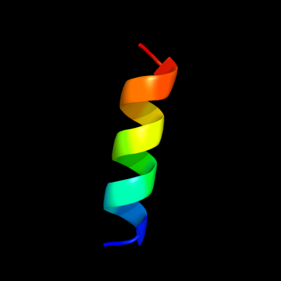
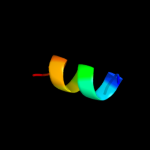
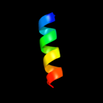
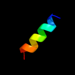
PDB 1eys chain H domain 2
Region: 21 - 35
Aligned: 15
Modelled: 15
Confidence: 22.1%
Identity: 27%
Fold: Single transmembrane helix
Superfamily: Photosystem II reaction centre subunit H, transmembrane region
Family: Photosystem II reaction centre subunit H, transmembrane region
Phyre2
| 2 |
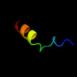
|
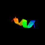
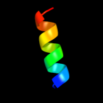
PDB 3qq5 chain A
Region: 21 - 33
Aligned: 13
Modelled: 13
Confidence: 21.8%
Identity: 31%
PDB header:oxidoreductase
Chain: A: PDB Molecule:small gtp-binding protein;
PDBTitle: crystal structure of the [fefe]-hydrogenase maturation protein hydf
Phyre2
| 3 |
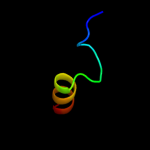
|
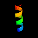
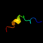
PDB 1v54 chain L
Region: 19 - 48
Aligned: 30
Modelled: 30
Confidence: 18.4%
Identity: 27%
Fold: Single transmembrane helix
Superfamily: Mitochondrial cytochrome c oxidase subunit VIIc (aka VIIIa)
Family: Mitochondrial cytochrome c oxidase subunit VIIc (aka VIIIa)
Phyre2
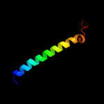
| 4 |
|
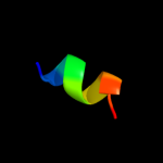
PDB 1rzh chain H domain 2
Region: 25 - 35
Aligned: 11
Modelled: 11
Confidence: 18.1%
Identity: 45%
Fold: Single transmembrane helix
Superfamily: Photosystem II reaction centre subunit H, transmembrane region
Family: Photosystem II reaction centre subunit H, transmembrane region
Phyre2
| 5 |
|
PDB 1l9b chain H domain 2
Region: 25 - 35
Aligned: 11
Modelled: 11
Confidence: 16.5%
Identity: 45%
Fold: Single transmembrane helix
Superfamily: Photosystem II reaction centre subunit H, transmembrane region
Family: Photosystem II reaction centre subunit H, transmembrane region
Phyre2
| 6 |
|
PDB 2rcr chain H domain 2
Region: 25 - 35
Aligned: 11
Modelled: 11
Confidence: 13.1%
Identity: 45%
Fold: Single transmembrane helix
Superfamily: Photosystem II reaction centre subunit H, transmembrane region
Family: Photosystem II reaction centre subunit H, transmembrane region
Phyre2
| 7 |
|
PDB 1adu chain B
Region: 25 - 38
Aligned: 14
Modelled: 14
Confidence: 11.5%
Identity: 29%
PDB header:dna-binding protein
Chain: B: PDB Molecule:adenovirus single-stranded dna-binding protein;
PDBTitle: early e2a dna-binding protein
Phyre2
| 8 |
|
PDB 1afo chain B
Region: 20 - 28
Aligned: 9
Modelled: 9
Confidence: 8.1%
Identity: 44%
PDB header:integral membrane protein
Chain: B: PDB Molecule:glycophorin a;
PDBTitle: dimeric transmembrane domain of human glycophorin a, nmr,2 20 structures
Phyre2
| 9 |
|
PDB 1k6n chain H
Region: 21 - 35
Aligned: 15
Modelled: 15
Confidence: 7.9%
Identity: 33%
PDB header:photosynthesis
Chain: H: PDB Molecule:photosynthetic reaction center h subunit;
PDBTitle: e(l212)a,d(l213)a double mutant structure of photosynthetic reaction2 center from rhodobacter sphaeroides
Phyre2
| 10 |
|
PDB 1p2z chain A domain 1
Region: 38 - 56
Aligned: 19
Modelled: 19
Confidence: 7.5%
Identity: 26%
Fold: Nucleoplasmin-like/VP (viral coat and capsid proteins)
Superfamily: Group II dsDNA viruses VP
Family: Adenovirus hexon
Phyre2
| 11 |
|
PDB 2klu chain A
Region: 15 - 30
Aligned: 16
Modelled: 16
Confidence: 6.9%
Identity: 25%
PDB header:immune system, membrane protein
Chain: A: PDB Molecule:t-cell surface glycoprotein cd4;
PDBTitle: nmr structure of the transmembrane and cytoplasmic domains2 of human cd4
Phyre2
| 12 |
|
PDB 1p30 chain A domain 1
Region: 38 - 56
Aligned: 19
Modelled: 19
Confidence: 6.9%
Identity: 26%
Fold: Nucleoplasmin-like/VP (viral coat and capsid proteins)
Superfamily: Group II dsDNA viruses VP
Family: Adenovirus hexon
Phyre2
| 13 |
|
PDB 3o0r chain C
Region: 13 - 54
Aligned: 42
Modelled: 42
Confidence: 6.8%
Identity: 14%
PDB header:immune system/oxidoreductase
Chain: C: PDB Molecule:nitric oxide reductase subunit c;
PDBTitle: crystal structure of nitric oxide reductase from pseudomonas2 aeruginosa in complex with antibody fragment
Phyre2
| 14 |
|
PDB 1eys chain H
Region: 21 - 35
Aligned: 15
Modelled: 15
Confidence: 6.6%
Identity: 27%
PDB header:electron transport
Chain: H: PDB Molecule:photosynthetic reaction center;
PDBTitle: crystal structure of photosynthetic reaction center from a2 thermophilic bacterium, thermochromatium tepidum
Phyre2
| 15 |
|
PDB 1adt chain A domain 1
Region: 25 - 38
Aligned: 14
Modelled: 14
Confidence: 6.6%
Identity: 29%
Fold: Domain of early E2A DNA-binding protein, ADDBP
Superfamily: Domain of early E2A DNA-binding protein, ADDBP
Family: Domain of early E2A DNA-binding protein, ADDBP
Phyre2
| 16 |
|
PDB 1l4t chain A
Region: 24 - 30
Aligned: 7
Modelled: 7
Confidence: 5.8%
Identity: 57%
PDB header:hormone/growth factor receptor
Chain: A: PDB Molecule:gastrin/cholecystokinin type b receptor;
PDBTitle: solution nmr structure of the cck2e3
Phyre2
| 17 |
|
PDB 2fef chain B
Region: 20 - 32
Aligned: 13
Modelled: 13
Confidence: 5.5%
Identity: 8%
PDB header:structural genomics, unknown function
Chain: B: PDB Molecule:hypothetical protein pa2201;
PDBTitle: the crystal structure of protein pa2201 from pseudomonas aeruginosa
Phyre2
| 18 |
|
PDB 2iny chain A
Region: 38 - 56
Aligned: 19
Modelled: 19
Confidence: 5.4%
Identity: 26%
PDB header:viral protein
Chain: A: PDB Molecule:hexon protein;
PDBTitle: nanoporous crystals of chicken embryo lethal orphan (celo) adenovirus2 major coat protein, hexon
Phyre2