Phyre Home Retrieve Phyre Job Id
Emaill.a.kelley@imperial.ac.uk
DescriptionP77162
DateThu Jan 5 12:25:48 GMT 2012
Unique Job ID79685878889f15f9

Summary 

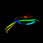
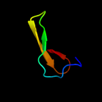
Top model
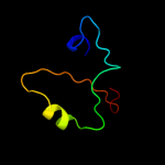
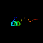
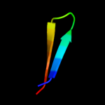
Image coloured by rainbow N → C terminus
Model (left) based on template d1ylxa1
Top template information
Fold:N domain of copper amine oxidase-like
Superfamily:GK1464-like
Family:GK1464-like
Confidence and coverage
Confidence: 63.6% Coverage: 31%
48 residues ( 31% of your sequence) have been modelled with 63.6% confidence by the single highest scoring template.
You may wish to submit your sequence to Phyrealarm. This will automatically scan your sequence every week for new potential templates as they appear in the Phyre2 library.
Please note: You must be registered and logged in to use Phyrealarm.
3D viewing
Interactive 3D view in Jmol

Sequence analysis 

Secondary structure and disorder prediction 

   1........10.........20.........30.........40.........50.........60
Sequence  MTILSLSRFMLAGVLLASFNASAIPGFWQQGYGQGNTEYSVTEASGKTFTINCTGNPDQN
Secondary structure 


SS confidence 


Disorder  ???????


??????????

?


?
Disorder confidence 


   .........70.........80.........90.........100.........110.........120
Sequence  GFYQHSVFLTLADDKMVSSHDDDTTITVVMDHQQYIIPSSLGWRNGDNAWFDFISNISEA
Secondary structure 


SS confidence 


Disorder 


?
?


????


Disorder confidence 


   .........130.........140.........150.....
Sequence  GQFDVYVNDHKAGTFTADRKNAEKVLSTLGDCSND
Secondary structure 


SS confidence 


Disorder 


??
???


???????
Disorder confidence 


Confidence Key
High(9)                    Low (0)
?Disordered
Alpha helix
Beta strand

Domain analysis 

Hover over an aligned region to see model and summary info

Please note, only up to the top 20 hits are modelled to reduce computer load

RankAligned region

PDB 1ylx chain A domain 1

3D model

Region: 44 - 94
Aligned: 48
Modelled: 51
Confidence: 63.6%
Identity: 19%
Fold: N domain of copper amine oxidase-like
Superfamily: GK1464-like
Family: GK1464-like

Phyre2

PDB 2eg9 chain B

3D model

Region: 102 - 153
Aligned: 52
Modelled: 52
Confidence: 26.4%
Identity: 21%
PDB header:hydrolase
Chain: B: PDB Molecule:adp-ribosyl cyclase 1;
PDBTitle: crystal structure of the truncated extracellular domain of2 mouse cd38

Phyre2

PDB 1uzv chain A

3D model

Region: 122 - 136
Aligned: 15
Modelled: 15
Confidence: 25.5%
Identity: 40%
Fold: Calcium-mediated lectin
Superfamily: Calcium-mediated lectin
Family: Calcium-mediated lectin

Phyre2

PDB 1wmx chain B

3D model

Region: 33 - 134
Aligned: 92
Modelled: 92
Confidence: 21.6%
Identity: 18%
Fold: Galactose-binding domain-like
Superfamily: Galactose-binding domain-like
Family: Family 30 carbohydrate binding module, CBM30 (PKD repeat)

Phyre2

PDB 3eit chain B

3D model

Region: 86 - 134
Aligned: 48
Modelled: 49
Confidence: 20.0%
Identity: 17%
PDB header:unknown function
Chain: B: PDB Molecule:putative atp/gtp binding protein;
PDBTitle: the 2.6 angstrom crystal structure of chbp, the cif homologue from2 burkholderia pseudomallei

Phyre2

PDB 2chh chain A domain 1

3D model

Region: 122 - 136
Aligned: 15
Modelled: 15
Confidence: 18.9%
Identity: 33%
Fold: Calcium-mediated lectin
Superfamily: Calcium-mediated lectin
Family: Calcium-mediated lectin

Phyre2

PDB 3hbk chain A

3D model

Region: 37 - 138
Aligned: 102
Modelled: 102
Confidence: 18.6%
Identity: 17%
PDB header:hydrolase
Chain: A: PDB Molecule:putative glycosyl hydrolase;
PDBTitle: crystal structure of putative glycosyl hydrolase, was domain of2 unknown function (duf1080) (yp_001302580.1) from parabacteroides3 distasonis atcc 8503 at 2.36 a resolution

Phyre2

PDB 1r4x chain A domain 2

3D model

Region: 104 - 149
Aligned: 38
Modelled: 46
Confidence: 17.4%
Identity: 16%
Fold: Subdomain of clathrin and coatomer appendage domain
Superfamily: Subdomain of clathrin and coatomer appendage domain
Family: Coatomer appendage domain

Phyre2

PDB 2khg chain A

3D model

Region: 109 - 120
Aligned: 12
Modelled: 12
Confidence: 16.8%
Identity: 33%
PDB header:antimicrobial protein
Chain: A: PDB Molecule:plnj;
PDBTitle: plantaricin j in tfe

Phyre2

PDB 2anu chain A

3D model

Region: 106 - 130
Aligned: 25
Modelled: 25
Confidence: 16.6%
Identity: 8%
PDB header:metal binding protein
Chain: A: PDB Molecule:hypothetical protein tm0559;
PDBTitle: crystal structure of predicted metal-dependent phosphoesterase (php2 family) (tm0559) from thermotoga maritima at 2.40 a resolution

Phyre2

PDB 2anu chain A domain 1

3D model

Region: 106 - 130
Aligned: 25
Modelled: 25
Confidence: 16.6%
Identity: 8%
Fold: 7-stranded beta/alpha barrel
Superfamily: PHP domain-like
Family: PHP domain

Phyre2

PDB 2khf chain A

3D model

Region: 109 - 120
Aligned: 12
Modelled: 12
Confidence: 16.5%
Identity: 33%
PDB header:antimicrobial protein
Chain: A: PDB Molecule:plnj;
PDBTitle: plantaricin j in dpc-micelles

Phyre2

PDB 3efy chain B

3D model

Region: 86 - 134
Aligned: 49
Modelled: 49
Confidence: 14.8%
Identity: 16%
PDB header:cell cycle
Chain: B: PDB Molecule:cif (cell cycle inhibiting factor);
PDBTitle: structure of the cyclomodulin cif from pathogenic2 escherichia coli

Phyre2

PDB 2r0f chain A

3D model

Region: 119 - 145
Aligned: 27
Modelled: 27
Confidence: 14.0%
Identity: 11%
PDB header:sugar binding protein
Chain: A: PDB Molecule:cgl3 lectin;
PDBTitle: ligand free structure of fungal lectin cgl3

Phyre2

PDB 2boi chain A

3D model

Region: 122 - 136
Aligned: 15
Modelled: 15
Confidence: 13.7%
Identity: 13%
PDB header:lectin
Chain: A: PDB Molecule:cv-iil lectin;
PDBTitle: 1.1a structure of chromobacterium violaceum lectin cv2l in2 complex with alpha-methyl-fucoside

Phyre2

PDB 3dtd chain I

3D model

Region: 36 - 151
Aligned: 109
Modelled: 116
Confidence: 12.7%
Identity: 9%
PDB header:structural genomics, unknown function
Chain: I: PDB Molecule:invasion-associated protein b;
PDBTitle: crystal structure of invasion associated protein b from bartonella2 henselae

Phyre2

PDB 2zw7 chain A

3D model

Region: 35 - 54
Aligned: 20
Modelled: 20
Confidence: 11.8%
Identity: 25%
PDB header:transferase
Chain: A: PDB Molecule:bleomycin acetyltransferase;
PDBTitle: crystal structure of bleomycin n-acetyltransferase complexed2 with bleomycin a2 and coenzyme a

Phyre2

PDB 3tdq chain B

3D model

Region: 88 - 101
Aligned: 14
Modelled: 14
Confidence: 11.7%
Identity: 43%
PDB header:cell adhesion
Chain: B: PDB Molecule:pily2 protein;
PDBTitle: crystal structure of a fimbrial biogenesis protein pily22 (pily2_pa4555) from pseudomonas aeruginosa pao1 at 2.10 a resolution

Phyre2

PDB 3n3f chain B

3D model

Region: 78 - 104
Aligned: 27
Modelled: 27
Confidence: 11.6%
Identity: 19%
PDB header:protein binding
Chain: B: PDB Molecule:collagen alpha-1(xv) chain;
PDBTitle: crystal structure of the human collagen xv trimerization domain: a2 potent trimerizing unit common to multiplexin collagens

Phyre2

PDB 2nn8 chain A domain 1

3D model

Region: 119 - 145
Aligned: 27
Modelled: 27
Confidence: 11.5%
Identity: 26%
Fold: Concanavalin A-like lectins/glucanases
Superfamily: Concanavalin A-like lectins/glucanases
Family: Galectin (animal S-lectin)

Phyre2
1

d1ylxa1
2

c2eg9B_
3

d1uzva_
4

d1wmxb_
5

c3eitB_
6

d2chha1
7

c3hbkA_
8

d1r4xa2
9

c2khgA_
10

c2anuA_
11

d2anua1
12

c2khfA_
13

c3efyB_
14

c2r0fA_
15

c2boiA_
16

c3dtdI_
17

c2zw7A_
18

c3tdqB_
19

c3n3fB_
20

d2nn8a1
21


22


23


24


25


26


27


28


29


30


31


32


33


34


35


36


37


38


39


40


41


42


43


44


45


46


47


48


49


50


51


52


53


54


55


56


57


58


59


Detailed template information 

#
Template Alignment Coverage3D Model Confidence
% i.d. Template Information
1d1ylxa1


63.6 19 Fold:N domain of copper amine oxidase-like
Superfamily:GK1464-like
Family:GK1464-like
2c2eg9B_


26.4 21 PDB header:hydrolase
Chain: B: PDB Molecule:adp-ribosyl cyclase 1;
PDBTitle: crystal structure of the truncated extracellular domain of2 mouse cd38
3d1uzva_


25.5 40 Fold:Calcium-mediated lectin
Superfamily:Calcium-mediated lectin
Family:Calcium-mediated lectin
4d1wmxb_


21.6 18 Fold:Galactose-binding domain-like
Superfamily:Galactose-binding domain-like
Family:Family 30 carbohydrate binding module, CBM30 (PKD repeat)
5c3eitB_


20.0 17 PDB header:unknown function
Chain: B: PDB Molecule:putative atp/gtp binding protein;
PDBTitle: the 2.6 angstrom crystal structure of chbp, the cif homologue from2 burkholderia pseudomallei
6d2chha1


18.9 33 Fold:Calcium-mediated lectin
Superfamily:Calcium-mediated lectin
Family:Calcium-mediated lectin
7c3hbkA_


18.6 17 PDB header:hydrolase
Chain: A: PDB Molecule:putative glycosyl hydrolase;
PDBTitle: crystal structure of putative glycosyl hydrolase, was domain of2 unknown function (duf1080) (yp_001302580.1) from parabacteroides3 distasonis atcc 8503 at 2.36 a resolution
8d1r4xa2


17.4 16 Fold:Subdomain of clathrin and coatomer appendage domain
Superfamily:Subdomain of clathrin and coatomer appendage domain
Family:Coatomer appendage domain
9c2khgA_


16.8 33 PDB header:antimicrobial protein
Chain: A: PDB Molecule:plnj;
PDBTitle: plantaricin j in tfe
10c2anuA_


16.6 8 PDB header:metal binding protein
Chain: A: PDB Molecule:hypothetical protein tm0559;
PDBTitle: crystal structure of predicted metal-dependent phosphoesterase (php2 family) (tm0559) from thermotoga maritima at 2.40 a resolution
11d2anua1


16.6 8 Fold:7-stranded beta/alpha barrel
Superfamily:PHP domain-like
Family:PHP domain
12c2khfA_


16.5 33 PDB header:antimicrobial protein
Chain: A: PDB Molecule:plnj;
PDBTitle: plantaricin j in dpc-micelles
13c3efyB_


14.8 16 PDB header:cell cycle
Chain: B: PDB Molecule:cif (cell cycle inhibiting factor);
PDBTitle: structure of the cyclomodulin cif from pathogenic2 escherichia coli
14c2r0fA_


14.0 11 PDB header:sugar binding protein
Chain: A: PDB Molecule:cgl3 lectin;
PDBTitle: ligand free structure of fungal lectin cgl3
15c2boiA_


13.7 13 PDB header:lectin
Chain: A: PDB Molecule:cv-iil lectin;
PDBTitle: 1.1a structure of chromobacterium violaceum lectin cv2l in2 complex with alpha-methyl-fucoside
16c3dtdI_


12.7 9 PDB header:structural genomics, unknown function
Chain: I: PDB Molecule:invasion-associated protein b;
PDBTitle: crystal structure of invasion associated protein b from bartonella2 henselae
17c2zw7A_


11.8 25 PDB header:transferase
Chain: A: PDB Molecule:bleomycin acetyltransferase;
PDBTitle: crystal structure of bleomycin n-acetyltransferase complexed2 with bleomycin a2 and coenzyme a
18c3tdqB_


11.7 43 PDB header:cell adhesion
Chain: B: PDB Molecule:pily2 protein;
PDBTitle: crystal structure of a fimbrial biogenesis protein pily22 (pily2_pa4555) from pseudomonas aeruginosa pao1 at 2.10 a resolution
19c3n3fB_


11.6 19 PDB header:protein binding
Chain: B: PDB Molecule:collagen alpha-1(xv) chain;
PDBTitle: crystal structure of the human collagen xv trimerization domain: a2 potent trimerizing unit common to multiplexin collagens
20d2nn8a1


11.5 26 Fold:Concanavalin A-like lectins/glucanases
Superfamily:Concanavalin A-like lectins/glucanases
Family:Galectin (animal S-lectin)
21d1wmxa_


not modelled 10.5 18 Fold:Galactose-binding domain-like
Superfamily:Galactose-binding domain-like
Family:Family 30 carbohydrate binding module, CBM30 (PKD repeat)
22d1q56a_


not modelled 10.3 11 Fold:Concanavalin A-like lectins/glucanases
Superfamily:Concanavalin A-like lectins/glucanases
Family:Laminin G-like module
23c3nv4A_


not modelled 10.1 19 PDB header:sugar binding protein
Chain: A: PDB Molecule:galectin 9 short isoform variant;
PDBTitle: crystal structure of human galectin-9 c-terminal crd in complex with2 sialyllactose
24d1isia_


not modelled 9.8 19 Fold:Flavodoxin-like
Superfamily:N-(deoxy)ribosyltransferase-like
Family:ADP ribosyl cyclase-like
25d1fyxa_


not modelled 9.1 25 Fold:Flavodoxin-like
Superfamily:Toll/Interleukin receptor TIR domain
Family:Toll/Interleukin receptor TIR domain
26d1e5ba_


not modelled 8.8 24 Fold:Common fold of diphtheria toxin/transcription factors/cytochrome f
Superfamily:Carbohydrate-binding domain
Family:Cellulose-binding domain family II
27d1d2sa_


not modelled 8.1 11 Fold:Concanavalin A-like lectins/glucanases
Superfamily:Concanavalin A-like lectins/glucanases
Family:Laminin G-like module
28c2eg5C_


not modelled 8.0 22 PDB header:transferase
Chain: C: PDB Molecule:xanthosine methyltransferase;
PDBTitle: the structure of xanthosine methyltransferase
29c3hshA_


not modelled 7.9 11 PDB header:protein binding
Chain: A: PDB Molecule:collagen alpha-1(xviii) chain;
PDBTitle: crystal structure of human collagen xviii trimerization domain2 (tetragonal crystal form)
30d1kkea2


not modelled 7.9 13 Fold:Virus attachment protein globular domain
Superfamily:Virus attachment protein globular domain
Family:Reovirus attachment protein sigma 1 head domain
31c2wjsA_


not modelled 7.7 11 PDB header:cell adhesion
Chain: A: PDB Molecule:laminin subunit alpha-2;
PDBTitle: crystal structure of the lg1-3 region of the laminin alpha22 chain
32c3b9cB_


not modelled 7.5 20 PDB header:unknown function
Chain: B: PDB Molecule:hspc159;
PDBTitle: crystal structure of human grp crd
33d1fyva_


not modelled 7.2 25 Fold:Flavodoxin-like
Superfamily:Toll/Interleukin receptor TIR domain
Family:Toll/Interleukin receptor TIR domain
34d1uypa1


not modelled 7.1 24 Fold:Concanavalin A-like lectins/glucanases
Superfamily:Concanavalin A-like lectins/glucanases
Family:Glycosyl hydrolases family 32 C-terminal domain
35c3kb8A_


not modelled 6.9 22 PDB header:transferase
Chain: A: PDB Molecule:hypoxanthine phosphoribosyltransferase;
PDBTitle: 2.09 angstrom resolution structure of a hypoxanthine-guanine2 phosphoribosyltransferase (hpt-1) from bacillus anthracis str. 'ames3 ancestor' in complex with gmp
36d2ef1a1


not modelled 6.9 17 Fold:Flavodoxin-like
Superfamily:N-(deoxy)ribosyltransferase-like
Family:ADP ribosyl cyclase-like
37c3mpbA_


not modelled 6.8 21 PDB header:isomerase
Chain: A: PDB Molecule:sugar isomerase;
PDBTitle: z5688 from e. coli o157:h7 bound to fructose
38c3b5iB_


not modelled 6.8 19 PDB header:transferase
Chain: B: PDB Molecule:s-adenosyl-l-methionine:salicylic acid carboxyl
PDBTitle: crystal structure of indole-3-acetic acid methyltransferase
39d1wdja_


not modelled 6.7 12 Fold:Restriction endonuclease-like
Superfamily:Restriction endonuclease-like
Family:Hypothetical protein TT1808 (TTHA1514)
40d2f20a1


not modelled 6.6 11 Fold:BB1717-like
Superfamily:BB1717-like
Family:BB1717-like
41c1vjqB_


not modelled 6.6 21 PDB header:structural genomics, de novo protein
Chain: B: PDB Molecule:designed protein;
PDBTitle: designed protein based on backbone conformation of2 procarboxypeptidase-a (1aye) with sidechains chosen for maximal3 predicted stability.
42c1kkeA_


not modelled 6.5 13 PDB header:viral protein
Chain: A: PDB Molecule:sigma 1 protein;
PDBTitle: crystal structure of reovirus attachment protein sigma12 trimer
43d2gala_


not modelled 6.5 22 Fold:Concanavalin A-like lectins/glucanases
Superfamily:Concanavalin A-like lectins/glucanases
Family:Galectin (animal S-lectin)
44d1qhoa3


not modelled 6.4 19 Fold:Glycosyl hydrolase domain
Superfamily:Glycosyl hydrolase domain
Family:alpha-Amylases, C-terminal beta-sheet domain
45c3gqjA_


not modelled 6.3 16 PDB header:unknown function
Chain: A: PDB Molecule:cell inhibiting factor (cif);
PDBTitle: crystal structure of cell inhibiting factor (cif) from photorhabdus2 luminescens
46c2yroA_


not modelled 6.2 20 PDB header:sugar binding protein
Chain: A: PDB Molecule:galectin-8;
PDBTitle: solution structure of the c-terminal gal-bind lectin2 protein from human galectin-8
47d2aw2a1


not modelled 6.2 14 Fold:Immunoglobulin-like beta-sandwich
Superfamily:Immunoglobulin
Family:V set domains (antibody variable domain-like)
48c1qu0D_


not modelled 6.2 9 PDB header:metal binding protein
Chain: D: PDB Molecule:laminin alpha2 chain;
PDBTitle: crystal structure of the fifth laminin g-like module of the2 mouse laminin alpha2 chain
49c1qu0A_


not modelled 6.2 9 PDB header:metal binding protein
Chain: A: PDB Molecule:laminin alpha2 chain;
PDBTitle: crystal structure of the fifth laminin g-like module of the2 mouse laminin alpha2 chain
50d2axwa1


not modelled 6.1 18 Fold:Common fold of diphtheria toxin/transcription factors/cytochrome f
Superfamily:Bacterial adhesins
Family:Pilus subunits
51d2ftxb1


not modelled 6.1 0 Fold:Kinetochore globular domain-like
Superfamily:Kinetochore globular domain
Family:Spc24-like
52c2fv4B_


not modelled 6.1 0 PDB header:structural protein, protein binding
Chain: B: PDB Molecule:hypothetical 24.6 kda protein in ilv2-ade17
PDBTitle: nmr solution structure of the yeast kinetochore spc24/spc252 globular domain
53d1xaua_


not modelled 5.8 15 Fold:Immunoglobulin-like beta-sandwich
Superfamily:Immunoglobulin
Family:I set domains
54c1x50A_


not modelled 5.5 22 PDB header:sugar binding protein
Chain: A: PDB Molecule:galectin-4;
PDBTitle: solution structure of the c-terminal gal-bind lectin domain2 from human galectin-4
55d2uz9a1


not modelled 5.4 29 Fold:Composite domain of metallo-dependent hydrolases
Superfamily:Composite domain of metallo-dependent hydrolases
Family:SAH/MTA deaminase-like
56d1c1la_


not modelled 5.3 17 Fold:Concanavalin A-like lectins/glucanases
Superfamily:Concanavalin A-like lectins/glucanases
Family:Galectin (animal S-lectin)
57c2ealB_


not modelled 5.2 25 PDB header:sugar binding protein
Chain: B: PDB Molecule:galectin-9;
PDBTitle: crystal structure of human galectin-9 n-terminal crd in complex with2 forssman pentasaccharide
58d1wdjb_


not modelled 5.2 12 Fold:Restriction endonuclease-like
Superfamily:Restriction endonuclease-like
Family:Hypothetical protein TT1808 (TTHA1514)
59d1fltx_


not modelled 5.1 15 Fold:Immunoglobulin-like beta-sandwich
Superfamily:Immunoglobulin
Family:I set domains

Binding site prediction 

Due to computational demand, binding site predictions are not run for batch jobs

If you want to predict binding sites, please manually submit your model of choice to 3DLigandSite


Phyre is for academic use only

Please cite: Protein structure prediction on the web: a case study using the Phyre server
Kelley LA and Sternberg MJE. Nature Protocols 4, 363 - 371 (2009) [pdf] [Import into BibTeX]
 
If you use the binding site predictions from 3DLigandSite, please also cite:
3DLigandSite: predicting ligand-binding sites using similar structures.
Wass MN, Kelley LA and Sternberg MJ Nucleic Acids Research 38, W469-73 (2010) [PubMed]
 
© Structural Bioinformatics Group
Imperial College London
Lawrence Kelley, Benjamin Jefferys
Disclaimer
Terms and Conditions
Component software
Template detection: HHpred 1.51
Secondary structure prediction: Psi-pred 2.5
Disorder prediction: Disopred 2.4
Transmembrane prediction: Memsat_SVM
Multi-template modelling and ab initio: Poing 1.0