Phyre Home Retrieve Phyre Job Id
Emaill.a.kelley@imperial.ac.uk
DescriptionP75992
DateThu Jan 5 12:17:05 GMT 2012
Unique Job ID70bc9700fcfceeb5

Summary 

Top model
Image coloured by rainbow N → C terminus
Model (left) based on template c2oxlA_
Top template information
PDB header:gene regulation
Chain: A: PDB Molecule:hypothetical protein ymgb;
PDBTitle: structure and function of the e. coli protein ymgb: a protein critical2 for biofilm formation and acid resistance
Confidence and coverage
Confidence: 42.6% Coverage: 54%
49 residues ( 54% of your sequence) have been modelled with 42.6% confidence by the single highest scoring template.
You may wish to submit your sequence to Phyrealarm. This will automatically scan your sequence every week for new potential templates as they appear in the Phyre2 library.
Please note: You must be registered and logged in to use Phyrealarm.
3D viewing
Interactive 3D view in Jmol

Sequence analysis 

Secondary structure and disorder prediction 

   1........10.........20.........30.........40.........50.........60
Sequence  MKTSDNERIKYEITGQAVLQILRMKINFSLQTLIKQLLVMKSAEEDAFRRDLIDSIIRDF
Secondary structure 


SS confidence 


Disorder  ????


?????


Disorder confidence 


   .........70.........80.........90
Sequence  SNSDSGGPNRRTATADNKSMFNGKKINRIH
Secondary structure 


SS confidence 


Disorder  ??????????????????????????????
Disorder confidence 


Confidence Key
High(9)                    Low (0)
?Disordered
Alpha helix
Beta strand

Domain analysis 

Hover over an aligned region to see model and summary info

Please note, only up to the top 20 hits are modelled to reduce computer load

RankAligned region

PDB 2oxl chain A

3D model

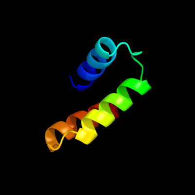
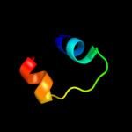
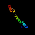
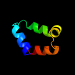
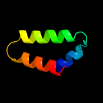
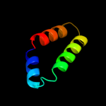
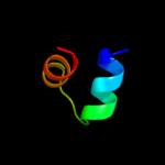
Region: 12 - 60
Aligned: 49
Modelled: 49
Confidence: 42.6%
Identity: 20%
PDB header:gene regulation
Chain: A: PDB Molecule:hypothetical protein ymgb;
PDBTitle: structure and function of the e. coli protein ymgb: a protein critical2 for biofilm formation and acid resistance

Phyre2

PDB 1z05 chain A domain 1

3D model

Region: 16 - 37
Aligned: 22
Modelled: 22
Confidence: 31.6%
Identity: 23%
Fold: DNA/RNA-binding 3-helical bundle
Superfamily: "Winged helix" DNA-binding domain
Family: ROK associated domain

Phyre2

PDB 3a7o chain B

3D model

Region: 2 - 15
Aligned: 14
Modelled: 14
Confidence: 29.8%
Identity: 29%
PDB header:protein transport
Chain: B: PDB Molecule:autophagy protein 16;
PDBTitle: the crystal structure of the coiled-coil domain of2 saccharomyces cerevisiae atg16

Phyre2

PDB 1bia chain A domain 1

3D model

Region: 16 - 37
Aligned: 22
Modelled: 22
Confidence: 25.3%
Identity: 18%
Fold: DNA/RNA-binding 3-helical bundle
Superfamily: "Winged helix" DNA-binding domain
Family: Biotin repressor-like

Phyre2

PDB 1z6r chain A domain 1

3D model

Region: 17 - 43
Aligned: 27
Modelled: 27
Confidence: 23.6%
Identity: 15%
Fold: DNA/RNA-binding 3-helical bundle
Superfamily: "Winged helix" DNA-binding domain
Family: ROK associated domain

Phyre2

PDB 2acv chain A domain 1

3D model

Region: 12 - 62
Aligned: 51
Modelled: 51
Confidence: 23.1%
Identity: 6%
Fold: UDP-Glycosyltransferase/glycogen phosphorylase
Superfamily: UDP-Glycosyltransferase/glycogen phosphorylase
Family: UDPGT-like

Phyre2

PDB 2kzt chain B

3D model

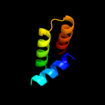
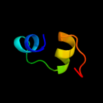
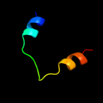
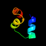
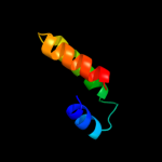
Region: 14 - 60
Aligned: 47
Modelled: 47
Confidence: 23.1%
Identity: 11%
PDB header:apoptosis
Chain: B: PDB Molecule:programmed cell death protein 4;
PDBTitle: structure of the tandem ma-3 region of pdcd4

Phyre2

PDB 3hbj chain A

3D model

Region: 5 - 61
Aligned: 57
Modelled: 57
Confidence: 20.5%
Identity: 16%
PDB header:transferase
Chain: A: PDB Molecule:flavonoid 3-o-glucosyltransferase;
PDBTitle: structure of ugt78g1 complexed with udp

Phyre2

PDB 2yud chain A

3D model

Region: 14 - 38
Aligned: 25
Modelled: 25
Confidence: 20.0%
Identity: 12%
PDB header:rna binding protein
Chain: A: PDB Molecule:yth domain-containing protein 1;
PDBTitle: solution structure of the yth domain in yth domain-2 containing protein 1 (putative splicing factor yt521)

Phyre2

PDB 1ub9 chain A

3D model

Region: 6 - 37
Aligned: 32
Modelled: 32
Confidence: 19.5%
Identity: 22%
Fold: DNA/RNA-binding 3-helical bundle
Superfamily: "Winged helix" DNA-binding domain
Family: MarR-like transcriptional regulators

Phyre2

PDB 2iol chain B

3D model

Region: 14 - 60
Aligned: 47
Modelled: 47
Confidence: 18.4%
Identity: 9%
PDB header:antitumor protein
Chain: B: PDB Molecule:programmed cell death 4, pdcd4;
PDBTitle: crystal structure of the c-terminal ma3 domain of pdcd4 (mouse); form2 1

Phyre2

PDB 2iol chain A

3D model

Region: 14 - 60
Aligned: 47
Modelled: 47
Confidence: 18.4%
Identity: 9%
PDB header:antitumor protein
Chain: A: PDB Molecule:programmed cell death 4, pdcd4;
PDBTitle: crystal structure of the c-terminal ma3 domain of pdcd4 (mouse); form2 1

Phyre2

PDB 1xmk chain A domain 1

3D model

Region: 14 - 47
Aligned: 34
Modelled: 34
Confidence: 15.8%
Identity: 18%
Fold: DNA/RNA-binding 3-helical bundle
Superfamily: "Winged helix" DNA-binding domain
Family: Z-DNA binding domain

Phyre2

PDB 2nsz chain A domain 1

3D model

Region: 14 - 60
Aligned: 47
Modelled: 47
Confidence: 15.6%
Identity: 11%
Fold: alpha-alpha superhelix
Superfamily: ARM repeat
Family: MIF4G domain-like

Phyre2

PDB 2nsz chain A

3D model

Region: 14 - 60
Aligned: 47
Modelled: 47
Confidence: 15.6%
Identity: 11%
PDB header:antitumor protein
Chain: A: PDB Molecule:programmed cell death protein 4;
PDBTitle: 1.15 angstrom crystal structure of the ma3 domain of pdcd4

Phyre2

PDB 2ion chain A

3D model

Region: 14 - 60
Aligned: 47
Modelled: 47
Confidence: 15.6%
Identity: 11%
PDB header:antitumor protein
Chain: A: PDB Molecule:programmed cell death 4, pdcd4;
PDBTitle: crystal structure of the c-terminal ma3 domain of pdcd4 (mouse); form2

Phyre2

PDB 1usd chain A

3D model

Region: 4 - 28
Aligned: 25
Modelled: 25
Confidence: 14.5%
Identity: 24%
PDB header:signaling protein
Chain: A: PDB Molecule:vasodilator-stimulated phosphoprotein;
PDBTitle: human vasp tetramerisation domain l352m

Phyre2

PDB 2hm8 chain A

3D model

Region: 14 - 60
Aligned: 47
Modelled: 47
Confidence: 14.2%
Identity: 11%
PDB header:apoptosis
Chain: A: PDB Molecule:pdcd4 c-terminal ma-3 domain;
PDBTitle: solution structure of the c-terminal ma-3 domain of pdcd4

Phyre2

PDB 3eit chain B

3D model

Region: 23 - 57
Aligned: 35
Modelled: 35
Confidence: 14.0%
Identity: 29%
PDB header:unknown function
Chain: B: PDB Molecule:putative atp/gtp binding protein;
PDBTitle: the 2.6 angstrom crystal structure of chbp, the cif homologue from2 burkholderia pseudomallei

Phyre2

PDB 3eyy chain A

3D model

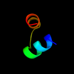
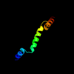
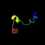
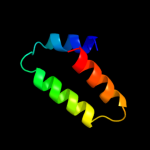
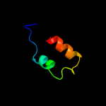
Region: 16 - 38
Aligned: 23
Modelled: 23
Confidence: 13.9%
Identity: 13%
PDB header:transport
Chain: A: PDB Molecule:putative iron uptake regulatory protein;
PDBTitle: structural basis for the specialization of nur, a nickel-2 specific fur homologue, in metal sensing and dna3 recognition

Phyre2
1

c2oxlA_
2

d1z05a1
3

c3a7oB_
4

d1biaa1
5

d1z6ra1
6

d2acva1
7

c2kztB_
8

c3hbjA_
9

c2yudA_
10

d1ub9a_
11

c2iolB_
12

c2iolA_
13

d1xmka1
14

d2nsza1
15

c2nszA_
16

c2ionA_
17

c1usdA_
18

c2hm8A_
19

c3eitB_
20

c3eyyA_
21


22


23


24


25


26


27


28


29


30


31


32


33


34


35


36


37


38


39


40


41


42


43


44


45


46


47


48


Detailed template information 

#
Template Alignment Coverage3D Model Confidence
% i.d. Template Information
1c2oxlA_


42.6 20 PDB header:gene regulation
Chain: A: PDB Molecule:hypothetical protein ymgb;
PDBTitle: structure and function of the e. coli protein ymgb: a protein critical2 for biofilm formation and acid resistance
2d1z05a1


31.6 23 Fold:DNA/RNA-binding 3-helical bundle
Superfamily:"Winged helix" DNA-binding domain
Family:ROK associated domain
3c3a7oB_


29.8 29 PDB header:protein transport
Chain: B: PDB Molecule:autophagy protein 16;
PDBTitle: the crystal structure of the coiled-coil domain of2 saccharomyces cerevisiae atg16
4d1biaa1


25.3 18 Fold:DNA/RNA-binding 3-helical bundle
Superfamily:"Winged helix" DNA-binding domain
Family:Biotin repressor-like
5d1z6ra1


23.6 15 Fold:DNA/RNA-binding 3-helical bundle
Superfamily:"Winged helix" DNA-binding domain
Family:ROK associated domain
6d2acva1


23.1 6 Fold:UDP-Glycosyltransferase/glycogen phosphorylase
Superfamily:UDP-Glycosyltransferase/glycogen phosphorylase
Family:UDPGT-like
7c2kztB_


23.1 11 PDB header:apoptosis
Chain: B: PDB Molecule:programmed cell death protein 4;
PDBTitle: structure of the tandem ma-3 region of pdcd4
8c3hbjA_


20.5 16 PDB header:transferase
Chain: A: PDB Molecule:flavonoid 3-o-glucosyltransferase;
PDBTitle: structure of ugt78g1 complexed with udp
9c2yudA_


20.0 12 PDB header:rna binding protein
Chain: A: PDB Molecule:yth domain-containing protein 1;
PDBTitle: solution structure of the yth domain in yth domain-2 containing protein 1 (putative splicing factor yt521)
10d1ub9a_


19.5 22 Fold:DNA/RNA-binding 3-helical bundle
Superfamily:"Winged helix" DNA-binding domain
Family:MarR-like transcriptional regulators
11c2iolB_


18.4 9 PDB header:antitumor protein
Chain: B: PDB Molecule:programmed cell death 4, pdcd4;
PDBTitle: crystal structure of the c-terminal ma3 domain of pdcd4 (mouse); form2 1
12c2iolA_


18.4 9 PDB header:antitumor protein
Chain: A: PDB Molecule:programmed cell death 4, pdcd4;
PDBTitle: crystal structure of the c-terminal ma3 domain of pdcd4 (mouse); form2 1
13d1xmka1


15.8 18 Fold:DNA/RNA-binding 3-helical bundle
Superfamily:"Winged helix" DNA-binding domain
Family:Z-DNA binding domain
14d2nsza1


15.6 11 Fold:alpha-alpha superhelix
Superfamily:ARM repeat
Family:MIF4G domain-like
15c2nszA_


15.6 11 PDB header:antitumor protein
Chain: A: PDB Molecule:programmed cell death protein 4;
PDBTitle: 1.15 angstrom crystal structure of the ma3 domain of pdcd4
16c2ionA_


15.6 11 PDB header:antitumor protein
Chain: A: PDB Molecule:programmed cell death 4, pdcd4;
PDBTitle: crystal structure of the c-terminal ma3 domain of pdcd4 (mouse); form2
17c1usdA_


14.5 24 PDB header:signaling protein
Chain: A: PDB Molecule:vasodilator-stimulated phosphoprotein;
PDBTitle: human vasp tetramerisation domain l352m
18c2hm8A_


14.2 11 PDB header:apoptosis
Chain: A: PDB Molecule:pdcd4 c-terminal ma-3 domain;
PDBTitle: solution structure of the c-terminal ma-3 domain of pdcd4
19c3eitB_


14.0 29 PDB header:unknown function
Chain: B: PDB Molecule:putative atp/gtp binding protein;
PDBTitle: the 2.6 angstrom crystal structure of chbp, the cif homologue from2 burkholderia pseudomallei
20c3eyyA_


13.9 13 PDB header:transport
Chain: A: PDB Molecule:putative iron uptake regulatory protein;
PDBTitle: structural basis for the specialization of nur, a nickel-2 specific fur homologue, in metal sensing and dna3 recognition
21d2pq6a1


not modelled 13.4 10 Fold:UDP-Glycosyltransferase/glycogen phosphorylase
Superfamily:UDP-Glycosyltransferase/glycogen phosphorylase
Family:UDPGT-like
22d1i1ga1


not modelled 13.2 24 Fold:DNA/RNA-binding 3-helical bundle
Superfamily:"Winged helix" DNA-binding domain
Family:Lrp/AsnC-like transcriptional regulator N-terminal domain
23c3nj2B_


not modelled 13.1 26 PDB header:unknown function
Chain: B: PDB Molecule:duf269-containing protein;
PDBTitle: crystal structure of cce_0566 from the cyanobacterium cyanothece2 51142, a protein associated with nitrogen fixation from the duf2693 family
24c3gqjA_


not modelled 11.8 31 PDB header:unknown function
Chain: A: PDB Molecule:cell inhibiting factor (cif);
PDBTitle: crystal structure of cell inhibiting factor (cif) from photorhabdus2 luminescens
25c1qeyD_


not modelled 10.4 40 PDB header:gene regulation
Chain: D: PDB Molecule:protein (regulatory protein mnt);
PDBTitle: nmr structure determination of the tetramerization domain2 of the mnt repressor: an asymmetric a-helical assembly in3 slow exchange
26c1qeyA_


not modelled 10.4 40 PDB header:gene regulation
Chain: A: PDB Molecule:protein (regulatory protein mnt);
PDBTitle: nmr structure determination of the tetramerization domain2 of the mnt repressor: an asymmetric a-helical assembly in3 slow exchange
27c1qeyC_


not modelled 10.4 40 PDB header:gene regulation
Chain: C: PDB Molecule:protein (regulatory protein mnt);
PDBTitle: nmr structure determination of the tetramerization domain2 of the mnt repressor: an asymmetric a-helical assembly in3 slow exchange
28c1qeyB_


not modelled 10.4 40 PDB header:gene regulation
Chain: B: PDB Molecule:protein (regulatory protein mnt);
PDBTitle: nmr structure determination of the tetramerization domain2 of the mnt repressor: an asymmetric a-helical assembly in3 slow exchange
29d2cfxa1


not modelled 10.2 14 Fold:DNA/RNA-binding 3-helical bundle
Superfamily:"Winged helix" DNA-binding domain
Family:Lrp/AsnC-like transcriptional regulator N-terminal domain
30d1b79a_


not modelled 10.0 13 Fold:N-terminal domain of DnaB helicase
Superfamily:N-terminal domain of DnaB helicase
Family:N-terminal domain of DnaB helicase
31c2veeC_


not modelled 9.6 18 PDB header:transport protein
Chain: C: PDB Molecule:protoglobin;
PDBTitle: structure of protoglobin from methanosarcina acetivorans2 c2a
32c2l4aA_


not modelled 8.6 21 PDB header:dna binding protein
Chain: A: PDB Molecule:leucine responsive regulatory protein;
PDBTitle: nmr structure of the dna-binding domain of e.coli lrp
33c4a1qB_


not modelled 8.6 38 PDB header:viral protein
Chain: B: PDB Molecule:orf e73;
PDBTitle: solution structure of e73 protein from sulfolobus spindle-2 shaped virus ragged hills, a hyperthermophilic3 crenarchaeal virus from yellowstone national park
34d1q1va_


not modelled 8.3 12 Fold:Another 3-helical bundle
Superfamily:DEK C-terminal domain
Family:DEK C-terminal domain
35c2ggfA_


not modelled 7.6 9 PDB header:translation
Chain: A: PDB Molecule:programmed cell death 4, isoform 1;
PDBTitle: solution structure of the ma3 domain of human programmed2 cell death 4
36d2gxba1


not modelled 7.5 22 Fold:DNA/RNA-binding 3-helical bundle
Superfamily:"Winged helix" DNA-binding domain
Family:Z-DNA binding domain
37d1qo0d_


not modelled 7.4 13 Fold:Flavodoxin-like
Superfamily:CheY-like
Family:Positive regulator of the amidase operon AmiR
38d2g7ga1


not modelled 7.3 21 Fold:DNA/RNA-binding 3-helical bundle
Superfamily:Homeodomain-like
Family:Tetracyclin repressor-like, N-terminal domain
39c1zrwA_


not modelled 7.3 50 PDB header:antimicrobial protein, antibiotic
Chain: A: PDB Molecule:spinigerin;
PDBTitle: solution structure of spinigerin in h20/tfe 10%
40c1zrvA_


not modelled 7.3 50 PDB header:antimicrobial protein, antibiotic
Chain: A: PDB Molecule:spinigerin;
PDBTitle: solution structure of spinigerin in h20/tfe 50%
41c2ewnA_


not modelled 7.1 20 PDB header:ligase, transcription
Chain: A: PDB Molecule:bira bifunctional protein;
PDBTitle: ecoli biotin repressor with co-repressor analog
42d2ev0a1


not modelled 6.9 9 Fold:DNA/RNA-binding 3-helical bundle
Superfamily:"Winged helix" DNA-binding domain
Family:Iron-dependent repressor protein
43d2iu5a1


not modelled 6.4 15 Fold:DNA/RNA-binding 3-helical bundle
Superfamily:Homeodomain-like
Family:Tetracyclin repressor-like, N-terminal domain
44d2cg4a1


not modelled 6.4 21 Fold:DNA/RNA-binding 3-helical bundle
Superfamily:"Winged helix" DNA-binding domain
Family:Lrp/AsnC-like transcriptional regulator N-terminal domain
45d1ixsa_


not modelled 6.1 35 Fold:RuvA C-terminal domain-like
Superfamily:DNA helicase RuvA subunit, C-terminal domain
Family:DNA helicase RuvA subunit, C-terminal domain
46c2l5yA_


not modelled 5.8 29 PDB header:signaling protein
Chain: A: PDB Molecule:stromal interaction molecule 2;
PDBTitle: nmr structure of calcium-loaded stim2 ef-sam.
47d2cyya1


not modelled 5.7 17 Fold:DNA/RNA-binding 3-helical bundle
Superfamily:"Winged helix" DNA-binding domain
Family:Lrp/AsnC-like transcriptional regulator N-terminal domain
48d1zoqc1


not modelled 5.3 40 Fold:Nuclear receptor coactivator interlocking domain
Superfamily:Nuclear receptor coactivator interlocking domain
Family:Nuclear receptor coactivator interlocking domain

Binding site prediction 

Due to computational demand, binding site predictions are not run for batch jobs

If you want to predict binding sites, please manually submit your model of choice to 3DLigandSite


Phyre is for academic use only

Please cite: Protein structure prediction on the web: a case study using the Phyre server
Kelley LA and Sternberg MJE. Nature Protocols 4, 363 - 371 (2009) [pdf] [Import into BibTeX]
 
If you use the binding site predictions from 3DLigandSite, please also cite:
3DLigandSite: predicting ligand-binding sites using similar structures.
Wass MN, Kelley LA and Sternberg MJ Nucleic Acids Research 38, W469-73 (2010) [PubMed]
 
© Structural Bioinformatics Group
Imperial College London
Lawrence Kelley, Benjamin Jefferys
Disclaimer
Terms and Conditions
Component software
Template detection: HHpred 1.51
Secondary structure prediction: Psi-pred 2.5
Disorder prediction: Disopred 2.4
Transmembrane prediction: Memsat_SVM
Multi-template modelling and ab initio: Poing 1.0