| Secondary structure and disorder prediction | |
 |
| | |
1 | . | . | . | . | . | . | . | . | 10 | . | . | . | . | . | . | . | . | . | 20 | . | . | . | . | . | . | . | . | . | 30 | . | . | . | . | . | . | . | . | . | 40 | . | . | . | . | . | . | . | . | . | 50 | . | . | . | . | . | . | . | . | . | 60 |
| Sequence | |
M | F | H | R | L | L | I | A | T | V | V | G | I | L | A | A | F | A | V | A | G | F | R | H | A | M | L | L | L | E | W | L | F | L | N | N | D | S | G | S | L | V | N | A | A | T | N | L | S | P | W | R | R | L | L | T | P | A | L | G |
| Secondary structure | |
|  | | | | | | | | | | | | | | | | | | | | | | | | | | | | | | | |
|
|
|
|
|
|
|
|
|
| | |
|
|
|
| | | | | | | | | | | |
| SS confidence | |
|
|
|
|
|
|
|
|
|
|
|
|
|
|
|
|
|
|
|
|
|
|
|
|
|
|
|
|
|
|
|
|
|
|
|
|
|
|
|
|
|
|
|
|
|
|
|
|
|
|
|
|
|
|
|
|
|
|
|
|
| Disorder | |
? |
|
|
|
|
|
|
|
|
|
|
|
|
|
|
|
|
|
|
|
|
|
|
|
|
|
|
|
|
|
|
|
|
|
|
|
|
| ? | ? | ? |
| ? |
|
|
|
|
|
|
|
|
|
|
|
|
|
|
|
|
|
| Disorder confidence | |
|
|
|
|
|
|
|
|
|
|
|
|
|
|
|
|
|
|
|
|
|
|
|
|
|
|
|
|
|
|
|
|
|
|
|
|
|
|
|
|
|
|
|
|
|
|
|
|
|
|
|
|
|
|
|
|
|
|
|
|
| |
| | |
. | . | . | . | . | . | . | . | . | 70 | . | . | . | . | . | . | . | . | . | 80 | . | . | . | . | . | . | . | . | . | 90 | . | . | . | . | . | . | . | . | . | 100 | . | . | . | . | . | . | . | . | . | 110 | . | . | . | . | . | . | . | . | . | 120 |
| Sequence | |
G | L | A | A | G | L | L | L | M | G | W | Q | K | F | T | Q | Q | R | P | H | A | P | T | D | Y | M | E | A | L | Q | T | D | G | Q | F | D | Y | A | A | S | L | V | K | S | L | A | S | L | L | V | V | T | S | G | S | A | I | G | R | E |
| Secondary structure | |
| | | | | | | | | | |
|
|
|
|
|
|
|
|
|
| | | | | | | | | |
|
|
|
|
|
| | | | | | | | | | | | | | | | | |
|
|
|
|
|
|
|
| SS confidence | |
|
|
|
|
|
|
|
|
|
|
|
|
|
|
|
|
|
|
|
|
|
|
|
|
|
|
|
|
|
|
|
|
|
|
|
|
|
|
|
|
|
|
|
|
|
|
|
|
|
|
|
|
|
|
|
|
|
|
|
|
| Disorder | |
|
|
|
|
|
|
|
|
|
|
|
| ? | ? | ? | ? | ? | ? | ? | ? | ? |
|
|
|
|
|
|
|
|
|
|
|
|
|
|
|
|
|
|
|
|
|
|
|
|
|
|
|
|
|
|
|
|
|
|
|
|
|
|
|
| Disorder confidence | |
|
|
|
|
|
|
|
|
|
|
|
|
|
|
|
|
|
|
|
|
|
|
|
|
|
|
|
|
|
|
|
|
|
|
|
|
|
|
|
|
|
|
|
|
|
|
|
|
|
|
|
|
|
|
|
|
|
|
|
|
| |
| | |
. | . | . | . | . | . | . | . | . | 130 | . | . | . | . | . | . | . | . | . | 140 | . | . | . | . | . | . | . | . | . | 150 | . | . | . | . | . | . | . | . | . | 160 | . | . | . | . | . | . | . | . | . | 170 | . | . | . | . | . | . | . | . | . | 180 |
| Sequence | |
G | A | M | I | L | L | A | A | L | A | A | S | C | F | A | Q | R | F | T | P | R | Q | E | W | K | L | W | I | A | C | G | A | A | A | G | M | A | A | A | Y | R | A | P | L | A | G | S | L | F | I | A | E | V | L | F | G | T | M | M | L |
| Secondary structure | |
| | | | | | | | | | | | | | | | | |
|
| | | | | | | | | | | | | | | | | | | |
|
|
|
| | | | | | | | | | | | | | |
|
| |
| SS confidence | |
|
|
|
|
|
|
|
|
|
|
|
|
|
|
|
|
|
|
|
|
|
|
|
|
|
|
|
|
|
|
|
|
|
|
|
|
|
|
|
|
|
|
|
|
|
|
|
|
|
|
|
|
|
|
|
|
|
|
|
|
| Disorder | |
|
|
|
|
|
|
|
|
|
|
|
|
|
|
|
|
|
|
|
|
|
|
|
|
|
|
|
|
|
|
|
|
|
|
|
|
|
|
|
|
|
|
|
|
|
|
|
|
|
|
|
|
|
|
|
|
|
|
|
|
| Disorder confidence | |
|
|
|
|
|
|
|
|
|
|
|
|
|
|
|
|
|
|
|
|
|
|
|
|
|
|
|
|
|
|
|
|
|
|
|
|
|
|
|
|
|
|
|
|
|
|
|
|
|
|
|
|
|
|
|
|
|
|
|
|
| |
| | |
. | . | . | . | . | . | . | . | . | 190 | . | . | . | . | . | . | . | . | . | 200 | . | . | . | . | . | . | . | . | . | 210 | . | . | . | . | . | . | . | . | . | 220 | . | . | . | . | . | . | . | . | . | 230 | . | . | . | . | . | . | . | . | . | 240 |
| Sequence | |
A | S | L | G | P | V | I | I | S | A | V | V | A | L | L | V | S | N | L | I | N | H | S | D | A | L | L | Y | N | V | Q | L | S | V | T | V | Q | A | R | D | Y | A | L | I | I | S | T | G | V | L | A | G | L | C | G | P | L | L | L | T |
| Secondary structure | |
| | | | | | | | | | | | | | | | | | | |
|
|
|
|
|
|
|
|
|
|
|
|
|
|
|
|
| | | | | | | | | | | | | | | | | | | | | | | |
| SS confidence | |
|
|
|
|
|
|
|
|
|
|
|
|
|
|
|
|
|
|
|
|
|
|
|
|
|
|
|
|
|
|
|
|
|
|
|
|
|
|
|
|
|
|
|
|
|
|
|
|
|
|
|
|
|
|
|
|
|
|
|
|
| Disorder | |
|
|
|
|
|
|
|
|
|
|
|
|
|
|
|
|
|
|
|
|
|
|
|
|
|
|
|
|
|
|
| ? |
|
|
|
|
|
|
|
|
|
|
|
|
|
|
|
|
|
|
|
|
|
|
|
|
|
|
|
|
| Disorder confidence | |
|
|
|
|
|
|
|
|
|
|
|
|
|
|
|
|
|
|
|
|
|
|
|
|
|
|
|
|
|
|
|
|
|
|
|
|
|
|
|
|
|
|
|
|
|
|
|
|
|
|
|
|
|
|
|
|
|
|
|
|
| |
| | |
. | . | . | . | . | . | . | . | . | 250 | . | . | . | . | . | . | . | . | . | 260 | . | . | . | . | . | . | . | . | . | 270 | . | . | . | . | . | . | . | . | . | 280 | . | . | . | . | . | . | . | . | . | 290 | . | . | . | . | . | . | . | . | . | 300 |
| Sequence | |
L | M | N | A | C | H | R | G | F | V | S | L | K | L | A | P | P | W | Q | L | A | L | G | G | L | I | V | G | L | L | S | L | F | T | P | A | V | W | G | N | G | Y | S | T | V | Q | S | F | L | T | A | P | P | L | L | M | I | I | A | G |
| Secondary structure | |
| | | | | | | | | | |
|
|
|
|
|
| | | | | | | | | | | | | | | | | | | | | |
|
|
| | | | | | | | |
|
|
|
|
| | | | | | |
| SS confidence | |
|
|
|
|
|
|
|
|
|
|
|
|
|
|
|
|
|
|
|
|
|
|
|
|
|
|
|
|
|
|
|
|
|
|
|
|
|
|
|
|
|
|
|
|
|
|
|
|
|
|
|
|
|
|
|
|
|
|
|
|
| Disorder | |
|
|
|
|
|
|
|
|
|
|
|
|
|
|
|
|
|
|
|
|
|
|
|
|
|
|
|
|
|
|
|
|
|
|
|
|
|
|
|
|
|
|
|
|
|
|
|
|
|
|
|
|
|
|
|
|
|
|
|
|
| Disorder confidence | |
|
|
|
|
|
|
|
|
|
|
|
|
|
|
|
|
|
|
|
|
|
|
|
|
|
|
|
|
|
|
|
|
|
|
|
|
|
|
|
|
|
|
|
|
|
|
|
|
|
|
|
|
|
|
|
|
|
|
|
|
| |
| | |
. | . | . | . | . | . | . | . | . | 310 | . | . | . | . | . | . | . | . | . | 320 | . | . | . | . | . | . | . | . | . | 330 | . | . | . | . | . | . | . | . | . | 340 | . | . | . | . | . | . | . | . | . | 350 | . | . | . | . | . | . | . | . | . | 360 |
| Sequence | |
I | F | L | C | K | L | C | A | V | L | A | S | S | G | S | G | A | P | G | G | V | F | T | P | T | L | F | I | G | L | A | I | G | M | L | Y | G | R | S | L | G | L | W | F | P | D | G | E | E | I | T | L | L | L | G | L | T | G | M | A |
| Secondary structure | |
| | | | | | | | | | | | | |
|
|
|
|
| | | | | | | | | | | | | | | | | | | | | | | | |
|
|
|
|
|
|
| | | | | | | | | | |
| SS confidence | |
|
|
|
|
|
|
|
|
|
|
|
|
|
|
|
|
|
|
|
|
|
|
|
|
|
|
|
|
|
|
|
|
|
|
|
|
|
|
|
|
|
|
|
|
|
|
|
|
|
|
|
|
|
|
|
|
|
|
|
|
| Disorder | |
|
|
|
|
|
|
|
|
|
|
|
|
|
|
|
|
|
|
|
|
|
|
|
|
|
|
|
|
|
|
|
|
|
|
|
|
|
|
|
|
|
|
|
|
|
|
|
|
|
|
|
|
|
|
|
|
|
|
|
|
| Disorder confidence | |
|
|
|
|
|
|
|
|
|
|
|
|
|
|
|
|
|
|
|
|
|
|
|
|
|
|
|
|
|
|
|
|
|
|
|
|
|
|
|
|
|
|
|
|
|
|
|
|
|
|
|
|
|
|
|
|
|
|
|
|
| |
| | |
. | . | . | . | . | . | . | . | . | 370 | . | . | . | . | . | . | . | . | . | 380 | . | . | . | . | . | . | . | . | . | 390 | . | . | . | . | . | . | . | . | . | 400 | . | . | . | . | . | . | . | . | . | 410 | . | . | . | . | . | . | . | . |
| Sequence | |
T | L | L | A | A | T | T | H | A | P | I | M | S | T | L | M | I | C | E | M | T | G | E | Y | Q | L | L | P | G | L | L | I | A | C | V | I | A | S | V | I | S | R | T | L | H | R | D | S | I | Y | R | Q | H | T | A | Q | H | S |
| Secondary structure | |
| | | | | | |
|
| | | | | | | | | | | | |
|
| | | | | | | | | | | | | | | | | | | | | |
|
|
|
|
|
| | | | | | |
|
|
| SS confidence | |
|
|
|
|
|
|
|
|
|
|
|
|
|
|
|
|
|
|
|
|
|
|
|
|
|
|
|
|
|
|
|
|
|
|
|
|
|
|
|
|
|
|
|
|
|
|
|
|
|
|
|
|
|
|
|
|
|
|
| Disorder | |
|
|
|
|
|
|
|
|
|
|
|
|
|
|
|
|
|
|
|
|
|
|
|
|
|
|
|
|
|
|
|
|
|
|
|
|
|
|
|
|
|
|
|
|
|
|
|
|
|
|
|
|
|
| ? | ? | ? | ? |
| Disorder confidence | |
|
|
|
|
|
|
|
|
|
|
|
|
|
|
|
|
|
|
|
|
|
|
|
|
|
|
|
|
|
|
|
|
|
|
|
|
|
|
|
|
|
|
|
|
|
|
|
|
|
|
|
|
|
|
|
|
|
|
| |
| Confidence Key |
| High(9) | |
|
|
|
|
|
|
|
|
|
Low (0) |
| ? | Disordered |
| Alpha helix |
  | Beta strand |
Hover over an aligned region to see model and summary info
Please note, only up to the top 20 hits are modelled to reduce computer load
|
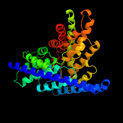
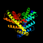
| 1 |
|
PDB 2ht2 chain B
Region: 1 - 416
Aligned: 411
Modelled: 416
Confidence: 100.0%
Identity: 24%
PDB header:membrane protein
Chain: B: PDB Molecule:h(+)/cl(-) exchange transporter clca;
PDBTitle: structure of the escherichia coli clc chloride channel2 y445h mutant and fab complex
Phyre2
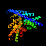
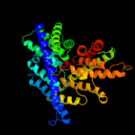
| 2 |
|
PDB 1ots chain A
Region: 1 - 417
Aligned: 412
Modelled: 417
Confidence: 100.0%
Identity: 24%
Fold: Clc chloride channel
Superfamily: Clc chloride channel
Family: Clc chloride channel
Phyre2
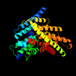
| 3 |
|
PDB 1kpl chain A
Region: 1 - 417
Aligned: 412
Modelled: 417
Confidence: 100.0%
Identity: 24%
Fold: Clc chloride channel
Superfamily: Clc chloride channel
Family: Clc chloride channel
Phyre2
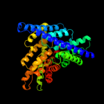
| 4 |
|
PDB 3nd0 chain A
Region: 3 - 416
Aligned: 409
Modelled: 414
Confidence: 100.0%
Identity: 24%
PDB header:transport protein
Chain: A: PDB Molecule:sll0855 protein;
PDBTitle: x-ray crystal structure of a slow cyanobacterial cl-/h+ antiporter
Phyre2
| 5 |
|
PDB 3org chain B
Region: 3 - 417
Aligned: 384
Modelled: 400
Confidence: 100.0%
Identity: 20%
PDB header:transport protein
Chain: B: PDB Molecule:cmclc;
PDBTitle: crystal structure of a eukaryotic clc transporter
Phyre2
| 6 |
|
PDB 2k47 chain A
Region: 116 - 145
Aligned: 30
Modelled: 30
Confidence: 13.8%
Identity: 17%
PDB header:replication
Chain: A: PDB Molecule:phosphoprotein;
PDBTitle: solution structure of the c-terminal n-rna binding domain2 of the vesicular stomatitis virus phosphoprotein
Phyre2
| 7 |
|
PDB 2knc chain A
Region: 222 - 256
Aligned: 35
Modelled: 35
Confidence: 10.6%
Identity: 23%
PDB header:cell adhesion
Chain: A: PDB Molecule:integrin alpha-iib;
PDBTitle: platelet integrin alfaiib-beta3 transmembrane-cytoplasmic2 heterocomplex
Phyre2
|
| Detailed template information | |
Due to computational demand, binding site predictions are not run for batch jobs
If you want to predict binding sites, please manually submit your model of choice to 3DLigandSite
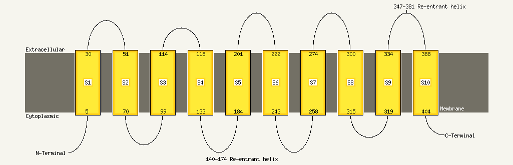
| Transmembrane helix prediction | |
Transmembrane helices have been predicted in your sequence to adopt the topology shown below
Phyre is for academic use only
| Please cite: Protein structure prediction on
the web: a case study using the Phyre server |
| Kelley LA and Sternberg MJE. Nature Protocols
4, 363 - 371 (2009) [pdf] [Import into BibTeX] |
| |
| If you use the binding site
predictions from 3DLigandSite, please also cite: |
| 3DLigandSite: predicting ligand-binding sites using similar structures. |
| Wass MN, Kelley LA and Sternberg
MJ Nucleic Acids Research 38, W469-73 (2010) [PubMed] |
| |
|
|
|
|